20 June 2023: Clinical Research
Impact of Otosclerosis on Auditory Ossicle Remodeling: A Scanning Electron Microscopy Analysis of Stapes Head Overloads
Agnieszka WiatrDOI: 10.12659/MSM.939679
Med Sci Monit 2023; 29:e939679


Abstract
BACKGROUND: Otosclerosis is a pathology that interferes with the conduction of vibrations to the inner ear, triggering changes in the auditory ossicles and their associated joints due to mechanical overload. This study primarily aims to evaluate these overload-induced modifications in the stapes head resulting from the immobilization of the base of the third auditory ossicle in otosclerosis patients.
MATERIAL AND METHODS: We conducted a comparative analysis of patients undergoing their first surgery for otosclerosis. The test group consisted of 31 patients who underwent stapedotomy between 2020-2021. For comparison, we utilized a control group comprising stapes samples extracted during vestibular schwannoma surgeries via a transcochlear approach. A prospective analysis of bone tissue surface topography and chemical composition was executed using scanning electron microscopy (SEM).
RESULTS: SEM analysis of the stapes head in otosclerosis patients relative to the control group displayed no significant differences in chemical composition or the presence of otosclerotic foci. Nonetheless, various forms of bone tissue surface damage were noted on the stapes head in all otosclerosis patients. Mild changes were evident in 90% of the samples, while small linear bone tissue fractures were observed in 58% of the samples. Furthermore, minor osteophytic changes were detected in 16% of the samples.
CONCLUSIONS: The immobilization of the stapes base by otosclerotic foci instigates overloads in the incus-stapes joint, leading to the eventual remodeling of the stapes head articular surface.
Keywords: Cartilage, Articular, Microscopy, Electron, Scanning, Otosclerosis, Photoelectron Spectroscopy, Stapes, Humans, Ear Ossicles, Stapes Surgery
Background
Hearing loss is a disease of heterogeneous etiology. More than 6% of the world’s population lives with hearing impairment, which can adversely affect development, education, and general health [1]. One of the related causes of acquired hearing impairment is otosclerosis.
Otosclerosis is a primary disease of multifactorial etiology, mostly involving the process of bone tissue remodeling in various areas of the temporal bone. Vascular proliferation, bone resorption, and the formation of new bone tissue observed histologically most often concern the area of the oval window in the immediate vicinity of the fissula ante fenestram. As the disease progresses, the pathological process causes the base of the stapes to become immobilized in the oval window, disrupting the process of conducting vibrations to the inner ear and changing the distribution of forces during vibrations, leading to overload changes in the ossicles and in the joints between the auditory ossicles. As a result of this process, conductive hearing loss occurs clinically. The coexistence of the disease process in the area of the oval window and the inner ear leads to mixed hearing loss. The presence of the disease process only in the structures of the inner ear causes sensorineural hearing loss.
The main clinical symptom in patients with otosclerosis is progressive hearing loss, which may be accompanied by tinnitus and vertigo [2–4].
Under normal conditions, sound waves reaching the eardrum causes its vibration, which transforms the acoustic wave into mechanical vibrations. Vibrations of the eardrum are transmitted to the ossicular complex and then through the stapes footplate to the oval window. The main component of mechanical transmission consists of 2 surfaces – the eardrum and the foot of the stapes – thus the sound pressure is proportional to the force per unit area. An important element of the auditory pathway is the ossicular chain.
The auditory ossicles connect with the walls of the tympanic cavity by means of connective-tissue ligament strands, while they are connected with each other through the incus-malleus joint and the incus-stapes joint. However, these are not joints in the strict sense of the word, but rather cartilage hyperplasia. Due to these connections, the mobility of the bones in relation to each other is very small. Disrupting the work of even a single element of the ossicles chain disrupts the distribution of forces between the auditory ossicles, leading to hearing loss [5,6].
The aim of the study was to analyze the degree of damage to the stapes head as a component of the incus-stapes joint in the course of window otosclerosis. Therefore, this prospective study of patients with otosclerosis aimed to assess overload changes in the stapes head secondary to immobilization of the base of the third auditory ossicle.
Material and Methods
ETHICS STATEMENT:
The study was approved by the Bioethics Committee (no. 1072.6120.311.2020). All participants gave their informed consent to participate.
MATERIAL:
A prospective analysis included 31 patients operated on for otosclerosis in the years 2020–2021 and 5 patients operated on for a vestibular schwannoma. Thirty-one women and 5 men participated in the study. The youngest patient was 18 and the oldest 57 years old. The average age was 35.
GROUPS OF PATIENTS:
The patients were divided into 2 groups:
METHODS:
The removed stapes suprastructures were examined by scanning electron microscopy (SEM). Healthy stapes that were surgical waste during surgery for a vestibular schwannoma were compared with stapes removed in the course of the otosclerotic process. The SEM assessment excluded damage to the articular surfaces resulting from the surgical methodology.
The surgical material for evaluation in the scanning electron microscope was prepared according to a standard protocol. The study was carried out using scanning electron microscopy (QUANTA 3D FEG SEM electron microscope) with a variable vacuum in the range of 0.3–0.6 torr. The study used electron beam accelerating voltages of 15 and 20 kV, electron beam currents of 5.66, 11.3, and 23 nA and working distances (WDs) (sample surface – objective lens pole) in the 8 to 14.6 mm range.
The LVSED (low-vacuum secondary electron detector) was used to record the bone tissue surface topography using secondary electrons (SE – secondary electron), and the backscattered electron (BSE) signal was recorded using a vCD (low voltage-high contrast detector).
The EDS Genesis X-ray detector was used for the preliminary qualitative analysis of the chemical composition in the microareas of the stapes bones, and the PhiRoZet software was used for quantitative analysis.
The chemical composition analysis was performed using the Raymond Gauvin (Variable Pressure method) correction procedure, measuring 2 spectra at 2 different CO2 pressures and then extrapolating the maximum values of individual peaks to zero gas pressure.
SEM using different magnifications revealed changes repetitively present only in the course of immobilization of the auditory ossicular chain. To assess the changes in the articular surface of the stapes observed in the course of otosclerosis, the authors proposed their division depending on the magnification used, taking as a criterion the observed changes up to and above magnification of 250x.
When assessing the extent of damage to the articular surface of the stapes head, lesions were characterized as benign or distinct:
All preparations were evaluated at 50×, 150×, 250×, 350×, and 500× magnification.
STATISTICAL ANALYSIS:
For statistical analysis, descriptive statistics were used to describe the characteristics of the studied group: mean, median, and standard deviation (SD), and the values of the first and third quartiles (IQR) and range were calculated.
The normality of the distribution of individual parameters assessed in the study was checked with the Shapiro-Wilk test.
The Mann-Whitney or Kruskal-Wallis U test was used for data with no normal distribution. The Dunn pairs test was used to compare individual groups.
To assess whether there were differences between the results of particular analyzed variables in particular periods (dependent variables) in the study group, the nonparametric Wilcoxon test or
To investigate the existence of monotonic relationships between the variables, the Spearman correlation coefficient was used. A
RESULTS:
Thirty-one stapes preparations with various degrees of advancement of the otosclerotic process were analyzed, as well as 5 stapes preparations without otosclerosis removed during the operation of the vestibular schwannoma, which constituted the control group. The analysis included 36 patients (31 women and 5 men). In patients operated on due to otosclerosis, the duration of disease symptoms ranged from 2 to 15 years, with a mean of 6.5 years. The average age was 35 years.
RESULTS OF THE ANALYSIS OF THE ELEMENTAL COMPOSITION OF THE STAPES HEAD IN THE STUDY GROUP AND THE CONTROL GROUP:
In the first stage, the chemical composition of the stapes head from the control group (Group 0) was analyzed, and then the identical procedure was performed on preparations from Group I. The results for the analyzed calcium, potassium, oxygen, carbon, sodium, and sulfur elements are presented in Table 1. A sample graph of the elemental composition obtained from preparations from the control group and Group I is shown in Figures 1 and 2.
The bone tissue constituting the stapes heads was identified in both groups as hydroxyapatite with a predominance of calcium hydroxyphosphate with a Ca/P ratio of 1.667–1.691. There was no significant difference in the Ca to P ratio calculated on the basis of the elemental composition between the control group and Group I. This proves an identical composition of the stapes head bone tissue both in the otosclerosis group (Group 1) and in the control group (Group 0).
The conducted analysis did not show a significant difference in the quantitative composition of the remaining elements (oxygen, carbon, sodium, and sulfur) between Group I and Group 0 (P<0.05) (Table 1).
Based on the assessment of the surface image in SEM and the elemental composition, the presence of otosclerotic foci in the preparations of the stapes head belonging to Group I was excluded.
THE RESULTS OF OBSERVATION OF THE SURFACE OF THE STAPES HEAD OBTAINED IN THE SCANNING ELECTRON MICROSCOPE IN THE STUDY GROUP AND THE CONTROL GROUP:
Then, the surface of the stapes head was analyzed with a scanning electron microscope at the magnification specified in the methodology. An example of the type of image obtained in the scanning electron microscope is shown in Figure 3 for the control group and Figures 4–6 for Group I.
SEM analysis of the surface of the stapes head obtained from the specimens belonging to the control group showed no damage to the bony surface of the head at any of the magnifications, and no exophytic changes in the articular surface of the head were observed (Figure 3).
In Group I, in all preparations obtained from patients diagnosed with otosclerosis, various types of damage to the surface of the bone tissue of the stapes head were observed (Figure 4).
In this group, mild changes, such as small exfoliations of the articular surface, were observed in 28 cases (which constitutes 90% of the preparations). Changes of this nature were observed at the highest magnification (500x). Additionally, small linear fractures of the bone tissue were observed in 18 patients, which constituted 58% of the preparations (Figure 5). In 5 (16%) preparations, in addition to the previously described changes, minor osteophytic changes were found (Figure 6).
We did not observe the presence of bacteria or fungi (as biofilm or free plankton) on the surface of the stapes suprastructure in any of the analyzed preparations.
Discussion
On the basis of the elemental analysis of the stapes head in Groups 0 and 1, no significant differences in the chemical composition were found, while the influence of bone remodeling as a result of the otosclerotic process on damage to the stapes head was excluded.
The immobilization of the base of the stapes in the oval window as a result of the development of otosclerotic foci in this area contributes to stiffness of the ossicular chain and inhibits effective vibration. The mechanics of forces spreading along the auditory ossicles chain at the immobilized base of the stapes cause overloads in the joints. According to our observations, overloading the joint, mainly in the area of the incus-stapes joint, causes damage to the surface of the stapes head in the form of small linear cracks, peeling, and minor changes [7].
The changes observed in our material concerned only the surface of the stapes head. The second surface of the incus-stapes joint (ie, the end of the long incus process) was not tested due to the methodology of the operating procedure.
Georg Bekesy described the movement of the stapes in 1936, presenting it as rotating along an axis passing through the rear part of the stapes plate, with the greatest deflection observed in the anterior part, which according to the anatomy of the annular ligament is wider but thinner in the front part of the stapes plate [8]. Kirikae observed that stapes movement is a combination of compression and hinge movement [9]. Goode and Nishihara examined and described movement of the stapes of up to 2 kHz as compressions, and anything above this frequency as complex with an unknown pattern [10]. In 1986, Vlaming and Feenstra, using the laser Doppler vibrometry method, measured the deflection of the stapes footplate, and determined the movement of the stapes footplate as compression [11]. In the following year, Goode’s research provided further interesting information on the mechanism of ossicular chain mobility, concluding that the navel of the tympanic membrane and the lenticular process of the incus vibrate like a piston up to 1 kHz, and at higher frequencies a rotational component also appears [12].
There is no advanced research on this topic in the literature. A study by Clarke-Brodber and Taxy described mild osteoarthritis involving the head of the stapes, which, according to the authors, probably occurs as a result of persistent oscillations of the auditory ossicles transferred to the immobilized base. The most common changes were fissures in the articular surface of the stapes head (65%), cartilage erosion (49%), and surface irregularities. Osteophytes have been observed sporadically [13]. These observations are in line with our research.
Our observations provide additional information on the depth of conductive hearing loss in patients with otosclerosis, pointing to factors other than immobilization of the stapes base due to the formation of an air-bone gap.
Regardless of the progressing otosclerotic process, disturbance of the mobility of the auditory ossicle chain through overloading leads to damage to the articular surfaces of the auditory ossicles, which may be a factor limiting improvement of hearing after surgical treatment.
The term “overload changes” proposed by the authors best describes the abnormalities of the stapes head articular surface observed in the SEM image. Damage to the articular surface of the stapes head is the result of increasing the pressure at the junction of the immobilized elements of the ossicular chain in the course of otosclerosis in the absence of other visible factors damaging these ossicles.
In further studies, it will be important to determine the effect of the duration of otosclerosis on damage to the articular surfaces. Planned scientific research will indicate whether the delay in surgical treatment adversely affects the joint connections between the auditory ossicles.
Due to the methodology of the surgical procedure in otosclerosis, there is no possibility of intravital assessment of the condition of the long process of the incus, which serves as a fastening for placing the prosthesis during stapedotomy.
A further limitation of the conducted research resulting from the methodology, the operated site, and the size of the assessed structures is the inability to assess the elements of the incus and stapes joint with other high-resolution imaging techniques and the histological assessment of the articular cartilage.
Conclusions
Analysis of the stapes head using SEM in patients with otosclerosis compared to the control group did not reveal differences in chemical composition or the presence of otosclerotic foci. Immobilization of the base of the stapes by otosclerotic foci leads to overloads in the incus and stapes joint and ultimately causes remodeling of the articular surface of the stapes head.
Figures
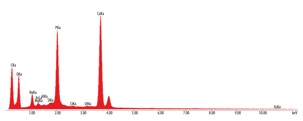 Figure 1. Control group. Result of the chemical composition of the stapes head. CaKa – calcium; PKa – phosphorus; OKa – oxygen; CKa – carbon; NaKa – sodium; SKa – sulfur. Prepared using CorelDraw9, Corel Corporation.
 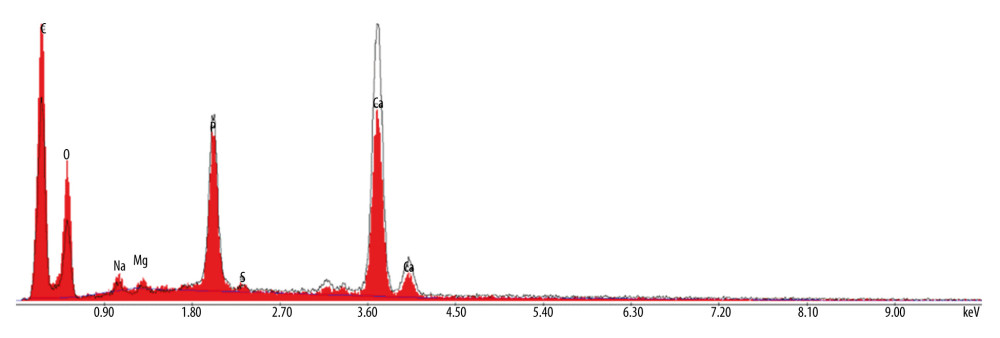 Figure 2. Otosclerosis. Result of the chemical composition of the stapes head. Ca – calcium; P – phosphorus; O – oxygen; C – carbon; Na – sodium; S – sulfur. Prepared using CorelDraw9, Corel Corporation.
 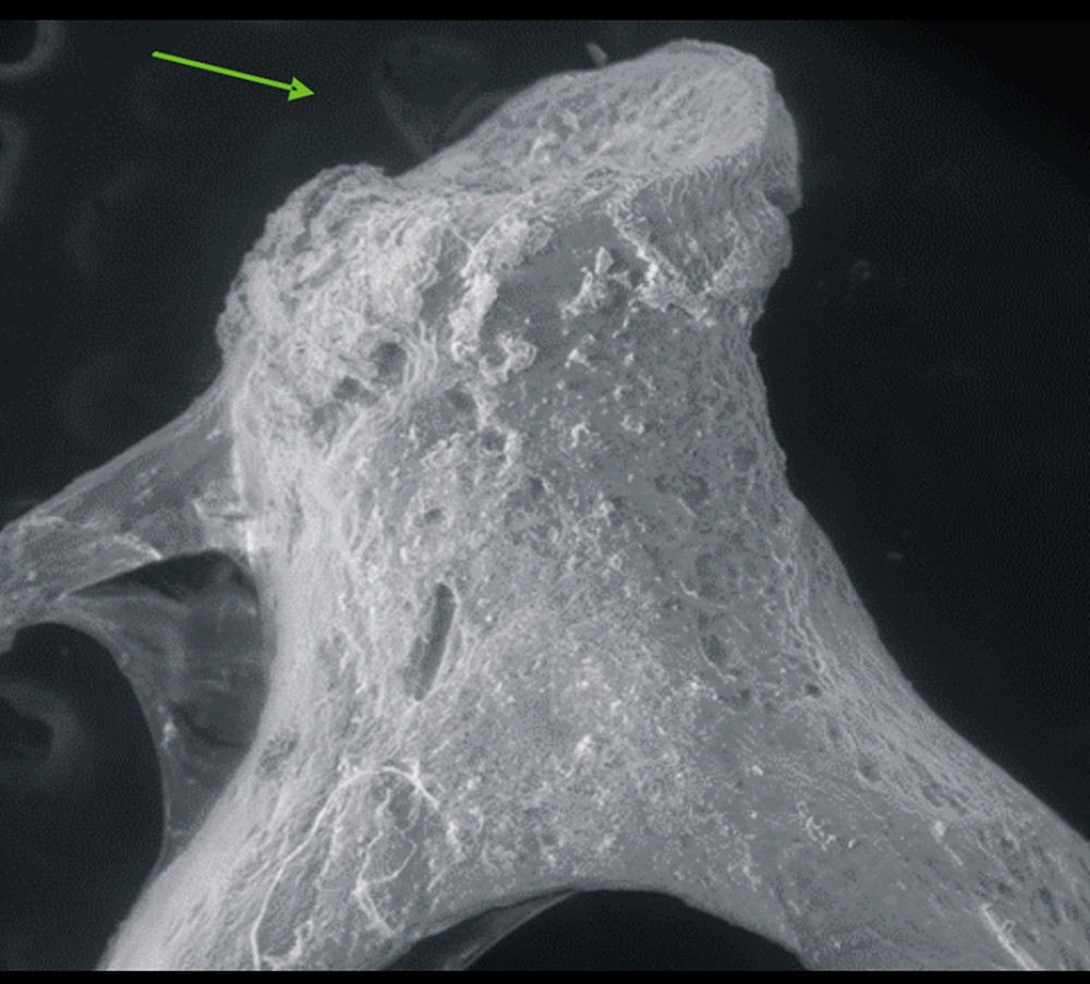 Figure 3. Control group. The head of the stapes (green arrow). Magnification 150×. LVSED (low vacuum secondary electron detector). Workshop of Scanning Microscopy. Prepared using CorelDraw9, Corel Corporation.
 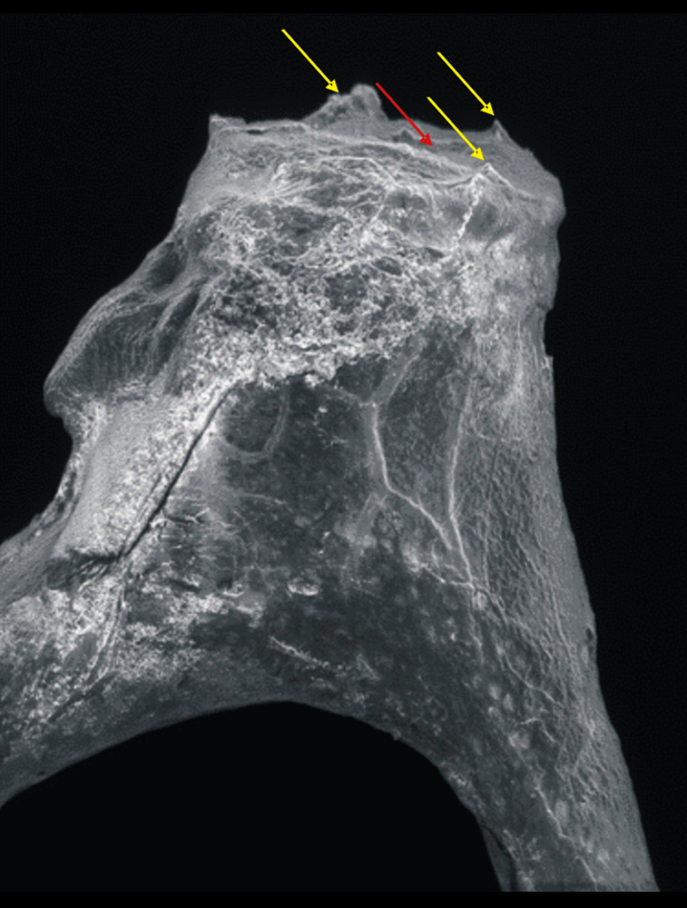 Figure 4. Otosclerosis. The head of the stapes. Magnification 150×. LVSED (low vacuum secondary electron detector). Workshop of Scanning Microscopy. Damage to the surface of the bone tissue of the stapes head: osteophytes (yellow arrow) and irregularity of the structure of the articular surface (red arrow). Prepared using CorelDraw9, Corel Corporation.
 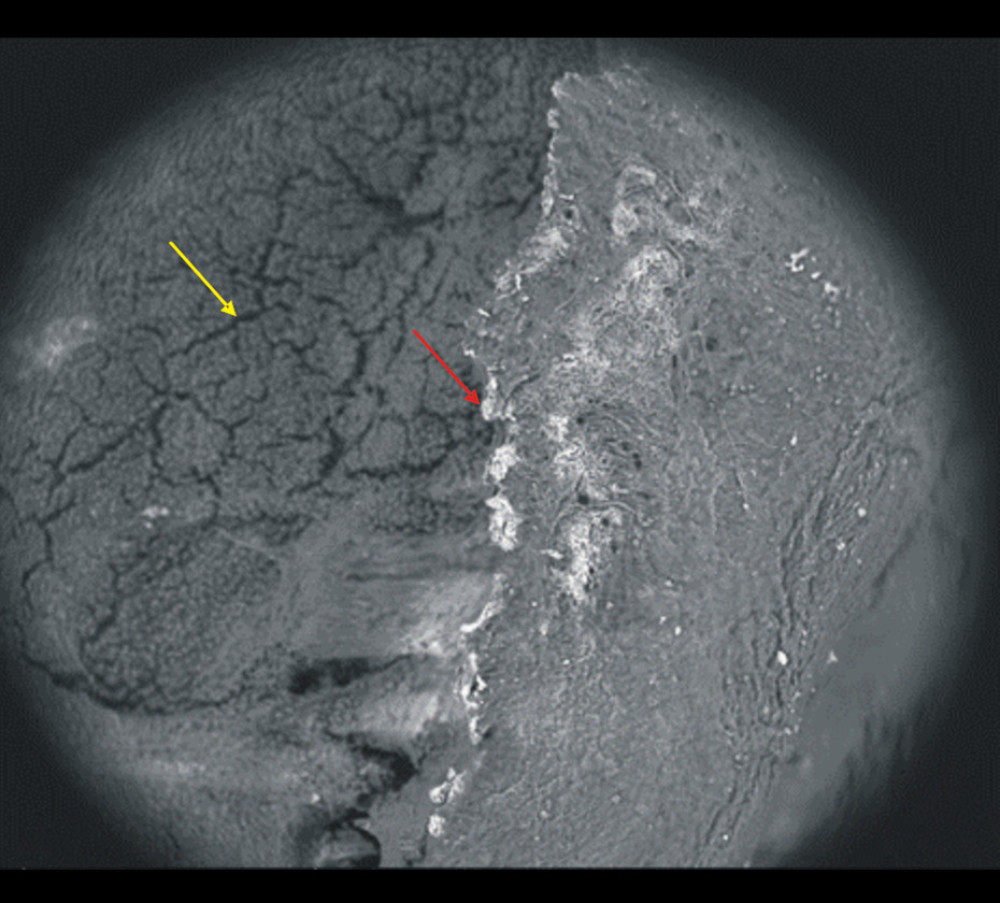 Figure 5. Otosclerosis. The head of the stapes. Magnification 500×. vCD (low voltage-high contrast detector). Workshop of Scanning Microscopy. Small exfoliation of the articular surface (red arrow) and small linear cracks (yellow arrow) of the stapes head. Prepared using CorelDraw9, Corel Corporation.
 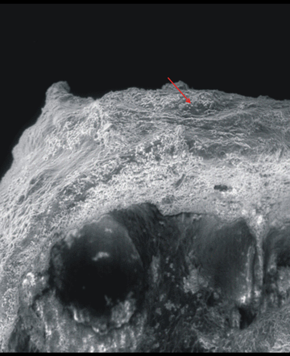 Figure 6. Otosclerosis. The head of the stapes. Magnification 350×. LVSED (low vacuum secondary electron detector). Workshop of Scanning Microscopy. Exfoliations of the articular surface of the stapes head (red arrow). Prepared using CorelDraw9, Corel Corporation.
 References
1. Chadha S, Kamenov K, Cieza A, The world report on hearing, 2021: Bull World Health Organ, 2021; 99(4); 242-242A
2. Job K, Wiatr A, Skladzien J, Wiatr M, The audiometric assessment of the effectiveness of surgical treatment of otosclerosis depending on the preoperative incidence of Carhart’s notch: Ear Nose Throat J, 2021 [Online ahead of print]
3. Zhang Y, Tang Q, Xue R, Analysis of the genetic characteristics of a Chinese family with otosclerosis: Ear Nose Throat J, 2021; 100; 774S-80S
4. Wiatr A, Składzień J, Świeży K, Wiatr M, A biochemical analysis of the stapes: Med Sci Monit, 2019; 25; 2679-86
5. Michels TC, Duffy MT, Rogers DJ, Hearing loss in adults: Differential diagnosis and treatment: Am Fam Physician, 2019; 100(2); 98-108
6. Szyfter W, Gawęcki W, Bartochowska A, Conductive hearing loss after surgical treatment of otosclerosis – long-term observations: Otolaryngol Pol, 2021; 75(1); 1-6
7. Palacios-Garcia J, Ropero-Romero F, Aguilar-Vera F, Sanchez-Gomez S, Short-term audiological outcomes of stapedotomy: microdrill at low revolutions versus manual perforator to perform a small footplate fenestra. A prospective observational study: Otolaryngol Pol, 2020; 74(5); 1-5
8. Manley GA, Narins PM, Fay RR, Experiments in comparative hearing: Georg von Békésy and beyond: Hear Res, 2012; 293(1–2); 44-50
9. Kirikae I, Physiology of the middle ear: Arch Otolaryngol, 1963; 78; 317-28
10. Goode RL, Nishihara S, Experimental models of ossiculoplasty: Otolaryngol Clin North Am, 1994; 27(4); 663-75
11. Vlaming MS, Feenstra L, Studies on the mechanics of the normal human middle ear: Clin Otolaryngol Allied Sci, 1986; 11(5); 353-63
12. Goode RL, Ball G, Nishihara S, Measurement of umbo vibration in human subjects – method and possible clinical applications: Am J Otol, 1993; 14(3); 247-51
13. Clarke-Brodber AL, Taxy JB, The stapes in otosclerosis: Osteoarthritis of an ear ossicle: Head Neck Pathol, 2021; 15(3); 737-42
Figures
Figure 1. Control group. Result of the chemical composition of the stapes head. CaKa – calcium; PKa – phosphorus; OKa – oxygen; CKa – carbon; NaKa – sodium; SKa – sulfur. Prepared using CorelDraw9, Corel Corporation.Figure 2. Otosclerosis. Result of the chemical composition of the stapes head. Ca – calcium; P – phosphorus; O – oxygen; C – carbon; Na – sodium; S – sulfur. Prepared using CorelDraw9, Corel Corporation.Figure 3. Control group. The head of the stapes (green arrow). Magnification 150×. LVSED (low vacuum secondary electron detector). Workshop of Scanning Microscopy. Prepared using CorelDraw9, Corel Corporation.Figure 4. Otosclerosis. The head of the stapes. Magnification 150×. LVSED (low vacuum secondary electron detector). Workshop of Scanning Microscopy. Damage to the surface of the bone tissue of the stapes head: osteophytes (yellow arrow) and irregularity of the structure of the articular surface (red arrow). Prepared using CorelDraw9, Corel Corporation.Figure 5. Otosclerosis. The head of the stapes. Magnification 500×. vCD (low voltage-high contrast detector). Workshop of Scanning Microscopy. Small exfoliation of the articular surface (red arrow) and small linear cracks (yellow arrow) of the stapes head. Prepared using CorelDraw9, Corel Corporation.Figure 6. Otosclerosis. The head of the stapes. Magnification 350×. LVSED (low vacuum secondary electron detector). Workshop of Scanning Microscopy. Exfoliations of the articular surface of the stapes head (red arrow). Prepared using CorelDraw9, Corel Corporation. Tables
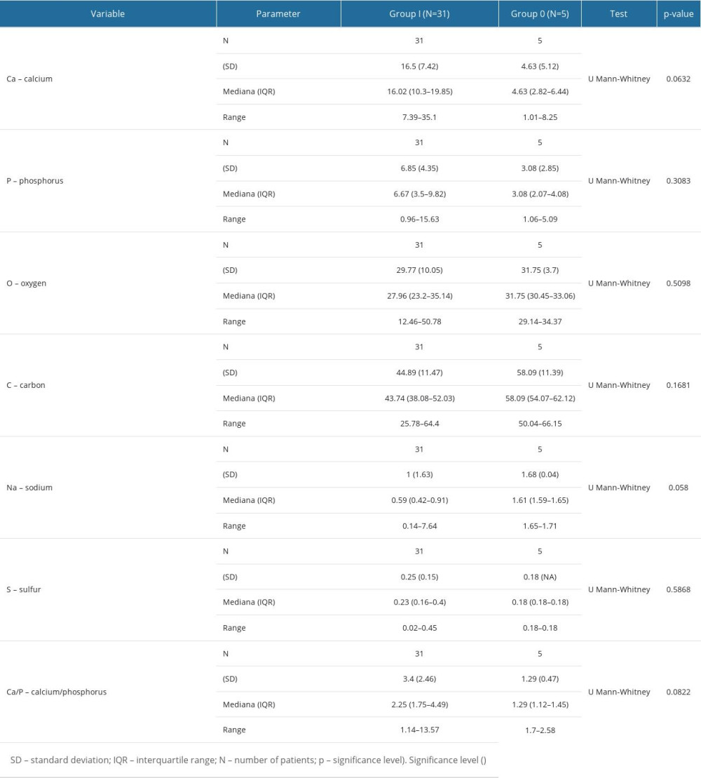 Table 1. Comparison of the results of the elemental composition in the area of the stapes head with the division into the test group (group I) and control group (group 0).Table 1. Comparison of the results of the elemental composition in the area of the stapes head with the division into the test group (group I) and control group (group 0).
 In Press
Clinical Research
Effects of Single-Bout Endurance Exercise Intensity on Peripheral Neurotrophic Factors in Patients With Isc...Med Sci Monit In Press; DOI: 10.12659/MSM.952089
Review article
Anisodus tanguticus in Cancer Research: A Review of Traditional Use, Phytochemistry, Extraction Methods, an...Med Sci Monit In Press; DOI: 10.12659/MSM.952999
Clinical Research
Nasal Mucociliary Clearance and Its Relationship With Disease Severity in Patients With Multiple SclerosisMed Sci Monit In Press; DOI: 10.12659/MSM.952850
Clinical Research
Modified Thoracoabdominal Nerves Block Through the Perichondrial Approach vs Subcostal Transversus Abdomini...Med Sci Monit In Press; DOI: 10.12659/MSM.953976
Most Viewed Current Articles
17 Jan 2024 : Review article 14,176,570
Vaccination Guidelines for Pregnant Women: Addressing COVID-19 and the Omicron VariantDOI :10.12659/MSM.942799
Med Sci Monit 2024; 30:e942799
13 Nov 2021 : Clinical Research 3,762,188
Acceptance of COVID-19 Vaccination and Its Associated Factors Among Cancer Patients Attending the Oncology ...DOI :10.12659/MSM.932788
Med Sci Monit 2021; 27:e932788
14 Dec 2022 : Clinical Research 2,466,310
Prevalence and Variability of Allergen-Specific Immunoglobulin E in Patients with Elevated Tryptase LevelsDOI :10.12659/MSM.937990
Med Sci Monit 2022; 28:e937990
16 May 2023 : Clinical Research 708,927
Electrophysiological Testing for an Auditory Processing Disorder and Reading Performance in 54 School Stude...DOI :10.12659/MSM.940387
Med Sci Monit 2023; 29:e940387