23 February 2022: Clinical Research
A Retrospective Study of the Safety and Efficacy of Endoscopic Radiofrequency Therapy Under Direct Vision in 59 Patients with Gastroesophageal Reflux Disease from 2 Centers in Beijing, China Using the Gastroesophageal Reflux Disease Questionnaire
Di Lu1ABCDEFG, Chun Shan Bi2ABCE, Xue Wei1BCDEF, Bao Na Guo2BC, Ying Xin Gao1BG, Jing Chen2B, Jie Qian1BG, Zi Hao Guo2B, Yan Bin Wang1B, Li Li2B, Chuan Zhang2A, Jian Yu Hao1AG*, Yan Gao1ABDDOI: 10.12659/MSM.933848
Med Sci Monit 2022; 28:e933848
Abstract
BACKGROUND: This retrospective study from 2 centers in Beijing, China aimed to assess the safety and efficacy of endoscopic radiofrequency therapy under direct vision in 59 patients with gastroesophageal reflux disease (GERD) using the gastroesophageal reflux disease questionnaire (GerdQ).
MATERIAL AND METHODS: Fifty-nine GERD patients who underwent endoscopic radiofrequency treatment were included. Patients were divided into 2 groups: the endoscopic radiofrequency therapy under direct vision group and the non-direct vision radiofrequency therapy group. Indicators such as GerdQ score, lower esophageal sphincter (LES) pressure, DeMeester score, acid exposure time, and proton pump inhibitors (PPIs) use were collected before and after radiofrequency treatment. Postoperative complications were also recorded. The efficacy and safety of endoscopic radiofrequency therapy under direct vision were evaluated by comparing the indicators of patients in the 2 groups.
RESULTS: At 3 months after radiofrequency treatment, patients in the endoscopic radiofrequency therapy under direct vision group improved significantly in GerdQ score, decreased from 11.0 (10.0, 12.0) to 6.0 (6.0, 8.0), better than patients in the non-direct vision radiofrequency therapy group, and the better improvements remained at 12 months after the procedure (P<0.05). At 6 months after treatment, patients in the endoscopic radiofrequency therapy under direct vision group had significant improvements in LES pressure, which increased from 8.15 (3.18, 12.88) mmHg to 15.20 (10.25, 27.03) mmHg (P<0.05). There were no severe complications in our trial.
CONCLUSIONS: When compared with non-visualized endoscopic radiofrequency therapy, treatment under direct vision was safer and improved the GerdQ score and LES pressure at up to 12 months.
Keywords: Gastroesophageal Reflux, Radiofrequency Therapy, Beijing, Endoscopy, Gastrointestinal, Female, Follow-Up Studies, Humans, Male, Prevalence, Surveys and Questionnaires
Background
Gastroesophageal reflux disease (GERD) is a series of symptoms and complications due to regurgitation of gastric contents to esophageal and oral cavity, even to the lungs. The incidence of GERD is about 13% worldwide, and is highest in Western countries [1]. In the United States, the incidence of GERD is over 20% [2], and economic spending for GERD has exceed $12 billion annually [3]. Acid regurgitation and heartburn are the classical clinical features of GERD. Without treatment, GERD can significantly influence patient quality of life [4,5]. As an important risk factor of esophageal adenocarcinoma, GERD can also lead to respiratory complication such as asthma and pulmonary interstitial fibrosis [6].
Several examinations can be used to diagnose and evaluate GERD. Among them, the gastroesophageal reflux disease questionnaire (GerdQ) is the most commonly used and concise tool for GERD diagnosis and treatment effectiveness evaluation [7]. Regardless of changing life style, treatment for GERD can be divided into drug therapy and non-drug therapy. Proton pump inhibitors (PPIs) therapy, a preferred alternative drug therapy for GERD, can alleviate symptoms and effectively reduce the risk of complications [8]. However, use of PPIs are limited in GERD treatment due to adverse reactions and drug dependence [9]. For patients who are not suitable for drug therapy, there are non-drug therapies such as surgery and endoscopic treatments. Fundoplication, the most common anti-reflux surgery, which treats GERD through creating a barrier to resist gastric material, has a similar effect as PPIs [10]. In addition, considering the high risk and non-repeatability of surgery, endoscopic treatment seems to be a safer and more acceptable choice for GERD patients.
Endoscopic radiofrequency therapy is one of the most advanced technologies in endoscopic treatment. In past decades, this minimally invasive treatment has been used in more than 20 000 patients [11]. A meta-analysis published in 2017 that included 28 controlled and prospective trials, with an average follow-up of 25 months, indicated that radiofrequency treatment can dramatically alleviate the symptoms of GERD, decrease acid exposure time (AET), and reduce PPIs usage [12].
However, traditional non-direct vision endoscopic radiofrequency therapy still needs further improvement because of its limitation in accurately locating the correct location. Based on this, we assume that endoscopic radiofrequency therapy under direct vision, which can locate the lower esophageal sphincter (LES) more accurately via gastroscopy, may have a better curative effect than traditional non-direct vision endoscopic radiofrequency therapy. Thus, our study aimed to evaluate the efficacy and safety of endoscopic radiofrequency therapy under direct vision by comparing it with traditional non-direct vision endoscopic radiofrequency to assess endoscopic radiofrequency therapy under direct vision for GERD treatment.
Therefore, this retrospective study from 2 centers in Beijing, China assessed the safety and efficacy of endoscopic radiofrequency therapy under direct vision in 59 patients with GERD using the GerdQ.
Material and Methods
ETHICS STATEMENT:
All the patient signed the informed consent form. This trial was approved by the local Ethics Committees.
PATIENT POPULATION:
A total of 59 GERD patients who received endoscopic radiofrequency therapy from February 2017 to December 2019 in Beijing Chaoyang Hospital and Beijing Tongren Hospital were included in this study. Patients were divided into 2 groups: a traditional non-direct vision endoscopic radiofrequency therapy group (N=30) and an endoscopic radiofrequency therapy under direct vision group (N=29).
INCLUSION AND EXCLUSION CRITERIA:
Inclusion criteria were: (a) age ≥18 years old; (b) diagnosed with GERD; (c) received a standardized PPIs treatment (take PPIs 30 min before meals twice a day) for 3 months, GERD symptom relieved but still need long-term medication therapy; (d) 24-h esophageal multichannel intraluminal impedance (MII)–pH monitoring indicated evident of pathological acid reflux or reflux events related to symptoms (off PPIs), including any criterion following: (1) AET >4.2%; (2) DeMeester score >14.72; (3) number of reflux events >73; (4) symptom index (SI) >50%; (5) symptom association probability >95% [13].
Exclusion criteria were: large esophageal hiatus hernia (>2 cm); Barrett’s esophagus; dysphagia; achalasia [14]; autoimmune diseases (such as scleroderma); coagulation disorder; surgery history of neck, chest, and abdomen; anesthetic intolerance; severe organ dysfunction; pregnancy.
ENDOSCOPIC RADIOFREQUENCY TREATMENT PROCEDURE:
Each patient had finished examinations such as esophageal manometry, 24-h esophageal MII–pH monitoring, and endoscopy before radiofrequency treatment to evaluate objective indicators such as LES pressure, DeMeester score, and AET.
Endoscopic radiofrequency treatments were performed by experienced endoscopists under intravenous general anesthesia. Specific steps were as follows:
ENDOSCOPIC RADIOFREQUENCY THERAPY UNDER DIRECT VISION GROUP:
1. Measure the distance between dentate line and incisor. 2. Place the guidewire into the duodenum through the endoscopic biopsy orifice. 3. Withdraw the gastroscope. 4. Introduce the radiofrequency catheter into the esophagus over a guidewire. 5. Insert the gastroscope again and place the gastroscope probe at the mouth side of the radiofrequency device balloon. 6. Inflate the radiofrequency equipment balloon at the level of the dentate line, then activate the generator by pushing out 4 electrode needles. 7. Deflate the balloon after finishing this set of treatments (4 points). 8. Finish this layer treatment by rotating 45 degrees to the right and conducting another set of treatments. 9. Similarly, conduct the treatment at the 6 other layers (0.5 cm, 1.0 cm, 1.5 cm above the dentate line, and 0.5 cm, 1.0 cm, 1.5 cm below the dentate line). 10. Introduce the radiofrequency catheter into the stomach. Inflate 25 ml air and pull the balloon back at the cardia, then activate the generator by pushing out the electrode needle. 11. Finish 2 other sets of treatment by rotating 30 degrees to the right and to the left. 12. Deflate the air to 22 ml and pull the balloon back to conduct another layer treatment [15].
Observe the white or red radiofrequency treatment point at the lower esophagus and cardia by gastroscopy while conducting radiofrequency treatment, meanwhile, suctioning excess liquid coolant in the esophagus. The whole radiofrequency treatment procedure lasts about 45 min.
NON-DIRECT VISION RADIOFREQUENCY THERAPY GROUP:
Traditional radiofrequency therapy does not insert the gastroscope again before the radiofrequency procedure (without step 5 in the endoscopic radiofrequency therapy under direct vision group).
All patients received a 4-week treatment of PPIs after the endoscopic radiofrequency procedure to protect the esophageal mucosa.
FOLLOW-UP AND OUTCOMES:
The patients were followed up at 3 months, 6 months, and 12 months after the procedure. We evaluated the improvement of GerdQ score and the reduction of PPIs usage. Esophageal manometry, 24-hesophageal MII–pH monitoring, endoscopy examinations, and post-procedure complications were also performed.
PRIMARY OUTCOMES:
The improvement of GERD symptom which can be evaluated by pre- and post-procedural GerdQ score.
SECONDARY OUTCOMES:
The improvement of indicators such as LES pressure, DeMeester score, AET, and improvement by reduction of PPIs use.
EQUIPMENT:
We used the electronic gastroscope system GIH-Q260H (Olympus Corporation, Tokyo, Japan), EG-L600ZW7 (Fujifilm Corporation, Tokyo, Japan), high-resolution esophageal manometric system ZAN-S61C01E (Sandhill Scientific, Inc., Colorado, USA), 24-h esophageal MII–pH monitoring system A089022B (Sandhill Scientific, Inc., Colorado, USA), and endoscopic radiofrequency equipment MER-200GA (Medi Corporation, Heilongjiang, China).
DATA COLLECTION AND STATISTICAL ANALYSIS:
We exported relevant data from the electronic medical records. To ensure the accuracy of the data, 2 experts checked data manually. The study included variables related to patient demographic characteristics, GerdQ score, PPIs usage, and pre- and post-procedural indicators including: LES pressure by esophageal manometry, DeMeester score, and AET by 24-h esophageal MII–pH monitoring.
Data were analyzed with SPSS 24.0 software (IBM SPSS Statistics, United States). Continuous variables are expressed as mean±SD and counting variables are expressed as percentages and proportions. Data normality was assessed by Shapiro-Wilk test. The independent-samples
Results
BASELINE CHARACTERISTICS:
At the first follow-up time (3 months after the procedure), 2 patients were lost to follow-up in the endoscopic radiofrequency therapy under direct vision group and 1 patient was lost to follow-up in the traditional non-direct vision radiofrequency group. Data on a total of 56 patients were analyzed, with 27 patients in the endoscopic radiofrequency therapy under direct vision group and 29 patients in the non-direct vision radiofrequency group. Twenty-one patients in the endoscopic radiofrequency therapy under direct vision group and 24 patients in the non-direct vision radiofrequency group finished the 12-month follow-up (Figure 1).
Among all patients analyzed, the mean age was 57.9 (±8.7) years and 55.4% were males. As a result, there were no statistically significant differences in the gender, age, GerdQ score, reflux esophagitis, LES pressure, or DeMeester score before radiofrequency treatment between the 2 groups (Table 1).
EFFICACY OF ENDOSCOPIC RADIOFREQUENCY THERAPY UNDER DIRECT VISION:
During the endoscopic radiofrequency therapy under direct vision procedure, we could position the balloon and locate the treatment points more precisely (Figure 2).
At 3 months after the radiofrequency procedure, GerdQ scores decreased from 11.0 (10.0, 12.0) to 6.0 (6.0, 8.0) in the endoscopic radiofrequency therapy under direct vision group, while in the traditional non-direct vision radiofrequency group, GerdQ scores only decreased from 10.0 (8.5, 11.5) to 8.0 (7.0, 9.0), which showed that patients’ GerdQ score improved better in the endoscopic radiofrequency therapy under direct vision group (P<0.05). GerdQ scores remained better in the endoscopic radiofrequency therapy under direct vision group at 12 months after radiofrequency procedure (P<0.01) (Table 2, Figure 3).
All the patients had to take full dose PPIs before the radiofrequency procedure. At 6 months after radiofrequency treatment, 88.9% (24/27) of patients had reduced PPIs use in the endoscopic radiofrequency therapy under direct vision group and 96.6% (28/29) of patients had reduces PPIs use in the traditional non-direct radiofrequency group. At 12 months after the procedure, 95% of patients in each group had reduced PPIs use. There were no significant differences between the 2 group in PPIs use reduction at 3-month follow-up (Table 3, Figure 4).
Most of the patients who underwent endoscopic radiofrequency treatment were satisfied with the curative effect, so they were unwilling to undergo post-procedural examinations such as esophageal manometry and 24-h esophageal MII–pH monitoring. Only 6 patients underwent evaluation of LES pressure, DeMeester score, and AET again at 6 months after radiofrequency procedure.
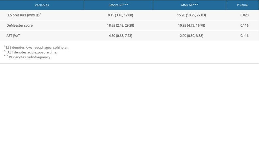
At 6 months after the radiofrequency procedure LES pressure increased significantly in the endoscopic radiofrequency therapy under direct vision group, from 8.15 (3.18, 12.88) mmHg to 15.20 (10.25, 27.03) mmHg (P<0.05). However, indicators such as DeMeester score and AET had improved slightly compared to the pre-procedural situation, and the difference was not statistically significant (Table 4).
SAFETY OF ENDOSCOPIC RADIOFREQUENCY THERAPY UNDER DIRECT VISION:
There were no severe complications in our trial. In the traditional non-direct vision radiofrequency therapy group, 2 patients had esophageal stenosis after the radiofrequency procedure, which was relieved by endoscopic balloon dilation and metal-coated stent placement; 1 patient reported post-procedural bleeding, which was cured by using PPIs. In contrast, there were no complications in the endoscopic radiofrequency therapy under direct vision group.
Discussion
In our study we found that, compared with the traditional non-direct vision radiofrequency therapy, endoscopic radiofrequency therapy under direct vision achieved a better therapeutic effect in improvements of indicators such as GerdQ score in GERD patients, and the better curative effect could remain at 12 months after the radiofrequency procedure. Meanwhile, endoscopic radiofrequency therapy under direct vision significantly increased LES pressure, and it also made the contraction function of residual LES more uniform and coordinated by precisely locating the treatment site.
As one of the most common chronic gastrointestinal diseases, GERD dramatically influences patients’ daily lives. Nowadays, medication therapy like PPIs remains the major treatment of GERD. Besides medications, a number of endoscopic therapies have been developed to treat GERD. Among all the endoscopic therapies, endoscopic radiofrequency remains the most advanced.
In our study, we integrated the latest domestic and international diagnosis and treatment consensus to determine the diagnostic criteria for GERD in the inclusion criteria. Typical symptoms, positive PPIs test, and esophageal reflux monitoring examination were selected as the preferred diagnostic criteria [16]. For patients with atypical symptoms, negative endoscopic examination and ineffective drug treatment, esophageal reflux monitoring should be done to evaluate the property and cause of the reflux [17]. Compared with simple esophageal pH monitoring, which can only detect acid reflux, 24-h esophageal impedance pH monitoring can also detect non-acid reflux, making the diagnosis more sensitive [18,19]. All patients in this study had completed 24-h esophageal multichannel intraluminal impedance (MII)–pH monitoring before radiofrequency treatment to assess DeMeester score and AET. In contrast to the positive standard of AET >6% in the Lyon Consensus [16], studies in China suggest that AET >4.2% is sufficient to indicate the presence of pathological acid reflux for Chinese patients [20, 21]. Therefore, the included criteria in our study set 4.2% as the threshold of AET in the diagnostic criteria.
Patients included in our study had received standard PPIs treatment (half an hour before meals) for 3 months before radiofrequency therapy. Their symptoms were relieved during the medication period but recurred after drug withdrawal; thus, they still needed long-term drug therapy. However, for patients who need to take PPIs for a long period of time, there may be an increased risk of fractures or
Before undergoing endoscopic radiofrequency therapy, all patients had completed routine examinations, such as gastroscopy and high-resolution manometric examination of the esophagus, in order to clarify the diagnosis and evaluate whether the patients were suitable for endoscopic therapy. Due to the high incidence of upper digestive tract tumors in China, for patients with typical symptoms such as heartburn and reflux, gastroscopy should be done to exclude upper digestive tract malignant tumors and other conditions. Meanwhile, the situation of esophageal mucosa for reflux esophagitis can be evaluated under endoscopy [24,25]. High-resolution manometric examination of the esophagus can detect hiatal hernia and severe esophageal motility disorders that are not suitable for endoscopic treatment [14,26].
In our study, the improvement of patients’ GerdQ score was taken as one of the main observation indicators to evaluate the efficacy. Studies have shown that the sensitivity of GerdQ score for diagnosis is about 65%, and the specificity is up to 71% [27]. The GerdQ score can not only assist in determining the diagnosis, but can also be used to evaluate the impact of GERD symptoms on quality of life and to evaluate the treatment effect. Therefore, in our study, improvement of GerdQ score was used as the main outcome indicator to evaluate the efficacy of endoscopic radiofrequency therapy under direct vision.
Our study found that both endoscopic radiofrequency therapy under direct vision and non-direct vision radiofrequency therapy can significantly improve patients’ symptoms and evaluation indicators. The GerdQ scores of patients in both groups were significantly lower than those before endoscopic radiofrequency treatment, and the therapeutic effect lasted up to 12 months after treatment. About 90% of patients could reduce the dose or frequency of PPIs after radiofrequency treatment. It is worth mentioning that the efficacy of endoscopic radiofrequency therapy in this study could last up to 12 months, showing good medium- and long-term efficacy, which is consistent with the results of previous studies.
In the past decades, many studies had proved the long-term efficacy of endoscopic radiofrequency. Endoscopic radiofrequency can have a significant effect not only on subjective indicators such as symptom score and PPIs usage reduction, but also on objective indicators such as esophagitis, LES pressure, DeMeester score, and AET.
However, traditional non-direct endoscopic radiofrequency therapy has limitation in GERD treatment, as it cannot locate the LES accurately. Therefore, we performed endoscopic radiofrequency therapy under direct vision for GERD patients, which had not been done worldwide, to explore whether the new technique is more effective.
In our study, we found that patients’ GerdQ scores improved more in the endoscopic radiofrequency therapy under direct vision group than in the traditional non-direct radiofrequency group, and the better curative effect remained at 12 months after radiofrequency treatment. These results suggest that endoscopic radiofrequency therapy under direct vision had better therapeutic effects in relieving GERD symptoms such as heartburn and reflux, showing a good medium- and long-term curative effect.
What is the reason for the better improvement of GerdQ score in the endoscopic radiofrequency therapy under direct vision group compared with the non-direct vision group? We suppose that this may be related to the mechanism of endoscopic radiofrequency therapy under direct vision. As shown in Figure 5A, endoscopic radiofrequency therapy under direct vision can be observed under an endoscope, which can better locate the treatment sites and make the distribution of treatment sites more uniform so as to make the contraction function of residual LES more coordinated after radiofrequency therapy and better enhance its anti-reflux effect. In our study, patients’ LES pressure in the endoscopic radiofrequency therapy under direct vision group was significantly higher than before, which also confirmed this to a certain extent. However, as for non-direct vision radiofrequency therapy, as shown in Figure 5B, because the radiofrequency operation is not carried out under direct vision of the endoscope, the positioning of treatment sites only based on the experience of the endoscopic physician will result in the uneven distribution of treatment sites, which will lead to the uncoordinated contraction function of LES and poor anti-reflux effect. Therefore, we believe that is why endoscopic radiofrequency therapy under direct vision had better therapeutic effects.
In addition, although patients in the 2 group had a similar reduction of PPIs use, it is worth noting that more than 95% of patients in both groups were able to reduce the dose or frequency of PPIs at 12 months after radiofrequency therapy. We suppose that this may be related to the pathogenesis of GERD and the mechanism of endoscopic radiofrequency therapy. Currently, the most recognized pathogenesis of GERD includes structural damage or dysfunction of the gastroesophageal junction. Besides, increased sensitivity of esophageal organs is also a common pathogenic mechanism of GERD. The mechanism of endoscopic radiofrequency therapy is that it cannot only cause coagulated necrosis of the muscular tissue at the treatment site through heat conduction, so as to improve the resting pressure of LES, but also can reduce the frequency of transient LES relaxation by blocking nerve blocks such as the vagus nerve, and reduce the sensitivity of the gastroesophageal junction to acid, so as to relieve the symptoms of GERD [28,29]. We believe that this may be the reason why the majority of patients could reduce the dosage of PPIs.
Our study also analyzed the improvement of objective indicators such as LES pressure, DeMeester score, and AET of GERD patients after endoscopic radiofrequency therapy under direct vision. It showed a significant improvement in LES pressure at 6 months after radiofrequency therapy and continued to 12 months after radiofrequency therapy. This is similar to previous research. A prospective study found that patients’ average LES resting pressure improved significantly at both 4 and 8 years after radiofrequency therapy compared with pre-treatment [30]. However, objective indicators, such as DeMeester score and AET, were improved in the endoscopic radiofrequency group, but the differences were not statistically significant. The improvement of these indicators in previous studies has varied over the decades. An open-label multicenter prospective study in the United States suggested a significant improvement in esophageal acid exposure after radiofrequency therapy compared with pre-treatment [31]. However, another randomized, double-blind, controlled trial showed that after 6 months of radiofrequency treatment, there was no significant difference in duration of esophageal acid exposure between patients in the radiofrequency group and those in the sham group [32]. A prospective study with a follow-up period of up to 8 years also found that patients’ esophageal acid exposure improved significantly at 4 years after radiofrequency therapy compared with pre-treatment, but returned to pre-treatment levels at 8 years [30]. Chinese studies also have reported different results in terms of improvement of objective indicators. A single-center prospective study found that radiofrequency therapy significantly increased LES resting pressure, lowered DeMeester scores, and reduced AET [33]. However, a Toupet fundoplication-controlled study found that 12 months after radiofrequency therapy, there were no significant changes in indicators such as reflux time, reflux frequency, and reflux time percentage compared with those before treatment, and the improvement effect of radiofrequency therapy on DeMeester score and LES pressure was not better than that of Toupet fundoplication [34]. In other words, endoscopic radiofrequency therapy may not be effective in improving DeMeester score and AET.
We suppose the inconspicuous improvement of DeMeester score and AET is related to the characteristics of endoscopic radiofrequency therapy. As a minimally invasive treatment, the electrode length of endoscopic radiofrequency is only 5.5 mm, which means the treatment depth is relatively shallow. Therefore, the improvement of the above indicators may not be as good as surgical treatment such as fundoplication. However, it is precisely because of the shallow depth of endoscopic radiofrequency therapy that the radiofrequency operation can be performed again in case of poor efficacy, so as to gradually achieve the expected efficacy. More importantly, due to the shallow depth of endoscopic radiofrequency therapy, it can significantly reduce the complications of esophageal stenosis, perforation, and other complications caused by deep treatment, which is safer than surgical treatment. Another possible reason is that the sample size used to evaluate the improvement of objective indicators is relatively small and there may have been selection bias. Since most patients in the endoscopic radiofrequency therapy group were satisfied with the treatment effect, they were not willing to undergo repeat examinations used for post-treatment evaluation, such as 24-h esophageal multichannel impedance pH monitoring, during follow-up. As a result, only a few patients were re-examined after radiofrequency therapy. Moreover, almost all the patients who received relevant examinations again after radiofrequency therapy were not satisfied with the treatment effect, so the objective indicators collected for evaluation may have a certain degree of selection bias. In addition, it may also be related to the mechanism of endoscopic radiofrequency therapy; that is, whether endoscopic radiofrequency therapy achieves therapeutic effect by reducing pathological acid reflux in the esophagus. It has been suggested that radiofrequency therapy alleviates GERD symptoms through esophageal desensitization rather than reducing acid exposure to the esophagus. At present, the relationship of radiofrequency therapy to relief of GERD symptoms and reduction of the duration of esophageal acid exposure, as well as the mechanism involved, are still under further investigation.
No serious complications occurred in this study. For traditional non-direct radiofrequency therapy, 2 patients had esophageal stenosis and 1 patient had bleeding, which were all relieved after treatment. This is similar to the results of published studies. So far, more than 25 000 radiofrequency treatments have been done globally [35]. In general, endoscopic radiofrequency therapy has good safety. Only a few mild complications, such as short-term chest pain, low fever, and dysphagia, have been reported, which are transient and can improve spontaneously without special treatment [36]. According to a prospective study with 10 years of follow-up, transient chest pain was the most common adverse event after radiofrequency therapy, and the overall safety was good [37].
It is worth mentioning that the direct vision endoscopic radiofrequency therapy can monitor the whole process of radiofrequency treatment under endoscopic vision, quickly deal with bleeding and other conditions during the radiofrequency operation, and reduce the possibility of intraoperative and postoperative complications to a greater extent. At the same time, endoscopic radiofrequency therapy under direct vision can flexibly determine the treatment level according to the distance between the dentate line and the cardia, so as to avoid esophageal stenosis caused by the proximity of adjacent treatment levels or the overlap of sites. In addition, endoscopic radiofrequency therapy under direct vision can also extract excess radiofrequency coolant through endoscopy in the radiofrequency process, which can better avoid the possibility of anesthesia aspiration and make the whole radiofrequency operation safer. In conclusion, we believe that endoscopic radiofrequency therapy under direct vision has better safety than non-direct vision radiofrequency therapy.
As a non-randomized controlled study, there are certain limitations in our study. First of all, the follow-up time was relatively short, only 12 months, which can only confirm the short- and medium-term efficacy of this new therapy. Secondly, our study lacked observation indicators such as DeMeester score and AET of patients in the non-direct vision radiofrequency therapy group, and failed to compare the improvement of these indicators between the endoscopic radiofrequency therapy under direct vision group and the non-direct vision radiofrequency therapy group. In the future, more prospective randomized controlled trials are needed to exclude relevant interfering factors, so as to evaluate the efficacy and safety of this new endoscopic radiofrequency therapy more comprehensively.
Conclusions
This study showed that, when compared with non-visualized endoscopic radiofrequency therapy, treatment under direct vision was safe and improved the GerdQ score and LES pressure at up to 12 months.
Figures
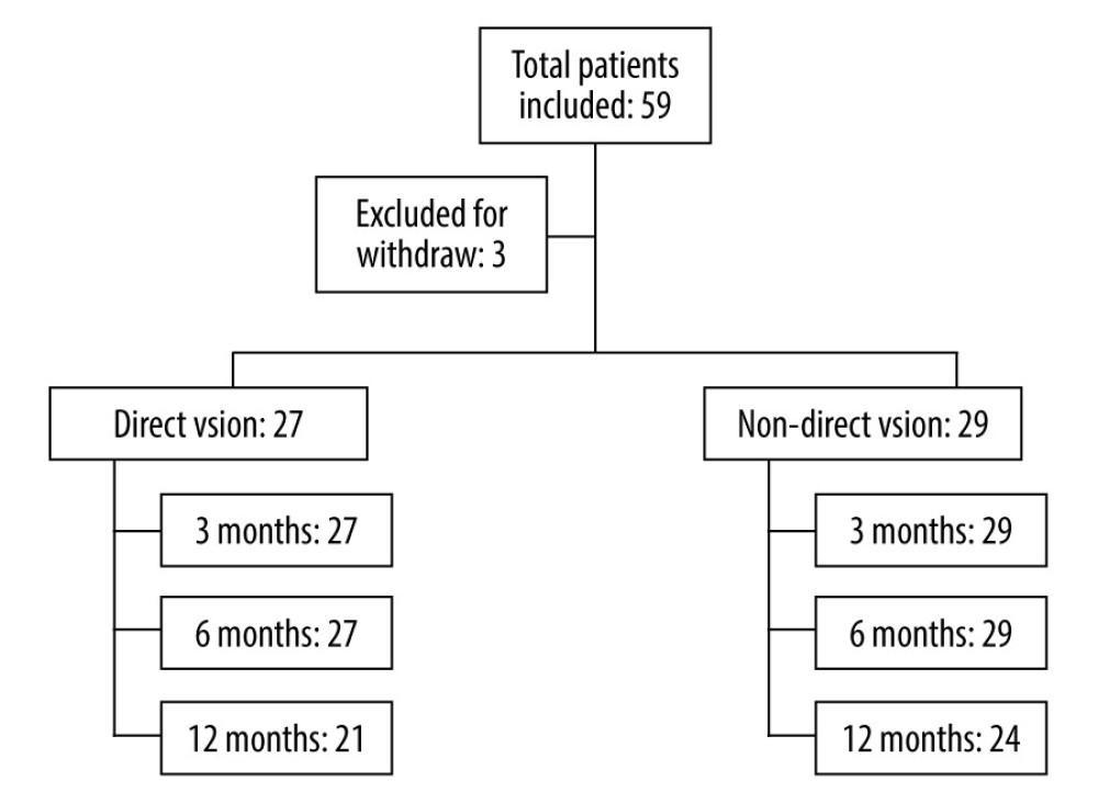 Figure 1. Flow chart on patients selected for the study (Office, 2021, Microsoft).
 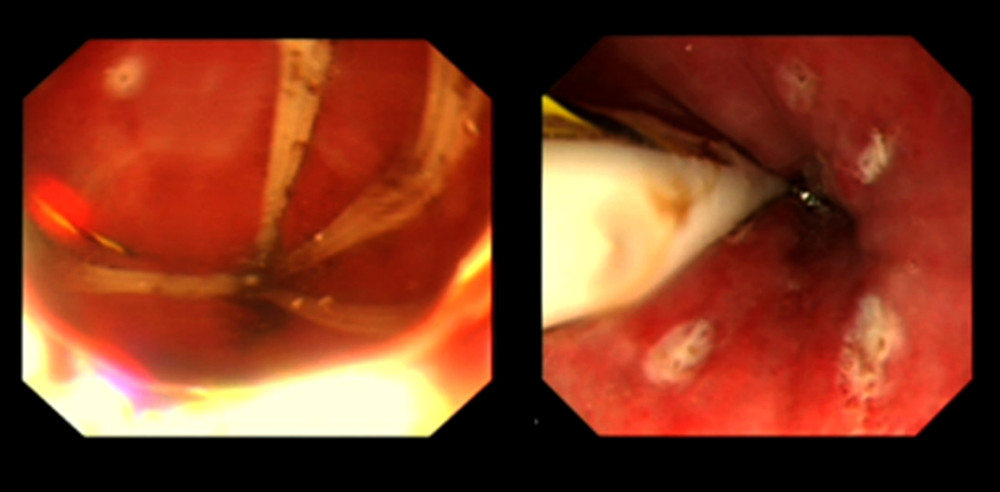 Figure 2. Images during and after endoscopic radiofrequency therapy under direct vision (Electronic gastroscope system GIH-Q260H, Olympus Corporation).
 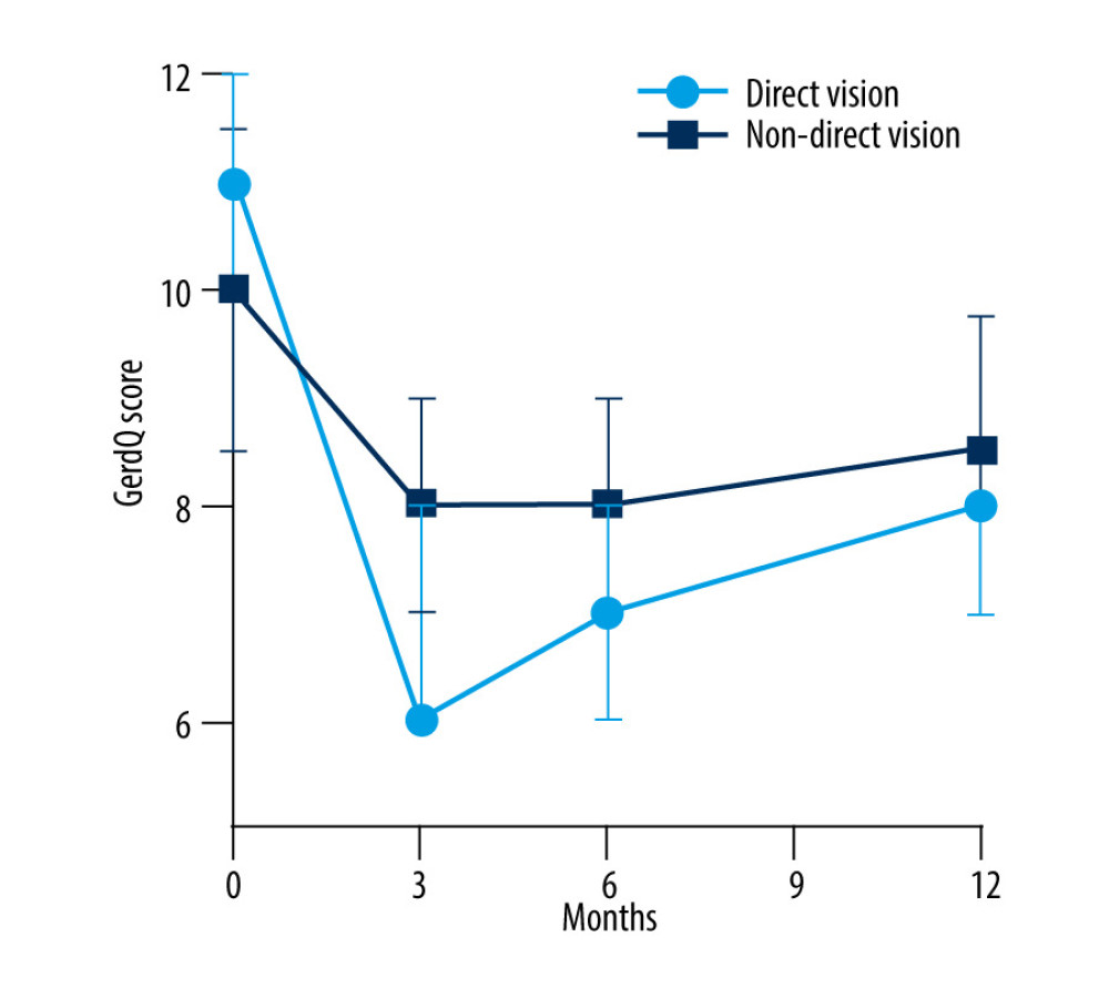 Figure 3. Comparison of GerdQ (gastroesophageal reflux disease questionnaire) score between the 2 groups (GraphPad Prism, 8.3.0, GraphPad Corporation).
 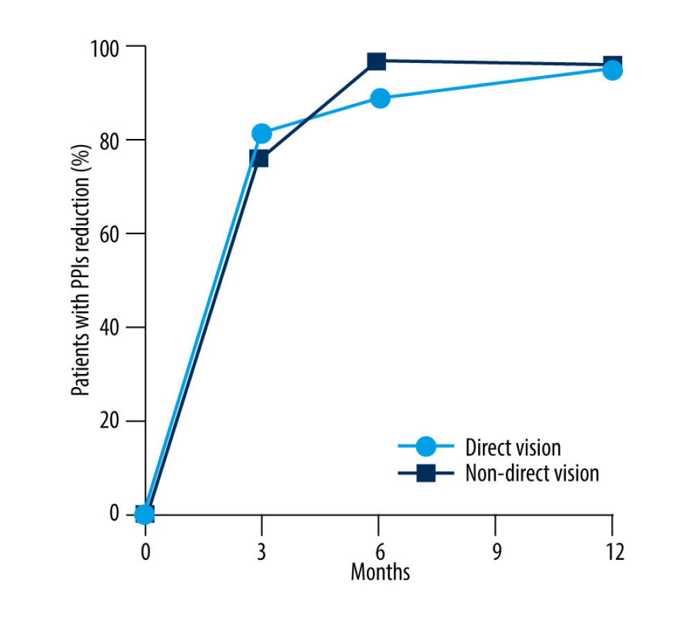 Figure 4. Comparison of PPIs (proton pump inhibitors) usage reduction between the 2 groups (GraphPad Prism, 8.3.0, GraphPad Corporation).
 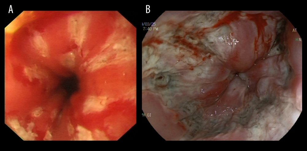 Figure 5. (A) Endoscopic images of endoscopic radiofrequency therapy under direct vision (Electronic gastroscope system GIH-Q260H, Olympus Corporation). (B) Endoscopic images of traditional non-direct radiofrequency therapy (Electronic gastroscope system EG-L600ZW7, Fujifilm Corporation).
 Tables
Table 1. Comparison of baseline characteristics between the 2 groups.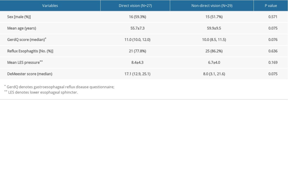 Table 2. Comparison of GerdQ* score between the 2 groups.
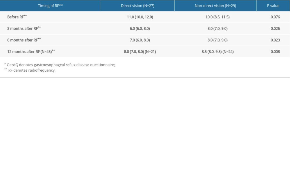 Table 3. Comparison of PPIs* usage reduction between the 2 groups.
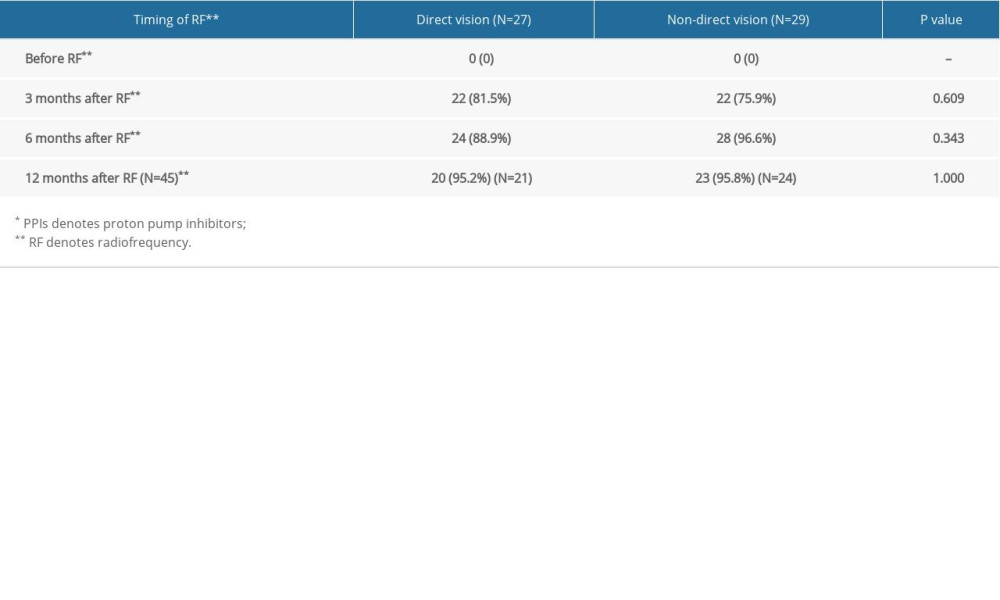 Table 4. Comparison of LES*, DeMeester score, and AET** before and after endoscopic radiofrequency procedure under direct vision (N=6).
References
1. Richter JE, Rubenstein JH, Presentation and epidemiology of gastroesophageal reflux disease: Gastroenterology, 2018; 154(2); 267-76
2. Locke GR, Talley NJ, Fett SL, Prevalence and clinical spectrum of gastroesophageal reflux: A population-based study in Olmsted County, Minnesota: Gastroenterology, 1997; 112(5); 1448-56
3. Everhart JE, Ruhl CE, Burden of digestive diseases in the United States part II: Lower gastrointestinal diseases: Gastroenterology, 2009; 136(3); 741-54
4. Dean BB, Crawley JA, Schmitt CM, The burden of illness of gastro-oesophageal reflux disease: impact on work productivity: Aliment Pharmacol Ther, 2003; 17(10); 1309-17
5. Farup C, Kleinman L, Sloan S, The impact of nocturnal symptoms associated with gastroesophageal reflux disease on health-related quality of life: Arch Intern Med, 2001; 161(1); 45-52
6. Ghisa M, Marinelli C, Savarino V, Savarino E, Idiopathic pulmonary fibrosis and GERD: Links and risks: Ther Clin Risk Manag, 2019; 15; 1081-93
7. Jones R, Junghard O, Dent J, Development of the GerdQ, a tool for the diagnosis and management of gastro-oesophageal reflux disease in primary care: Aliment Pharmacol Ther, 2009; 30(10); 1030-38
8. Katz PO, Gerson LB, Vela MF, Guidelines for the diagnosis and management of gastroesophageal reflux disease: Am J Gastroenterol, 2013; 108(3); 308-28
9. Scarpellini E, Ang D, Pauwels A, Management of refractory typical GERD symptoms: Nat Rev Gastroenterol Hepatol, 2016; 13(5); 281-94
10. Morgenthal CB, Lin E, Shane MD, Who will fail laparoscopic Nissen fundoplication? Preoperative prediction of long-term outcomes: Surg Endosc, 2007; 21(11); 1978-84
11. Sandhu DS, Fass R, Current trends in the management of gastroesophageal reflux disease: Gut Liver, 2018; 12(1); 7-16
12. Fass R, Cahn F, Scotti DJ, Gregory DA, Systematic review and meta-analysis of controlled and prospective cohort efficacy studies of endoscopic radiofrequency for treatment of gastroesophageal reflux disease: Surg Endosc, 2017; 31(12); 4865-82
13. Coron E, Sebille V, Cadiot G, Clinical trial: Radiofrequency energy delivery in proton pump inhibitor-dependent gastro-oesophageal reflux disease patients: Aliment Pharmacol Ther, 2008; 28(9); 1147-58
14. Chan WW, Haroian LR, Gyawali CP, Value of preoperative esophageal function studies before laparoscopic antireflux surgery: Surg Endosc, 2011; 25(9); 2943-49
15. Triadafilopoulos G, Dibaise JK, Nostrant TT, Radiofrequency energy delivery to the gastroesophageal junction for the treatment of GERD: Gastrointest Endosc, 2001; 53(4); 407-15
16. Gyawali CP, Kahrilas PJ, Savarino E, Modern diagnosis of GERD: The Lyon Consensus: Gut, 2018; 67(7); 1351-62
17. Roman S, Gyawali CP, Savarino E, Ambulatory reflux monitoring for diagnosis of gastro-esophageal reflux disease: Update of the Porto consensus and recommendations from an international consensus group: Neurogastroenterol Motil, 2017; 29(10); 1-15
18. Frazzoni M, Conigliaro R, Mirante VG, Melotti G, The added value of quantitative analysis of on-therapy impedance-pH parameters in distinguishing refractory non-erosive reflux disease from functional heartburn: Neurogastroenterol Motil, 2012; 24(2); 141-6 , e87
19. Zhou LY, Wang Y, Lu JJ, Accuracy of diagnosing gastroesophageal reflux disease by GerdQ, esophageal impedance monitoring and histology: J Dig Dis, 2014; 15(5); 230-38
20. Kahrilas PJ, Quigley EM, Clinical esophageal pH recording: A technical review for practice guideline development: Gastroenterology, 1996; 110(6); 1982-96
21. Zhang MY, Tan ND, Li YW, Esophageal physiologic profiles within erosive esophagitis in China: Predominantly low-grade esophagitis with low reflux burden: Neurogastroenterol Motil, 2019; 31(12); e13702
22. McDonald EG, Milligan J, Frenette C, Lee TC: JAMA Intern Med, 2015; 175(5); 784-91
23. Trifan A, Stanciu C, Girleanu I: World J Gastroenterol, 2017; 23(35); 6500-15
24. Lundell LR, Dent J, Bennett JR, Endoscopic assessment of oesophagitis: Clinical and functional correlates and further validation of the Los Angeles classification: Gut, 1999; 45(2); 172-80
25. Liu L, Li S, Zhu KX, Relationship between esophageal motility and severity of gastroesophageal reflux disease according to the Los Angeles classification: Medicine (Baltimore), 2019; 98(19); e15543
26. Weijenborg PW, van Hoeij FB, Smout AJ, Bredenoord AJ, Accuracy of hiatal hernia detection with esophageal high-resolution manometry: Neurogastroenterol Motil, 2015; 27(2); 293-99
27. Dent J, Vakil N, Jones R, Accuracy of the diagnosis of GORD by questionnaire, physicians and a trial of proton pump inhibitor treatment: The Diamond Study: Gut, 2010; 59(6); 714-21
28. Fry LC, Mönkemüller K, Malfertheiner P, Systematic review: Endoluminal therapy for gastro-oesophageal reflux disease: Evidence from clinical trials: Eur J Gastroenterol Hepatol, 2007; 19(12); 1125-39
29. Tam WC, Schoeman MN, Zhang Q, Delivery of radiofrequency energy to the lower oesophageal sphincter and gastric cardia inhibits transient lower oesophageal sphincter relaxations and gastro-oesophageal reflux in patients with reflux disease: Gut, 2003; 52(4); 479-85
30. Dughera L, Rotondano G, De Cento M, Durability of stretta radiofrequency treatment for GERD: Results of an 8-year follow-up: Gastroenterol Res Pract, 2014; 2014; 531907
31. Triadafilopoulos G, DiBaise JK, Nostrant TT, The Stretta procedure for the treatment of GERD: 6 and 12 month follow-up of the U.S. open label trial: Gastrointest Endosc, 2002; 55(2); 149-56
32. Corley DA, Katz P, Wo JM, Improvement of gastroesophageal reflux symptoms after radiofrequency energy: A randomized, sham-controlled trial: Gastroenterology, 2003; 125(3); 668-76
33. Liu PP, Meng QQ, Lin H, Radiofrequency ablation is safe and effective in the treatment of Chinese patients with gastroesophageal reflux disease: A single-center prospective study: J Dig Dis, 2019; 20(5); 229-34
34. Ma L, Li T, Liu G, Stretta radiofrequency treatment vs Toupet fundoplication for gastroesophageal reflux disease: A comparative study: BMC Gastroenterol, 2020; 20(1); 162
35. Perry KA, Banerjee A, Melvin WS, Radiofrequency energy delivery to the lower esophageal sphincter reduces esophageal acid exposure and improves GERD symptoms: A systematic review and meta-analysis: Surg Laparosc Endosc Percutan Tech, 2012; 22(4); 283-88
36. Chen D, Barber C, McLoughlin P, Systematic review of endoscopic treatments for gastro-oesophageal reflux disease: Br J Surg, 2009; 96(2); 128-36
37. Noar M, Squires P, Noar E, Lee M, Long-term maintenance effect of radiofrequency energy delivery for refractory GERD: A decade later: Surg Endosc, 2014; 28(8); 2323-33
Figures
 Figure 1. Flow chart on patients selected for the study (Office, 2021, Microsoft).
 Figure 2. Images during and after endoscopic radiofrequency therapy under direct vision (Electronic gastroscope system GIH-Q260H, Olympus Corporation).
 Figure 3. Comparison of GerdQ (gastroesophageal reflux disease questionnaire) score between the 2 groups (GraphPad Prism, 8.3.0, GraphPad Corporation).
 Figure 4. Comparison of PPIs (proton pump inhibitors) usage reduction between the 2 groups (GraphPad Prism, 8.3.0, GraphPad Corporation).
 Figure 5. (A) Endoscopic images of endoscopic radiofrequency therapy under direct vision (Electronic gastroscope system GIH-Q260H, Olympus Corporation). (B) Endoscopic images of traditional non-direct radiofrequency therapy (Electronic gastroscope system EG-L600ZW7, Fujifilm Corporation).
 Tables
 Table 1. Comparison of baseline characteristics between the 2 groups.
 Table 2. Comparison of GerdQ* score between the 2 groups.
 Table 3. Comparison of PPIs* usage reduction between the 2 groups.
 Table 4. Comparison of LES*, DeMeester score, and AET** before and after endoscopic radiofrequency procedure under direct vision (N=6).
 Table 1. Comparison of baseline characteristics between the 2 groups.
 Table 2. Comparison of GerdQ* score between the 2 groups.
 Table 3. Comparison of PPIs* usage reduction between the 2 groups.
 Table 4. Comparison of LES*, DeMeester score, and AET** before and after endoscopic radiofrequency procedure under direct vision (N=6).
 In Press
15 Apr 2024 : Laboratory Research
The Role of Copper-Induced M2 Macrophage Polarization in Protecting Cartilage Matrix in OsteoarthritisMed Sci Monit In Press; DOI: 10.12659/MSM.943738
07 Mar 2024 : Clinical Research
Knowledge of and Attitudes Toward Clinical Trials: A Questionnaire-Based Study of 179 Male Third- and Fourt...Med Sci Monit In Press; DOI: 10.12659/MSM.943468
08 Mar 2024 : Animal Research
Modification of Experimental Model of Necrotizing Enterocolitis (NEC) in Rat Pups by Single Exposure to Hyp...Med Sci Monit In Press; DOI: 10.12659/MSM.943443
18 Apr 2024 : Clinical Research
Comparative Analysis of Open and Closed Sphincterotomy for the Treatment of Chronic Anal Fissure: Safety an...Med Sci Monit In Press; DOI: 10.12659/MSM.944127
Most Viewed Current Articles
17 Jan 2024 : Review article
Vaccination Guidelines for Pregnant Women: Addressing COVID-19 and the Omicron VariantDOI :10.12659/MSM.942799
Med Sci Monit 2024; 30:e942799
14 Dec 2022 : Clinical Research
Prevalence and Variability of Allergen-Specific Immunoglobulin E in Patients with Elevated Tryptase LevelsDOI :10.12659/MSM.937990
Med Sci Monit 2022; 28:e937990
16 May 2023 : Clinical Research
Electrophysiological Testing for an Auditory Processing Disorder and Reading Performance in 54 School Stude...DOI :10.12659/MSM.940387
Med Sci Monit 2023; 29:e940387
01 Jan 2022 : Editorial
Editorial: Current Status of Oral Antiviral Drug Treatments for SARS-CoV-2 Infection in Non-Hospitalized Pa...DOI :10.12659/MSM.935952
Med Sci Monit 2022; 28:e935952


