18 January 2022: Clinical Research
Use of a Brain Navigator to Identify the Precentral Knob of the Precentral Gyrus in Normal Subjects
Sung Ho JangDOI: 10.12659/MSM.935181
Med Sci Monit 2022; 28:e935181
Abstract
BACKGROUND: [color=black]The precentral knob of the precentral gyrus is the original site for hand somatotopy in the corticospinal tract, and it is considered an important target for neuromodulation. However, little is known about the anatomical location of the precentral knob for easy clinical use. This study aimed to describe the use of an optical tracking brain navigator to identify the anatomical location of the precentral knob in the precentral gyrus in normal subjects. [/color]
MATERIAL AND METHODS: [color=black]Twenty healthy right-handed subjects were enrolled for this study. The locations of target and surface points in each subject were determined using a brain navigator. The target and surface points were defined as the precentral knob and the area of the scalp in the vertical direction from the target point, respectively. Then, by placing a marked 1-cm grid on each subject’s head, the horizontal and vertical distances from the midline central (Cz) were measured using the point marker.[/color]
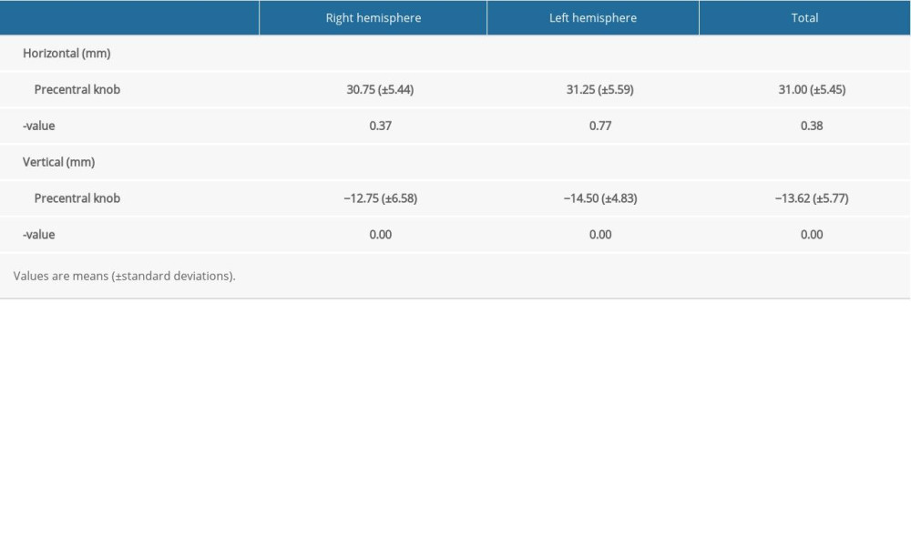
RESULTS: [color=black]The average distance from Cz to the location of the precentral knob in the horizontal direction was 30.75 mm in the right hemisphere, 31.25 mm in the left hemisphere, and 31.00 mm in both hemispheres. The average distance from Cz to the location of the precentral knob in the vertical direction was -12.75 mm in the right hemisphere, -14.50 mm in the left hemisphere, and -13.62 mm in both hemispheres. [/color]
CONCLUSIONS: [color=black]This study showed that the anatomical location of the precentral knob in normal subjects could be identified using a brain navigator and this method may be used clinically for patients requiring neuromodulation.[/color]
Keywords: Motor Cortex, Transcranial Direct Current Stimulation, Transcranial Magnetic Stimulation, Hand, Adult, Brain Mapping, Female, Humans, Male, Psychomotor Performance, Reference Values, young adult
Background
Non-invasive brain stimulation such as repetitive transcranial magnetic stimulation (rTMS) and transcranial direct-current electrical stimulation (tDCS) are often used for neuromodulation in patients with brain injury [1–5]. Accurate localization of the target neural structure is required for the precise use of these techniques. The precentral knob of the precentral gyrus, the original site for hand somatotopy in the corticospinal tract, is an important target for neuromodulation [6,7]. Hand function is essential for activities of daily living and hand disability is a serious disabling sequelae of some brain diseases; for example, a previous study reported that approximately half of stroke patients showed hand disability as a permanent sequela [6–9]. Various methods have been employed to locate the precentral knob: brain magnetic resonance image (MRI), intra-operative recording, electroencephalography, TMS, and functional MRI (fMRI) [6,10–23]. However, TMS and electroencephalography have poor spatial resolution [15,24]. By contrast, although fMRI has high spatial resolution, it should be used in combination with other methods such as a brain navigator to pinpoint the anatomical location on the scalp [11,12,25]. As a result, the detailed anatomical location of the precentral knob that can be easily used clinically has not clearly elucidated.
A brain navigator system allows an excellent three-dimensional orientation through real-time graphic anatomical interaction and visualizes any position on the scalp in relation to the anatomical brain structure below it [26–29]. Previous studies using a brain navigator have overlaid a brain region of interest on the scalp based on the results of fMRI and/or TMS for the precentral knob [11,12]. However, these studies involved complex and time-consuming experiments in a small number of subjects and did not present detailed data for a precise anatomical location that can be used clinically. By contrast, the brain navigator system, using an optical tracking method, allows precise localization of a target neural structure within a shorter time [26,28,29]. Regarding the anatomical location, a previous study reported that the distance between the C3 position on the international 10–20 system and the center of gravity on TMS for hand muscles was on average 19.2 mm using a brain navigator [23]. However, the results of that study were not easily translated into clinical practice because the C3 position is different in each subject.
The present study aimed to describe the use of an optical tracking brain navigator to identify the anatomical location of the precentral knob in the precentral gyrus in 20 normal right-handed subjects.
Material and Methods
ETHICS APPROVAL:
The retrospective study was performed in accordance with the requirements of the Declaration of Helsinki research guidelines and the study protocol was approved by the Institutional Review Board of Yeungnam University hospital (ethics approval number: YUMC-2021-03-014).
SUBJECTS:
The sample size of this study was calculated to be 16 (power=0.8, α=0.05) using power analysis software (G*Power v3.1.9.7; Heinrich-Heine-Universität Düsseldorf, Düsseldorf, Germany). Twenty subjects (13 males, 7 females; mean age 33.75±10.28 years, range 22–63) were enrolled in this study according to the following inclusion and exclusion criteria [28]: the inclusion criteria were right-handed subjects with a healthy normal state; the exclusion criteria were previous history of any neurological, psychiatric, or physical illness. All subjects understood the purpose of this study and provided written, informed consent prior to participation.
MAGNETIC RESONANCE IMAGING:
MRI images were acquired using a 3.0-T Siemens MR scanner (Siemens, Erlangen, Germany). A T2-weighted, three-dimensional gradient-echo sequence was used to produce 256 continuous axial slices of 1-mm thickness covering the entire cerebrum and the fatty head-surface markers in navigator mode.
LOCALITE TMS NAVIGATOR SYSTEM:
The Localite TMS navigator (Localite GmbH, Sankt Augustin, Germany) is an image-guided tool to assist in the positioning of a TMS coil over a subject’s brain. A small object called a tracker was attached to the subject and this tracker was monitored by an optical position sensor (optical camera). This information was sent to a computer that, after a calibration procedure, displayed the position and orientation of the point on the individual MRI of each subject. This Localite TMS navigator system was used for MR imaging, and nasion, left eye, and right eye were measured in each subject with a tracker to allow for correction for each subject. Then, we set target and surface points on the individual MRI of each subject [28]. The horizontal and vertical distances to the surface point were measured from the midline central (Cz) using the point marker; when the point marker pointed to a location on the scalp, the navigator system could indicate where that location is [28].
DETERMINATION OF THE LOCATION OF TARGET AND SURFACE POINT:
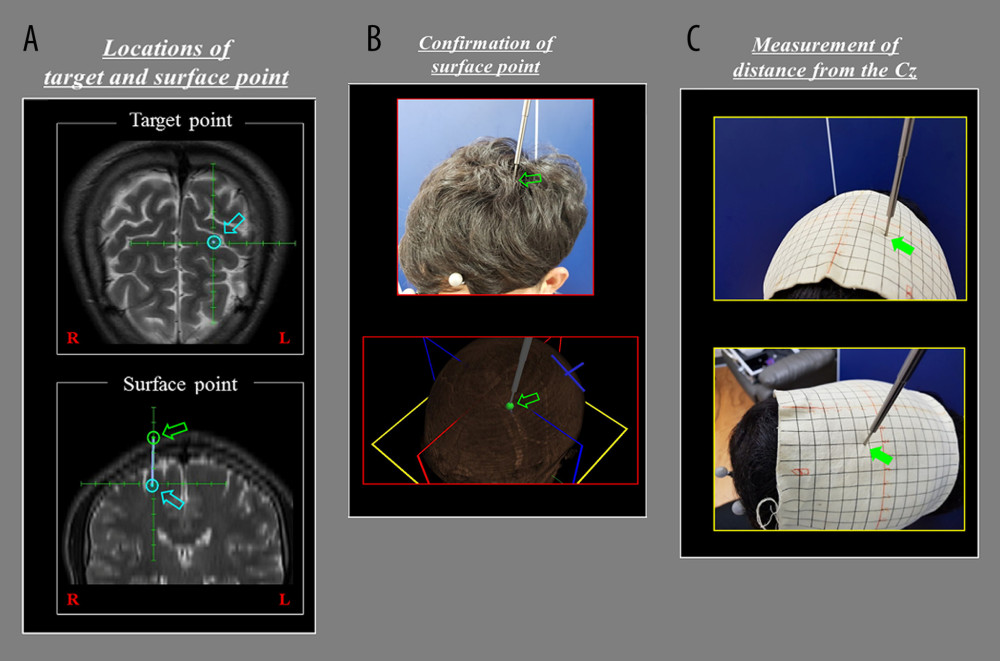
We determined the location of the target and surface points as follows [28]: 1) the target point: an omega-shaped area in the precentral knob in front of the precentral sulcus; we adjusted the round target point based on the posterior margin of the precentral knob [6,7] (Figure 1A), and 2) the surface point: the area of the scalp in the vertical direction from the target point that was parallel to the sagittal plane (Figure 1A). After we determined the locations of the target and surface points, the position of the surface point on the scalp in the brain navigator system was confirmed by the point marker (Figure 1B). A grid marked at 1-cm intervals was placed on each subject’s head to measure the horizontal and vertical distances from the midline central (Cz) to the obtained surface point (Figure 1C).
STATISTICAL ANALYSIS:
Statistical analysis was performed by using SPSS 21.0 for Windows (SPSS, Chicago, IL, USA). The independent
Result
Results of the distances between the precentral knob and the midline central (Cz) are summarized in Table 1. In the horizontal direction, the average distance from midline central (Cz) to the location of the precentral knob was 30.75 mm in the right hemisphere, 31.25 mm in the left hemisphere, and 31.00 mm in both hemispheres. Significant differences were not observed between right and left hemispheres in the horizontal direction for the precentral knob (
Discussion
In the current study, we used a brain navigator system to identify the anatomical location of the precentral knob on the scalp from the midline central (Cz) in 20 normal subjects. We found that the precentral knob on scalp was located, on average, 31.00 mm in the horizontal direction and −13.62 mm in the vertical direction from the midline central (Cz). Practically, for rTMS or tDCS, the anatomical location data in the tangential direction on the scalp is more useful for clinical application. However, we measured the anatomical location on the scalp in the vertical direction from a target point that was parallel to the sagittal plane because the vertical direction can be advantageous to reduce variability compared to the tangential direction. Thus, we think that our results provide useful reference data for clinical application in neuromodulation. A previous study reported that the average distance between the C3 position on the international 10–20 system and the center of gravity on TMS for hand muscles was 19.2 mm [14]. Because we measured the distance from the C3 position on the international 10–20 system, direct comparison of our results with the results of the above study is impossible. However, it appears that our results are easier to use clinically because localization of the midline central (Cz) is easier than localization of C3 in each subject.
Many studies have demonstrated the anatomical location of the precentral knob using brain MRI, intra-operative recording, electroencephalography, TMS, and fMRI [8,10–23]. By contrast, only a few studies have tried to determine the anatomical location of the precentral knob using a brain navigator [11,12,14]. In 1999, Boroojerdi et al demonstrated the center of gravity on TMS for the hand muscles lay within the precentral knob which was identified by fMRI and a brain navigator system in 4 normal subjects [11]. In 2004, Neggers et al measured the distances between the center of gravity on TMS and the fMRI activation area for a hand muscle using a brain navigator in 4 normal subjects [12]. The authors found that the distance between the center of gravity on TMS and the maximal fMRI activation area was less than 5 mm [12]. In 2011, Koenraadt et al measured the anatomical location of the center of gravity on TMS for hand muscles using a brain navigator in 11 normal subjects [14]. The authors reported that the average distance between the C3 position on the international 10–20 system and the center of gravity on TMS for hand muscles was 19.2 mm [14]. In addition, all the centers of gravity on TMS were situated more anterior compared to the C3 position and majority of the centers of gravity were located more medial [14].
In the current study we attempted to clarify the anatomical location of the precentral knob using a brain navigator system. However, the limitations of this study should be considered. First, we set the target and surface points without TMS or fMRI data. Although we tried to clarify the anatomical data using the brain navigator system because the precentral knob is already a well-known area for hand somatotopy of the corticospinal tract, further studies combined with TMS or/and fMRI are necessary. Second, as we mentioned above, in this study, we measured the data in the vertical direction to reduce the variability that can occur by measuring in the tangential direction. However, additional data in the tangential direction are also necessary because rTMS or tDCS are usually applied in the tangential direction.
Conclusions
In conclusion, this study showed that the detailed anatomical location of the precentral knob in normal subjects could be identified using an optical tracking brain navigator and this method may be used clinically for patients requiring neuromodulation.
References
1. Paulus W, Transcranial direct current stimulation (tDCS): Suppl Clin Neurophysiol, 2003; 56; 249-54
2. Griskova I, Hoppner J, Ruksenas O, Dapsys K, Transcranial magnetic stimulation: The method and application: Medicina (Kaunas), 2006; 42; 798-804
3. Paquette C, Thiel A, Rehabilitation interventions for chronic motor deficits with repetitive transcranial magnetic stimulation: J Neurosurg Sci, 2012; 56; 299-306
4. Etoh S, Noma T, Takiyoshi Y, Effects of repetitive facilitative exercise with neuromuscular electrical stimulation, vibratory stimulation and repetitive transcranial magnetic stimulation of the hemiplegic hand in chronic stroke patients: Int J Neurosci, 2016; 126; 1007-12
5. He Y, Li K, Chen Q, Repetitive transcranial magnetic stimulation on motor recovery for patients with stroke: A prisma compliant systematic review and meta-analysis: Am J Phys Med Rehabil, 2020; 99; 99-108
6. Yousry TA, Schmid UD, Alkadhi H, Localization of the motor hand area to a knob on the precentral gyrus. A new landmark: Brain, 1997; 120(Pt 1); 141-57
7. Banker L, Tadi P, Neuroanatomy, precentral gyrus. [Updated 2021 Jul 31]: StatPearls [Internet], 2021, Treasure Island (FL), StatPearls Publishing Available from: https://www.ncbi.nlm.nih.gov/books/NBK544218
8. Olsen TS, Arm and leg paresis as outcome predictors in stroke rehabilitation: Stroke, 1990; 21; 247-51
9. Kim CH, Kim SJ, Motor recovery after stroke: Journal of Korean Academy of Rehabilitation Medicine, 1995; 19; 8
10. Lagerlund TD, Sharbrough FW, Jack CR, Determination of 10–20 system electrode locations using magnetic resonance image scanning with markers: Electroencephalogr Clin Neurophysiol, 1993; 86; 7-14
11. Boroojerdi B, Foltys H, Krings T, Localization of the motor hand area using transcranial magnetic stimulation and functional magnetic resonance imaging: Clin Neurophysiol, 1999; 110; 699-704
12. Neggers SF, Langerak TR, Schutter DJ, A stereotactic method for image-guided transcranial magnetic stimulation validated with fMRI and motor-evoked potentials: Neuroimage, 2004; 21; 1805-17
13. Okamoto M, Dan H, Sakamoto K, Three-dimensional probabilistic anatomical cranio-cerebral correlation via the international 10–20 system oriented for transcranial functional brain mapping: Neuroimage, 2004; 21; 99-111
14. Koenraadt KL, Munneke MA, Duysens J, Keijsers NL, TMS: A navigator for NIRS of the primary motor cortex?: J Neurosci Methods, 2011; 201; 142-48
15. Rich TL, Lixandrão M, Hoefer A, Reliability of the location of primary motor cortex using the international 10/20 electroencephalogram system (10/20 EEG): J Pediatr Neurol Neurosci, 2017; 1; 6-7
16. Rich TL, Menk JS, Rudser KD, Determining electrode placement for transcranial direct current stimulation: A comparison of EEG- versus tms-guided methods: Clin EEG Neurosci, 2017; 48; 367-75
17. Viganò L, Fornia L, Rossi M, Anatomo-functional characterisation of the human “hand-knob” : A direct electrophysiological study: Cortex, 2019; 113; 239-54
18. Willett FR, Deo DR, Avansino DT, Hand knob area of premotor cortex represents the whole body in a compositional way: Cell, 2020; 181(2); 396-409.e26
19. DeJong SL, Bisson JA, Darling WG, Shields RK, Simultaneous recording of motor evoked potentials in hand, wrist and arm muscles to assess corticospinal divergence: Brain Topogr, 2021; 34; 415-29
20. Karabanov AN, Shindo K, Shindo Y, Multimodal assessment of precentral anodal tdcs: Individual rise in supplementary motor activity scales with increase in corticospinal excitability: Front Hum Neurosci, 2021; 15; 639274
21. Simone L, Viganò L, Fornia L, Distinct functional and structural connectivity of the human hand-knob supported by intraoperative findings: J Neurosci, 2021; 41(19); 4223-33
22. Sollmann N, Krieg SM, Säisänen L, Julkunen P, Mapping of motor function with neuronavigated transcranial magnetic stimulation: A review on clinical application in brain tumors and methods for ensuring feasible accuracy: Brain Sci, 2021; 11; 897
23. Sondergaard RE, Martino D, Kiss ZHT, Condliffe EG, TMS motor mapping methodology and reliability: A structured review: Front Neurosci, 2021; 15; 709368
24. Rossini PM, Barker AT, Berardelli A, Non-invasive electrical and magnetic stimulation of the brain, spinal cord and roots: Basic principles and procedures for routine clinical application. Report of an ifcn committee: Electroencephalogr Clin Neurophysiol, 1994; 91; 79-92
25. Choi SH, Lee M, Wang Y, Hong B, Estimation of optimal location of EEG reference electrode for motor imagery based bci using fMRI: Conf Proc IEEE Eng Med Biol Soc, 2006; 1; 1193-96
26. Spetzger U, Laborde G, Gilsbach JM, Frameless neuronavigation in modern neurosurgery: Minim Invasive Neurosurg, 1995; 38; 163-66
27. Spetzger U, Gilsbach JM, Mosges R, The computer-assisted localizer, a navigational help in microneurosurgery: Eur Surg Res, 1997; 29; 481-87
28. Jang SH, Lee HD, Choi KH, Anatomic location of precentral knob in precentral gyrus as measured by the brain navigator system
29. Köhlert K, Jähne K, Saur D, Meixensberger J, Neurophysiological examination combined with functional intraoperative navigation using TMS in patients with brain tumor near the central region – a pilot study: Acta Neurochir (Wien), 2019; 161(9); 1853-64
In Press
15 Apr 2024 : Laboratory Research
The Role of Copper-Induced M2 Macrophage Polarization in Protecting Cartilage Matrix in OsteoarthritisMed Sci Monit In Press; DOI: 10.12659/MSM.943738
07 Mar 2024 : Clinical Research
Knowledge of and Attitudes Toward Clinical Trials: A Questionnaire-Based Study of 179 Male Third- and Fourt...Med Sci Monit In Press; DOI: 10.12659/MSM.943468
08 Mar 2024 : Animal Research
Modification of Experimental Model of Necrotizing Enterocolitis (NEC) in Rat Pups by Single Exposure to Hyp...Med Sci Monit In Press; DOI: 10.12659/MSM.943443
18 Apr 2024 : Clinical Research
Comparative Analysis of Open and Closed Sphincterotomy for the Treatment of Chronic Anal Fissure: Safety an...Med Sci Monit In Press; DOI: 10.12659/MSM.944127
Most Viewed Current Articles
17 Jan 2024 : Review article
Vaccination Guidelines for Pregnant Women: Addressing COVID-19 and the Omicron VariantDOI :10.12659/MSM.942799
Med Sci Monit 2024; 30:e942799
14 Dec 2022 : Clinical Research
Prevalence and Variability of Allergen-Specific Immunoglobulin E in Patients with Elevated Tryptase LevelsDOI :10.12659/MSM.937990
Med Sci Monit 2022; 28:e937990
16 May 2023 : Clinical Research
Electrophysiological Testing for an Auditory Processing Disorder and Reading Performance in 54 School Stude...DOI :10.12659/MSM.940387
Med Sci Monit 2023; 29:e940387
01 Jan 2022 : Editorial
Editorial: Current Status of Oral Antiviral Drug Treatments for SARS-CoV-2 Infection in Non-Hospitalized Pa...DOI :10.12659/MSM.935952
Med Sci Monit 2022; 28:e935952


