01 May 2022: Clinical Research
Association Between Thromboelastography and Coronary Heart Disease
Liuqiao Yang123ACDEF, Lei Ruan4ABD, Yanping ZhaoDOI: 10.12659/MSM.935340
Med Sci Monit 2022; 28:e935340
Abstract
BACKGROUND: Thromboelastography (TEG) is a novel blood viscoelasticity detection method revealing blood coagulation status and has been reported to be helpful in predicting clinical outcomes in patients with cardiovascular diseases (CVD). In this study, we aimed to investigate the association between TEG and CVD.
MATERIAL AND METHODS: A single-center case-control study was performed. Individuals who took TEG tests at Tongji Hospital in Wuhan, China from 2015 to 2019 were included. The nearest-neighbor Mahalanobis matching with replacement, within propensity score calipers of 0.25 was used to control the covariate imbalance between CVD patients and controls. Logistic regression analyses were conducted to assess the relationship between TEG and CVD. Subgroup and sensitivity analyses were performed to evaluate the robustness of the association between TEG and CVD.
RESULTS: After matching, a total of 151 participants were included in this study, with 83 patients having CVD (49 patients having coronary heart disease [CHD] and 34 patients having an ischemic stroke). By comparison, CHD patients had a significantly higher maximum amplitude (MA) (P=0.02) than controls. After multivariable adjustment, MA (OR 1.11, 95% CI 1.01-1.24, P=0.04) was independently associated with CHD. The association between MA and CHD remained robust across subgroups and in sensitivity analyses.
CONCLUSIONS: The current study suggests that MA is significantly associated with CHD. Enhanced platelet reactivity as described by high MA might be associated with risk of CHD. The exact role of MA in the measurement of CHD risk needs to be further examined in large-scale prospective cohort studies.
Keywords: Blood Coagulation, Cardiovascular Diseases, Thrombelastography, Case-Control Studies, Coronary Disease, Humans, Prospective Studies
Background
Cardiovascular diseases (CVD) are the predominant cause of mortality globally, accounting for approximately 17.9 million deaths every year [1]. Over 40% of deaths in China are attributed to CVD, and about 330 million people have CVD [2]. Driven by the expanding aging population and lifestyle changes associated with urbanization, the prevalence of CVD keeps increasing [3]. The increasing burden of CVD is one of the most important public health issues in China [4].
The primary cause of CVD, including coronary heart disease (CHD) and ischemic stroke, is atherosclerosis [5]. Atherosclerosis is an inflammatory disorder of the vascular system. It was hypothesized that atherosclerosis is “coagulation in the wrong place” [6]. Atherothrombosis is a highly procoagulant process in arteries [7], and many procoagulant factors of the coagulation system are involved in this process [8]. Researchers have investigated the association between coagulation and CVD occurrence, morbidity, and mortality [9], and higher rates of hypercoagulation were observed in patients with CVD. Assessment of blood coagulation status and using effective antiplatelet therapy in people with hypercoagulability can prevent atherosclerosis and decrease the incidence of CVD [10,11].
Blood coagulation status can be evaluated by fibrinogen level, activated partial thromboplastin time (APTT), and thromboelastography (TEG). TEG is a blood viscoelasticity detection method that dynamically monitors the whole process of blood coagulation and fibrinolysis, which was developed in 1948 and was at first used as a research tool [12,13]. TEG is now being broadly applied as a guiding tool for clotting factor replacement, platelet transfusion, and fibrinolysis treatment [14]. Some studies have noted that TEG is useful in predicting clinical outcomes in patients with CVD [15,16]. Conventional test indicators assess isolated factors of the coagulation system, but they are unable to reveal the role of these factors in the whole process of hemostasis [17]. In contrast, TEG can reveal the interaction of all components of coagulation, including platelets, fibrin, clotting factors, and thrombin, and provides an overall assessment of blood coagulation status [12]. Furthermore, the TEG test is evaluated in whole blood and can provide additional information about platelet function [18]. Given that hypercoagulability will increase the risk of CVD and TEG can assess the blood coagulation status, we hypothesized that TEG parameters were independent CVD risk factors, and hypercoagulability measured by TEG would be associated with an increased risk of CVD. We aimed to assess the association of TEG with CVD among individuals requiring routine physical examinations.
Material and Methods
STUDY DESIGN:
This study was a single-center retrospective case-control study and was approved with a waiver of informed consent by the Ethics Committee of Tongji Medical College, Huazhong University of Science and Technology (TJ-IRB20191215).
Individuals requiring routine physical examinations at the physical examination center of Tongji Hospital in Wuhan, China from 2015 to 2019 were included. This is the largest physical examination center in Wuhan. TEG is always used to guide blood transfusion during cardiac surgery (such as percutaneous coronary intervention and coronary artery bypass grafting) and to monitor antiplatelet therapy. The most common indications for antiplatelet medications are cardiovascular and cerebrovascular diseases, atrial fibrillation, and peripheral arterial disease. Participants who were curious about their blood coagulation were also offered the TEG test.
The inclusion criteria were: (1) age >20 years old; (2) having TEG data. The exclusion criteria were: (1) presence of malignancy; (2) liver failure (Child-Pugh score ≥7) or kidney failure (eGFR <60 ml/[min·1.73 m2]); (3) other heart diseases, including congenital heart disease, rheumatic heart disease, and severe cardiac arrhythmia; (4) currently on anticoagulant therapy (those receiving anticoagulants might have venous thromboembolism or atrial fibrillation and have abnormal coagulation parameters); (5) missing data; and (6) surgery in the last 3 months. Clinical data, including demographic information, results of routine laboratory tests, and TEG test results, were collected by trained doctors.
Cases were participants who had CVD (CHD or ischemic stroke). CHD included coronary artery disease, ischemic heart disease, myocardial infarction, and coronary revascularization. Ischemic stroke was defined as focal neurological deficits lasting ≥24 h and in the absence of signs of cerebral hemorrhage on computed tomography. Controls were participants who had no CVD.
THROMBOELASTOGRAPHY:
Venous blood (3 ml) was collected and analyzed in the TEG 5000 device (Haemonetics® Corporation, Braintree, USA) using kaolin as the coagulation activator. TEG parameters were recorded, including reaction time (R, min), kinetic time (K, min), angle (α, degrees), and maximum amplitude (MA, mm).
STATISTICAL ANALYSIS:
Categorical variables were presented as counts (percentage). Continuous variables were presented as means±standard deviation (SD), if normally distributed; otherwise, they were presented as median (interquartile range). Statistical comparisons between case and control groups were performed using the chi-square or the Fisher exact test for categorical variables and using the
In observational studies, imbalance in covariates is an inevitable problem and is likely to introduce bias. The nearest-neighbor Mahalanobis matching with replacement, within propensity score calipers of 0.25 was used to control the covariate imbalance between cases and controls [19]. Variables that might affect the outcome of interest and that were unbalanced between cases and controls were chosen as matched factors. These covariates included age, sex, year of physical examination, and medication history (antiplatelet drugs and lipid-lowering drugs).
Four logistic regression models were performed to examine the association between TEG and CVD. Model 1 only included TEG values that were significantly different between cases and controls. Model 2 additionally included matched factors (age, sex, year of physical examination, and medication history), and model 3 additionally adjusted for laboratory indicators (low-density lipoprotein cholesterol [LDL-C] and platelet count). Model 4 included variables with a
To investigate whether the association between TEG and CVD were influenced by participants’ basic characteristics, we conducted subgroup analyses. Subgroups were defined according to age (<60 years or 60 years or older), sex, medication with an antiplatelet drug, and medication with a lipid-lowering drug.
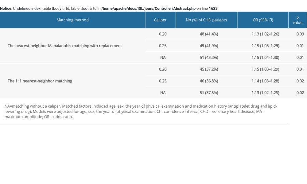
To evaluate the robustness of our results, several sensitivity analyses were performed based on different matching methods. Firstly, the nearest-neighbor Mahalanobis matching with replacement, within propensity score calipers of 0.20, 0.25, and without a caliper were performed. Secondly, the 1: 1 nearest-neighbor matching, within propensity score calipers of 0.20, 0.25, and without a caliper were performed.
Results
BASIC CHARACTERISTICS AND TEG PARAMETERS:
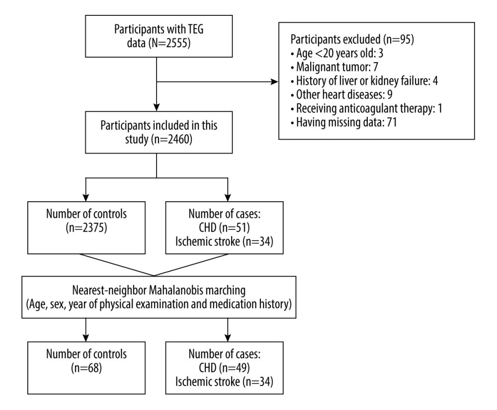
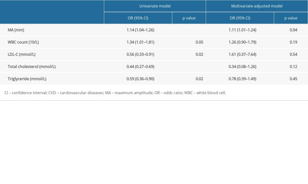
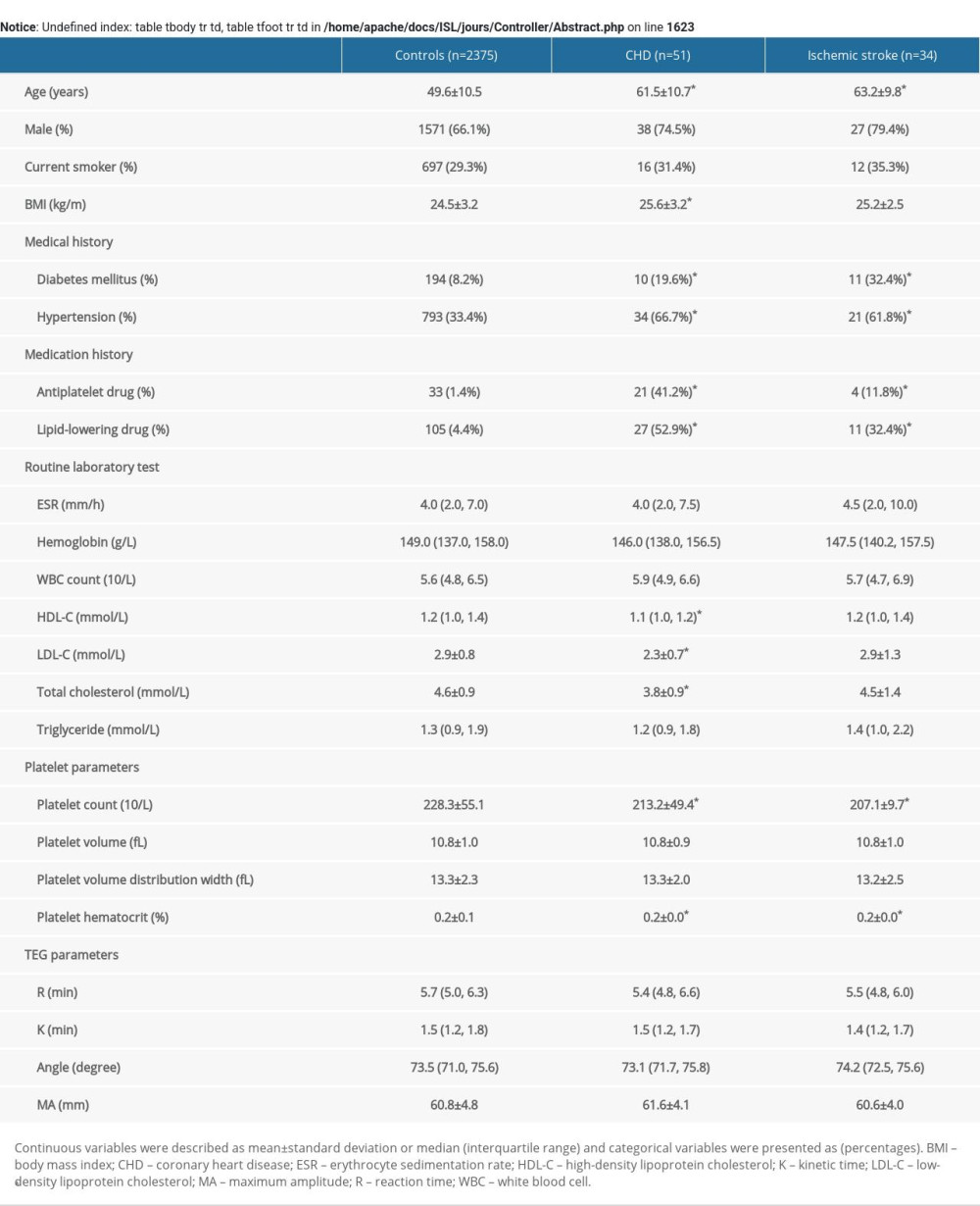
The study flow chart is shown in Figure 1. Altogether, 2555 participants met the inclusion criteria and 95 participants met the exclusion criteria, leaving 2460 participants enrolled in this study. After matching, we have had 68 controls and 83 cases (49 having CHD and 34 having an ischemic stroke). The basic characteristics of the 3 groups (control group, CHD group, and ischemic stroke group) are summarized in Table 1. Data before matching are summarized in Supplementary Table 1. Demographic background, routine laboratory data, and platelet parameters of the CHD group and the ischemic stroke group were compared with controls. Patients with CHD had lower LDL-C (P=0.02), lower total cholesterol (TC) (P<0.01), and lower triglyceride (P=0.03).
The TEG results are demonstrated in Table 2. In the comparison of TEG parameters, CHD patients had a significantly higher MA (P=0.02) than controls. However, other TEG parameters (including R, K, and angle) of patients were not significantly different from controls.
CORRELATION BETWEEN BASIC CHARACTERISTICS AND TEG PARAMETERS:
Spearman’s correlation analysis was performed to assess the association between basic characteristics and TEG parameters. The results of correlation analysis are displayed in Table 3. Our results show that both R and K values were positively correlated with hemoglobin (P<0.01) but negatively correlated with age (P<0.01). Angle and MA were negatively correlated with hemoglobin (P<0.01), but positively correlated with erythrocyte sedimentation rate (ESR) (P<0.01). Regarding the platelet parameters, our results show that MA was positively correlated with platelet count (P<0.01) and platelet hematocrit (P<0.01).
LOGISTIC REGRESSION:
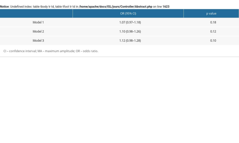
Logistic regression analysis was performed to examine the association between MA and CHD. In model 1, MA was positively associated with CHD (OR 1.14, 95% CI 1.04–1.26, P<0.01). After adjusting for matched factors (age, sex, year of physical examination, and medication history), the results were similar (MA: OR 1.17, 95% CI 1.04–1.32, P<0.01). After additionally adjusting for LDL-C and platelet count, the positive association between MA and CHD remained (OR 1.19, 95% CI 1.05–1.37, P=0.01). The results of model 4 are shown in Table 4. After adjusting for other factors (WBC count, LDL-C, TC, and triglyceride), MA was still associated with CHD (OR 1.11, 95% CI 1.01–1.24, P=0.04). Logistic regression results for ischemic stroke are shown in Supplementary Table 2. No significant association was found between TEG and ischemic stroke.
SUBGROUP ANALYSES AND SENSITIVITY ANALYSES:
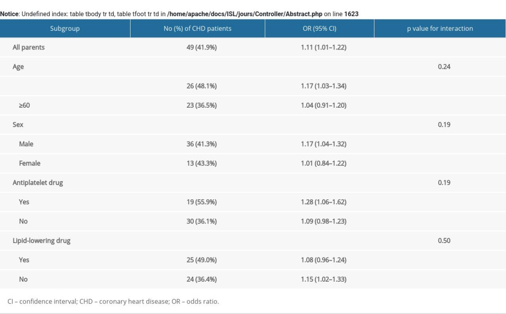
Subgroup analyses were conducted after stratification for age, sex, medication with an antiplatelet drug, and medication with a lipid-lowering drug. No significant interaction was observed for CHD in any subgroups (P value for interaction >0.05; Supplementary Table 3). Sensitivity analyses was performed to evaluate the robustness of our results, showing that the association between MA and CHD was been broadly consistent in sensitivity analyses (Supplementary Table 4).
Discussion
Our study demonstrates that MA was significantly higher in patients with CHD. Higher MA was associated with increased risk of CHD in different models. Furthermore, the association of MA with CHD remained robust across subgroups and in sensitivity analyses.
Several previous publications showed the good efficacy of TEG parameters in assessing the risk of CVD. For example, Yao et al [15] found that a higher MA level was related to the severity of acute stroke (r=0.205,
The present study also evaluated the relationship between TEG parameters and blood composition, including hemoglobin and platelets. A previous study suggested that when hemoglobin decreased, the plasma protein content increased and subsequently lead to an increase in the strength of blood clots [26]. Consistent with these findings, our results showed that both R and K values were positively correlated with hemoglobin, while angle and MA were negatively correlated with hemoglobin. Platelet aggregation is a critical step in the formation of clots [27]. Thus, it is reasonable to find a positive correlation between MA and platelet component (platelet count and platelet hematocrit).
Numerous studies have shown that lipid profile, including LDL-C, TC, and triglyceride, was a major risk factor for CVD. Higher levels of LDL-C may contribute to most clinical CVD events. The Chinese guidelines for the management of dyslipidemia recommend that the desirable level of LDL-C for primary prevention of CVD is <2.6 mmol/L [28]. Although LDL-C is a well-studied traditional risk factor for CVD, patients with normal blood lipid levels can also have CVD. In the current study, a majority of patients with CVD (66.3%) had LDL <2.6 mmol/L and 54.4% of controls had LDL <2.6 mmol/L. In addition, we observed that patients with CHD, even those not taking lipid-lowering medication, had lower LDL-C, TC, and triglyceride levels than controls, which was concordant with the findings described by Sachdeva et al [29]. The reason why people with lower lipid levels still have CVD might be changes in the prevalence of other CVD risk factors, such as smoking, diabetes, and hypertension [30]. Our study suggests that hypercoagulability as described by high MA might be one of the fundamental reasons. In a previous study, researchers also demonstrated in atherosclerosis animal models that platelet aggregation and activation preceded the occurrence of atherosclerosis and might play a dominant role in the development of CVD [31]. Alternatively, stronger platelet reactivity may contribute to the formation of atherosclerosis and eventually lead to CVD. Deeper understanding of biomarkers other than traditional risk factors may prompt development of more rapid and accurate interventions for CVD.
Our study showed that MA was significantly higher in patients with CHD. It is likely that CHD patients have higher platelet reactivity and have an increased risk of thrombosis. If these findings are verified in large-scale studies, the adjustment of MA to a proper level with personalized antiplatelet therapy in individuals with CHD might be able to improve their prognosis and reduce the risk of unfavorable outcomes.
There were some limitations in our study. First, it was a single-center, retrospective study. Second, the number of CVD patients (n=83) included in this study was relatively small. The study population did not include anyone with acute illness. Therefore, the patients in this study might not be representative of other populations. It should be noted that we also enrolled participants taking antiplatelet drugs, which might affect the TEG test, so these findings must be interpreted with caution. Third, a selection bias was possible because of the inclusion criteria (participants who underwent TEG). Fourth, a lack of traditional coagulation analysis, such as fibrinogen level and APTT, was a major limitation of the present study.
Conclusions
The results of the present study suggest that MA is significantly associated with CHD. Stronger platelet reactivity as described by high MA may be associated with the risk of CHD. Further large-scale studies are warranted to investigate the exact role of MA in the measurement of CHD risk.
Tables
Table 1. Basic characteristics of study participants.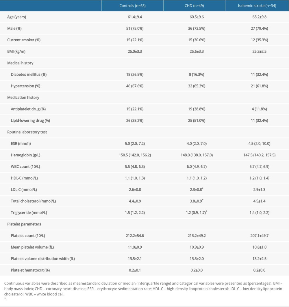 Table 2. TEG parameters of study participants.
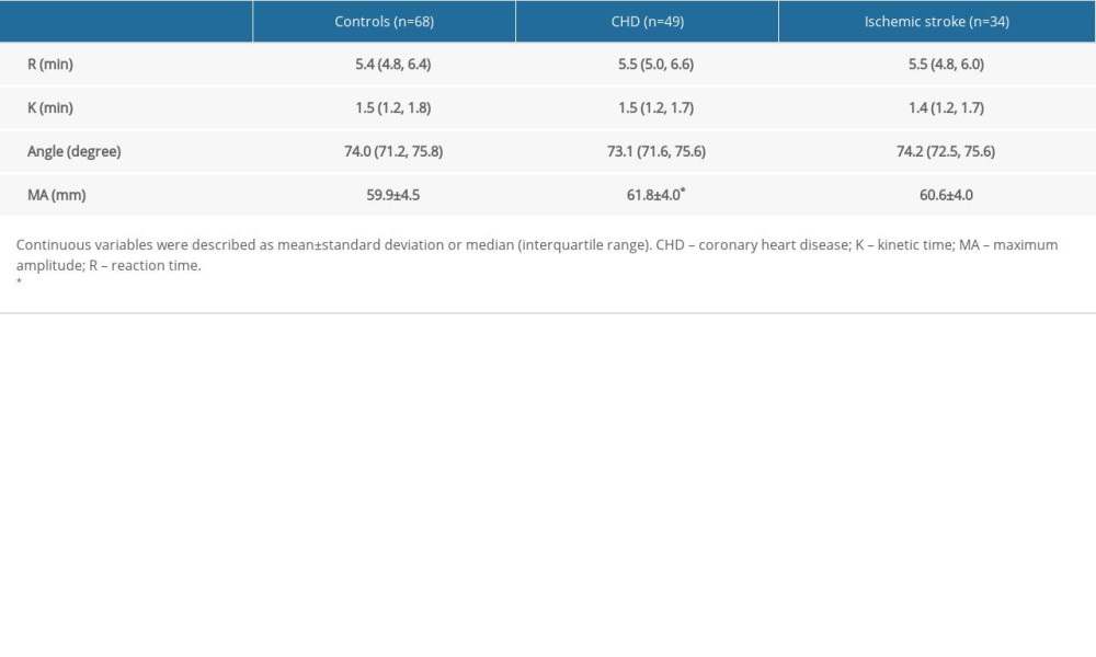 Table 3. The correlation between basic characteristics and TEG parameters.
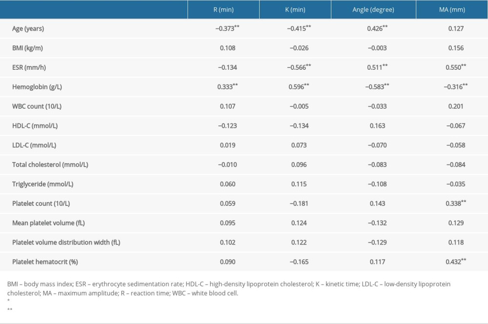 Table 4. Univariate and multivariate logistic regression of CVD.
 Supplementary Table 1. Basic characteristics and TEG parameters of study participants before matching.
 Supplementary Table 2. Logistic regression of MA for ischemic stroke.
 Supplementary Table 3. Results of subgroup analyses after stratification for age, sex, medication of antiplatelet drug, and medication of lipid-lowering drug.
 Supplementary Table 4. Results of sensitivity analyses examining association between MA and CHD.
References
1. GBD 2017 Causes of Death Collaborators, Global, regional, and national age-sex-specific mortality for 282 causes of death in 195 countries and territories, 1980–2017: A systematic analysis for the Global Burden of Disease Study 2017: Lancet, 2018; 392(10159); 1736-88
2. Zhao D, Liu J, Wang M, Epidemiology of cardiovascular disease in China: Current features and implications: Nat Rev Cardiol, 2019; 16(4); 203-12
3. The Writing Committee of the Report on Cardiovascular Health and Diseases in China, Report on cardiovascular health and diseases in China 2019: An updated summary: Chin Circ J, 2020; 35(09); 833-54
4. Zhou M, Wang H, Zeng X, Mortality, morbidity, and risk factors in China and its provinces, 1990–2017: A systematic analysis for the Global Burden of Disease Study 2017: Lancet, 2019; 394(10204); 1145-58
5. Hansson GK, Inflammation, atherosclerosis, and coronary artery disease: N Engl J Med, 2005; 352(16); 1685-95
6. Lowe G, Rumley A, The relevance of coagulation in cardiovascular disease: What do the biomarkers tell us?: Thromb Haemost, 2014; 112(5); 860-67
7. Ten Cate H, Hackeng TM, García de Frutos P, Coagulation factor and protease pathways in thrombosis and cardiovascular disease: Thromb Haemost, 2017; 117(7); 1265-71
8. Borissoff JI, Heeneman S, Kilinç E, Early atherosclerosis exhibits an enhanced procoagulant state: Circulation, 2010; 122(8); 821-30
9. Womack CJ, Nagelkirk PR, Coughlin AM, Exercise-induced changes in coagulation and fibrinolysis in healthy populations and patients with cardiovascular disease: Sports Med Auckl NZ, 2003; 33(11); 795-807
10. Xia S, Du X, Guo L, Sex differences in primary and secondary prevention of cardiovascular disease in China: Circulation, 2020; 141(7); 530-39
11. Borissoff JI, Spronk HMH, ten Cate H, The hemostatic system as a modulator of atherosclerosis: N Engl J Med, 2011; 364(18); 1746-60
12. Hobson AR, Agarwala RA, Swallow RA, Thrombelastography: Current clinical applications and its potential role in interventional cardiology: Platelets, 2006; 17(8); 509-18
13. Hartert H, Thrombelastography, a method for physical analysis of blood coagulation: Z Gesamte Exp Med, 1951; 117(2); 189-203
14. Sakai T, Comparison between thromboelastography and thromboelastometry: Minerva Anestesiol, 2019; 85(12); 1346-56
15. Yao X, Dong Q, Song Y, Thrombelastography maximal clot strength could predict one-year functional outcome in patients with ischemic stroke: Cerebrovasc Dis, 2014; 38(3); 182-90
16. Rafiq S, Johansson PI, Ostrowski SR, Hypercoagulability in patients undergoing coronary artery bypass grafting: Prevalence, patient characteristics and postoperative outcome: Eur J Cardiothorac Surg, 2012; 41(3); 550-55
17. Dias JD, Sauaia A, Achneck HE, Thromboelastography-guided therapy improves patient blood management and certain clinical outcomes in elective cardiac and liver surgery and emergency resuscitation: A systematic review and analysis: J Thromb Haemost, 2019; 17(6); 984-94
18. Ganter MT, Hofer CK, Coagulation monitoring: current techniques and clinical use of viscoelastic point-of-care coagulation devices: Anesth Analg, 2008; 106(5); 1366-75
19. Patorno E, Grotta A, Bellocco R, Schneeweiss S, Propensity score methodology for confounding control in health care utilization databases: Epidemiol Biostat Public Health, 2013; 10(3); e89401-16
20. Kreutz RP, Schmeisser G, Maatman B, Fibrin clot strength measured by thrombelastography and outcomes after percutaneous coronary intervention: Thromb Haemost, 2017; 117(2); 426-28
21. Yuan Q, Yu L, Wang F, Efficacy of using thromboelastography to detect coagulation function and platelet function in patients with acute cerebral infarction: Acta Neurol Belg, 2021; 121(6); 1661-67
22. Salooja N, Perry DJ, Thrombelastography: Blood Coagul Fibrinolysis, 2001; 12(5); 327-37
23. McCrath DJ, Cerboni E, Frumento RJ, Thromboelastography maximum amplitude predicts postoperative thrombotic complications including myocardial infarction: Anesth Analg, 2005; 100(6); 1576-83
24. Cheng D, Zhao S, Hao Y, Net platelet clot strength of thromboelastography platelet mapping assay for the identification of high on-treatment platelet reactivity in post-PCI patients: Biosci Rep, 2020; 40(7); BSR20201346
25. Gilbert BW, Bissell BD, Santiago RD, Rech MA, Tracing the lines: A review of viscoelastography for emergency medicine clinicians: J Emerg Med, 2020; 59(2); 201-15
26. Noorman F, Hess JR, The contribution of the individual blood elements to the variability of thromboelastographic measures: Transfusion, 2018; 58(10); 2430-36
27. Ranucci M, Di Dedda U, Baryshnikova E, Platelet contribution to clot strength in thromboelastometry: Count, function, or both?: Platelets, 2020; 31(1); 88-93
28. 2016 Chinese guideline for the management of dyslipidemia in adults: Zhonghua Xin Xue Guan Bing Za Zhi, 2016; 44(10); 833-53 [in Chinese]
29. Sachdeva A, Cannon CP, Deedwania PC, Lipid levels in patients hospitalized with coronary artery disease: An analysis of 136,905 hospitalizations in Get With The Guidelines: Am Heart J, 2009; 157(1); 111-117e2
30. Joseph P, Leong D, McKee M, Reducing the global burden of cardiovascular disease, Part 1: The epidemiology and risk factors: Circ Res, 2017; 121(6); 677-94
31. Lebas H, Yahiaoui K, Martos R, Boulaftali Y, Platelets are at the nexus of vascular diseases: Front Cardiovasc Med, 2019; 6; 132
Tables
 Table 1. Basic characteristics of study participants.
 Table 2. TEG parameters of study participants.
 Table 3. The correlation between basic characteristics and TEG parameters.
 Table 4. Univariate and multivariate logistic regression of CVD.
 Table 1. Basic characteristics of study participants.
 Table 2. TEG parameters of study participants.
 Table 3. The correlation between basic characteristics and TEG parameters.
 Table 4. Univariate and multivariate logistic regression of CVD.
 Supplementary Table 1. Basic characteristics and TEG parameters of study participants before matching.
 Supplementary Table 2. Logistic regression of MA for ischemic stroke.
 Supplementary Table 3. Results of subgroup analyses after stratification for age, sex, medication of antiplatelet drug, and medication of lipid-lowering drug.
 Supplementary Table 4. Results of sensitivity analyses examining association between MA and CHD.
 In Press
15 Apr 2024 : Laboratory Research
The Role of Copper-Induced M2 Macrophage Polarization in Protecting Cartilage Matrix in OsteoarthritisMed Sci Monit In Press; DOI: 10.12659/MSM.943738
07 Mar 2024 : Clinical Research
Knowledge of and Attitudes Toward Clinical Trials: A Questionnaire-Based Study of 179 Male Third- and Fourt...Med Sci Monit In Press; DOI: 10.12659/MSM.943468
08 Mar 2024 : Animal Research
Modification of Experimental Model of Necrotizing Enterocolitis (NEC) in Rat Pups by Single Exposure to Hyp...Med Sci Monit In Press; DOI: 10.12659/MSM.943443
18 Apr 2024 : Clinical Research
Comparative Analysis of Open and Closed Sphincterotomy for the Treatment of Chronic Anal Fissure: Safety an...Med Sci Monit In Press; DOI: 10.12659/MSM.944127
Most Viewed Current Articles
17 Jan 2024 : Review article
Vaccination Guidelines for Pregnant Women: Addressing COVID-19 and the Omicron VariantDOI :10.12659/MSM.942799
Med Sci Monit 2024; 30:e942799
14 Dec 2022 : Clinical Research
Prevalence and Variability of Allergen-Specific Immunoglobulin E in Patients with Elevated Tryptase LevelsDOI :10.12659/MSM.937990
Med Sci Monit 2022; 28:e937990
16 May 2023 : Clinical Research
Electrophysiological Testing for an Auditory Processing Disorder and Reading Performance in 54 School Stude...DOI :10.12659/MSM.940387
Med Sci Monit 2023; 29:e940387
01 Jan 2022 : Editorial
Editorial: Current Status of Oral Antiviral Drug Treatments for SARS-CoV-2 Infection in Non-Hospitalized Pa...DOI :10.12659/MSM.935952
Med Sci Monit 2022; 28:e935952


