22 March 2023: Clinical Research
Application of Musculoskeletal Ultrasound Guidance in Lumbar Transforaminal Endoscopic Surgery for Puncture and Catheterization
Xingen Zhang1ABCDEFG*, Xianjie Sun1ABCEDOI: 10.12659/MSM.937692
Med Sci Monit 2023; 29:e937692
Abstract
BACKGROUND: Foraminal puncture is a key step in foraminal endoscopic surgery, but the radiation dosage poses a clinical risk to patients. To reduce the radiation dosage, we investigated the feasibility and clinical effect of endoscopic transforaminal puncture through the use of the musculoskeletal ultrasound-guided lumbar percutaneous posterolateral approach.
MATERIAL AND METHODS: Retrospective data of 80 lumbar percutaneous posterolateral approach endoscopic surgery patients from March 2018 to June 2021 were analyzed. The clinical efficacy was assessed by visual analogue scale (VAS) and Oswestry disability index (ODI) during the follow-up.
RESULTS: Between the musculoskeletal ultrasound-guided group and the C-arm X-ray machine fluoroscopy group, the puncture time of the musculoskeletal ultrasound-guided group was significantly shorter than that of the C-arm X-ray machine fluoroscopy group (t=13.113, P=0.010). The radiation received in the ultrasound guidance group was significantly less than in the C-arm X-ray group. There was no difference in ODI values between the 2 groups before surgery (t=0.195, P=0.286), 3 months after surgery (t=0.235, P=0.092), and 1 year after surgery (t=0.168, P=0.173). There was no significant difference in VAS scores between the 2 groups before surgery (t=0.715, P=0.610), 3 months after surgery (t=0.367, P=0.192), and 1 year after surgery (t=0.496, P=0.390).
CONCLUSIONS: Our data demonstrate that musculoskeletal ultrasound can accurately guide the lumbar percutaneous posterolateral approach for endoscopic foraminal puncture, which can significantly reduce the puncture time and the amount of X-ray radiation.
Keywords: Lumbar Vertebrae, Magnetic Resonance Imaging, Ultrasonic Therapy, Humans, Intervertebral Disc Displacement, Diskectomy, Percutaneous, Spinal Puncture, Treatment Outcome, Catheterization
Background
Foraminal puncture is a key step in foraminal endoscopic surgery. The traditional puncture methods include blind detection and radiation-guided puncture such as X-ray and CT. X-ray and CT are better for imaging the bony structure of the spine, which can improve the accuracy of intervertebral foraminal puncture and reduce damage to surrounding tissues [1,2]. However, the unavoidable problem is that CT equipment is expensive and cannot obtain real-time images. It is complicated to operate, and it is difficult for both doctors and patients to avoid radiation damage, limiting its wide use [3,4]. In contrast, ultrasound imaging does not cause radiation injury, it is real-time, and can be visualized during the surgery. Ultrasonic imaging can dynamically generate images in real time without radiation hazards to operators or patients. However, ultrasound cannot penetrate bony tissue and cannot detail the internal structure of bony tissue. Only ultrasound can display the irregular structure of the spinal bone surface and the corresponding spinal structures such as intervertebral joints, spinous processes, and posterior tuberosities of cervical transverse processes, as well as showing the internal structures of the spinal canal, spinal cord, and cerebrospinal fluid. In the clinic, ultrasound can identify the bony landmarks of the spine and the internal structure of the spinal canal, and confirm the position, distance, and angle of related tissues to facilitate spinal-related anesthesia or analgesia [5–10]. In recent years, the use of ultrasound to guide intervertebral foramen puncture has greatly increased, but due to the deep tissue structure of the intervertebral foramen and the obstruction of surrounding bones, it is still difficult to image using ultrasound, so that the whole process of ultrasound imaging and guided puncture are carried out gradually. Ultrasound can show liquid diffusion in real time and clearly display the blood vessels, thereby avoiding blood vessel damage, preventing drugs from entering by mistake, and improving the accuracy and safety of intervertebral foraminal puncture.
In this study, 80 patients diagnosed with lumbar intervertebral disc herniation from March 2018 to June 2021 were treated by musculoskeletal ultrasound-guided foraminal puncture in the outpatient clinic. The curative effect was satisfactory. Musculoskeletal ultrasound imaging can accurately guide the lumbar percutaneous posterolateral approach for endoscopic foraminal puncture, which can significantly reduce the puncture time and the amount of X-ray radiation.
Material and Methods
GENERAL INFORMATION:
A total of 80 patients diagnosed with lumbar disc herniation in our hospital from March 2018 to June 2021 were selected. There were 40 males and 40 females, and the average age was 33.2 years. All the patients provided signed informed consent to the treatment used in this study.
Patient inclusion criteria included: (1) manifested as low back pain accompanied by lower-extremity radicular pain, and its localized segment L4/5; (2) traditional treatment was ineffective for more than 6 months; (3) imaging showed L4/5 single segment highlighted.
Patient exclusion criteria included: (1) a history of spinal trauma or surgery, resulting in structural defects or discontinuities of the spine; (2) a history of spinal tumors; (3) spinal deformity (vertebral body destruction, or rotation); (4) obvious spinal degenerative changes; (5) equipped with a pacemaker (electromagnetic tracking system that interferes with ultrasound).
The following routine examinations were required for each patient before the surgery, including: (1) anterior and lateral lumbar spine X-rays to determine the shape and size of the intervertebral foramen, the height of the iliac spine and the shape of the spine, and to determine the target whip point and range of puncture; (2) lumbar vertebrae MRI scan was performed to observe the position and degree of intervertebral disc herniation, whether it was accompanied by other diseases such as lumbar spinal stenosis, and determine the position and direction of the working channel.
The guiding methods of foraminal puncture (C-arm machine-guided, musculoskeletal ultrasound-guided) were randomly determined before the surgery, and the patients were divided into 2 groups, including 40 cases in the C-arm machine-guided group and 40 cases in the musculoskeletal ultrasound-guided group. During the surgery, the patient was prone on the Jackson spine operating table, and local anesthesia was performed with 1% lidocaine at the location to be punctured. Surgery was performed using the SPINENDOS Spinal Endoscopy System. The C-arm machine guidance group was operated in accordance with the standard procedure. The Philips C-arm machine was used for intraoperative fluoroscopy. The scanning parameters included: tube voltage 80 KVp, tube current 2.3 MA, and exposure time was 0.1 seconds each time (to ensure the same amount of radiation for each fluoroscopy). The puncture time and the number of fluoroscopies were recorded. Musculoskeletal ultrasound uses GE’s Logiq E9 ultrasound instrument, C1-5-D convex array probe (the surface of the human body is soft, and the convex array probe can closely fit the skin), and the probe frequency is about 4MHZ.
TREATMENT METHODS:
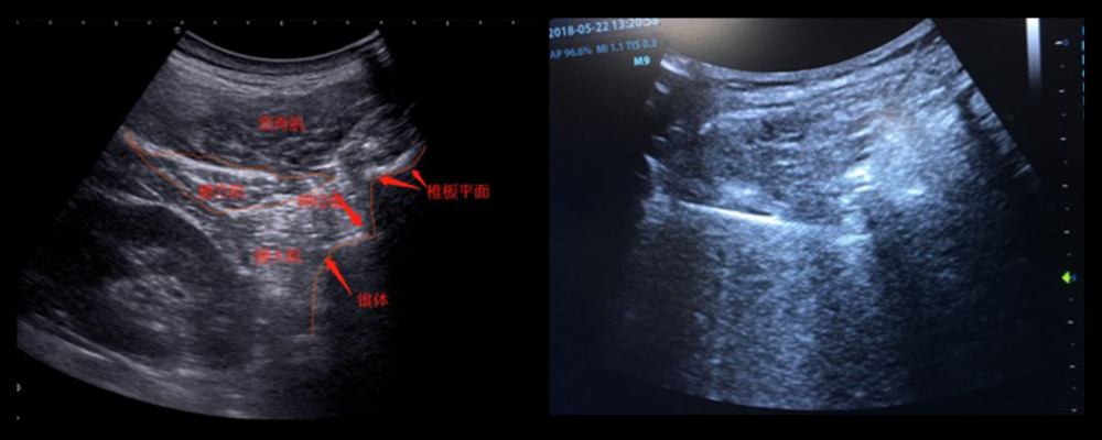
In the musculoskeletal ultrasound guidance group, the arc low-frequency probe was placed longitudinally on the spinous process, the corresponding intervertebral foramen vertebral segment in the patient was determined, and the vertebral segment of the intervertebral foramen was scanned by ultrasound, and then rotated by 90° degree to obtain lateral view, and re-confirmed the lateral view of spinous process, and traced the lamina forward according to the spinous process, and then determined the position of the lamina to move the probe down slowly to determine the lower boundary of the lamina, and to move the ultrasound probe to the side until the lower joint is displayed, and to use the in-plane needle insertion method to reach the facet joint. The anterior and inferior position was the intervertebral foramen. The location of the lumbar intervertebral foramen can be found by showing the spinous process, transverse process, inferior articular process, lamina, and lateral side of the vertebral body in sequence. The spinous process was scanned sagittal with the ultrasound probe to determine the puncture plane, and the probe was adjusted to the transverse axis, the lateral view of the lamina and the lateral surface of the vertebral body were displayed in the transverse view of the ultrasound, the lumbar intervertebral foramen was between the 2 highlighted bony landmarks, and the needle was inserted into the plane. For the puncture, the needle tip reaches the intervertebral foramen just before and below the facet joint (Figure 1). After the puncture was completed, a Philips “C”-arm X-ray machine was used, and the exposure time is 0.1 seconds each time to ensure the same amount of radiation for each fluoroscopy and to confirm the position of the puncture needle.
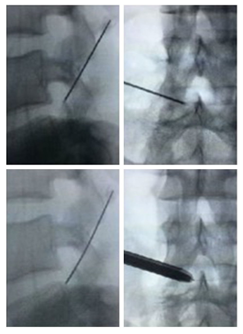
For C-arm X-ray machine fluoroscopy treatment, 40 patients underwent surgery under C-arm X-ray machine fluoroscopy according to the published method [11]. X-ray machine fluoroscopy was used to confirm the surgical segment, layer-by-layer infiltration anesthesia, positioning and puncturing to the needle entry point, and then placing the working cannula step by step, the trephine assists with foraminoplasty, and then the endoscope is placed, and the nucleus pulposus forceps will be protruded. The nucleus pulposus tissue was taken out, the working cannula and the endoscope were rotated, the nerve root and the dural sac were thoroughly decompressed, an annuloplasty was performed, and the endoscope and the working cannula were finally withdrawn (Figure 2). The completion time of ultrasound calibration, the total time of intervertebral foraminal puncture, the number of C-arm X-ray machine fluoroscopy, and complications were recorded. Both the groups of patients received standard surgical treatments in the compliance with the requirements for hospitalization.
OBSERVATION INDICATORS AND CURATIVE EFFECT EVALUATION CRITERIA:
Patients were followed up at 3 months and 1 year after surgery by specially trained researchers. The visual analogue scale (visual analogue scale, VAS) [12,13] was used to evaluate the degree of pain before and after the surgery. The full score is 10 points, of which 0 is no pain, 1–3 is tolerable mild pain, 4–6 is pain that interferes with sleep, and 7–10 is unbearable severe pain.
The Oswestry disability index (ODI) [14] was used to evaluate lumbar spine function. ODI index is divided into 5 grades: mild dysfunction (0~20%), moderate dysfunction (21~40%), severe dysfunction (41~60%), use crutches or limps (61~80%), and unable to get out of bed (81~100%). All the data were collected blindly.
STATISTICAL METHODS:
Statistical analysis was performed using SPSS 17.0 statistical software (SPSS, Inc., USA). The measurement data are expressed as (x±s), the total time for puncture in the musculoskeletal ultrasound-guided group and the C-arm X-ray machine fluoroscopy group, the number of C-arm X-ray machine fluoroscopy during the puncture process, the number of times before and after surgery. The independent samples
Results
SURGICAL OUTCOMES:
All 80 patients signed consent agreement forms and successfully completed the surgery, and none of them were changed to open surgery. The puncture accuracy of the musculoskeletal ultrasound-guided group and the C-arm X-ray machine fluoroscopy group were both 100% (80/80), and the puncture needle could eventually reach the target position and the inner side of the safety triangle with a length of 5 mm.
The preoperative ultrasound calibration time in the ultrasound-guided group was about 12–17 minutes, with an average of 14.7±2.1 minutes and the total time of the puncture process was about 17–29 minutes, with an average of 21.6±3.1 minutes. X-ray fluoroscopy was performed 5–8 times, with an average of 5.9±0.8 times.
The whole process of puncture in the C-arm X-ray machine fluoroscopy group took 26–33 minutes, with an average of 28.9±1.7 minutes. There were statistically significant differences between the ultrasound-guided group and the C-arm X-ray machine fluoroscopy group (t=13.113,
FOLLOW-UP AND EFFICACY EVALUATION:
All 80 patients were followed up for 12 to 24 months, with an average of 15.8 months. The ODI value of the ultrasound-guided group was 73.1±6.1% before the surgery, 17.3±3.9% at 3 months after the surgery, and 16.9±3.6% at 1 year after the surgery. The ODI value of the C-arm X-ray group was 73.3±4.7% before the surgery, 17.5±3.6% at 3 months after the surgery, and 16.2±2.8% at 1 year after the surgery. There was no difference in ODI values between the 2 groups before the surgery (t=0.195,
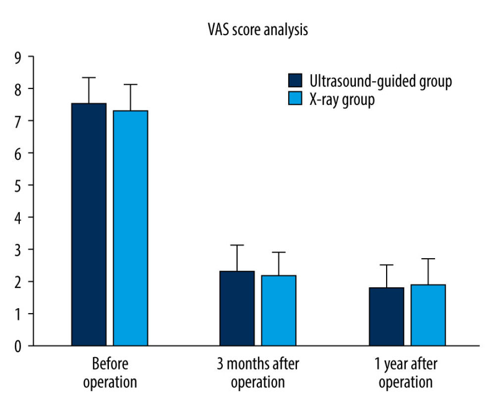
The VAS score in the ultrasound-guided group was 7.5±0.8 points before surgery, 2.3±0.8 points at 3 months after surgery, and 1.8±0.8 points at 1 year after surgery. The VAS score in C-arm X-ray group was 7.3±0.8 points before surgery, 2.2±0.8 points at 3 months after surgery, and 1.9±0.8 points at 1 year after surgery. There was no difference in VAS scores between the 2 groups before surgery (t=0.715, P=0.610), 3 months after surgery (t=0.367, P=0.192), and 1 year after surgery (t=0.496, P=0.390) (Figure 3).
Discussion
Although the operative time and blood loss of endoscopic-guided intervertebral discectomy were significantly lower than those of traditional posterior lumbar laminectomy, the postoperative excellent and satisfactory rates were comparable to about 90% [15–17], but it is difficult to establish a working channel to reach the target area (Kambin’s triangle) [18,19]. Therefore, how to establish a working channel accurately and quickly to avoid damage to the large blood vessels and nerve roots from the intervertebral foramen has important clinical significance. During the conventional C-arm X-ray machine fluoroscopy-guided foraminal puncture, both the patients and operators are exposed to large doses of radiation [20–22], which seriously affects their health. The surgeon needs to have extensive experience in open surgery and be familiar with the relevant anatomical structures near the lumbar spine surgery area [18,19]. Otherwise, the number of exposures under X-ray and the time of the entire surgery will be greatly increased, which will increase the risk of exposure to the large doses of radiation; at the same time, an inaccurate puncture position can also damage the large abdominal blood vessels, nerve roots, and abdominal organs, which makes the complete endoscopic technique have a very steep learning curve.
With the development of ultrasound technology, many researchers have conducted extensive research on the application of ultrasound in the lower lumbar spine and its adjacent structures [23–26]. Detection of lumbar spine-related structures that were previously thought to be undetectable has greatly improved [25], and accurate observation of the facet joints of the lumbar spine can guide lumbar spine surgery [27–29], such as displaying the inferior articular process and lumbar intervertebral foramen by ultrasound, and the location of the lumbar intervertebral foramen can be targeted at the spinous process, transverse process, inferior articular process, lamina, and lateral side of the vertebral body in sequence [30]. Compared with traditional navigation equipment such as C-arm machine, musculoskeletal ultrasound is a radiation-free, real-time, and visualized surgery platform with no radiation hazards to operators and patients [30]. Ultrasound can show the internal structures of the spinal canal, spinal cord, and cerebrospinal fluid [30]. Clinically, ultrasound can be used to identify the bony landmarks of the spine, and confirm the position, distance, and angle of the related tissue structures to facilitate the surgery of spinal-related anesthesia or analgesia [31]. Our data also demonstrated that ultrasonic had advantages in reducing radiation exposure and surgery duration, preventing drugs from entering by mistake, and improving the accuracy and safety of intervertebral foraminal puncture.
Conclusions
The musculoskeletal ultrasound guidance is accurate and real-time for endoscopic foraminal puncture, which can significantly reduce the puncture time and the amount of X-ray radiation, exhibiting great advantages in the clinic compared to the traditional navigation methods in puncture accuracy.
Figures
 Figure 1. For puncture, the needle tip reaches the intervertebral foramen just before and below the facet joint.
  Figure 2. X-ray machine fluoroscopy is used to confirm the surgical segment.
  Figure 3. The VAS score analysis in ultrasonic group and X-ray group, and showed no significant difference before surgery, 3 months after surgery, and 1 year after surgery.
 References
1. Bailey RS, Puryear A, Advances in minimally invasive techniques in pediatric orthopedics: Percutaneous spine fracture fixation: Orthop Clin North Am, 2020; 51(3); 339-43
2. Caruso G, Lombardi E, Andreotti M, Minimally invasive fixation techniques for thoracolumbar fractures: Comparison between percutaneous pedicle screw with intermediate screw (PPSIS) and percutaneous pedicle screw with kyphoplasty (PPSK): Eur J Orthop Surg Traumatol, 2018; 28(5); 849-58
3. Lofrese G, Mongardi L, Cultrera F, Surgical treatment of intraforaminal/extraforaminal lumbar disc herniations: Many approaches for few surgical routes: Acta Neurochir (Wien), 2017; 159(7); 1273-81
4. Quillo-Olvera J, Akbary K, Kim JS, Percutaneous endoscopic transpedicular approach for high-grade down-migrated lumbar disc herniations: Acta Neurochir (Wien), 2018; 160(8); 1603-7
5. Ikuta K, Tono O, Senba H, Translaminar microendoscopic herniotomy for cranially migrated lumbar disc herniations encroaching on the exiting nerve root in the preforaminal and foraminal zones: Asian Spine J, 2013; 7(3); 190-95
6. Shin BJ, Risk factors for recurrent lumbar disc herniations: Asian Spine J, 2014; 8(2); 211-15
7. Kjaer P, Tunset A, Boyle E, Jensen TS, Progression of lumbar disc herniations over an eight-year period in a group of adult Danes from the general population – a longitudinal MRI study using quantitative measures: BMC Musculoskelet Disord, 2016; 17; 26
8. Liao J, Huang Y, Wang Q, Gene regulatory network from cranial neural crest cells to osteoblast differentiation and calvarial bone development: Cell Mol Life Sci, 2022; 79(3); 158
9. Juma SN, Gong X, Hu S, Shark new antigen receptor (IgNAR): Structure, characteristics and potential biomedical applications: Cells, 2021; 10(5); 1140
10. Zhang X, Shi G, Sun X, Factors influencing the outcomes of artificial hip replacements: Cells Tissues Organs, 2018; 206(4–5); 254-62
11. Ruetten S, Komp M, Godolias G, An extreme lateral access for the surgery of lumbar disc herniations inside the spinal canal using the full-endoscopic uniportal transforaminal approach-technique and prospective results of 463 patients: Spine (Phila Pa 1976), 2005; 30(22); 2570-78
12. Kliger M, Stahl S, Haddad M, Measuring the intensity of chronic pain: Are the visual analogue scale and the verbal rating scale interchangeable?: Pain Pract, 2015; 15(6); 538-47
13. Collins SL, Moore RA, McQuay HJ, The visual analogue pain intensity scale: What is moderate pain in millimetres?: Pain, 1997; 72(1–2); 95-97
14. Kong YS, Jang GU, Park S, The effects of prone bridge exercise on the Oswestry disability index and proprioception of patients with chronic low back pain: J Phys Ther Sci, 2015; 27(9); 2749-52
15. Nellensteijn J, Ostelo R, Bartels R, Transforaminal endoscopic surgery for symptomatic lumbar disc herniations: A systematic review of the literature: Eur Spine J, 2010; 19(2); 181-204
16. Rao PJ, Thayaparan GK, Fairhall JM, Mobbs RJ, Minimally invasive percutaneous fixation techniques for metastatic spinal disease: Orthop Surg, 2014; 6(3); 187-95
17. Hoogland T, Schubert M, Miklitz B, Ramirez A, Transforaminal posterolateral endoscopic discectomy with or without the combination of a low-dose chymopapain: A prospective randomized study in 280 consecutive cases: Spine (Phila Pa 1976), 2006; 31(24); E890-97
18. Yokosuka J, Oshima Y, Kaneko T, Advantages and disadvantages of posterolateral approach for percutaneous endoscopic lumbar discectomy: J Spine Surg, 2016; 2(3); 158-66
19. Tenenbaum S, Arzi H, Herman A, Percutaneous posterolateral transforaminal endoscopic discectomy: Clinical outcome, complications, and learning curve evaluation: Surg Technol Int, 2011; 21; 278-83
20. Chen G, Yao Y, Xu G, Zhang X, Regional difference in microRNA regulation in the skull vault: Dev Dyn, 2019; 248(10); 1009-19
21. Chen G, Xu H, Yao Y, BMP signaling in the development and regeneration of cranium bones and maintenance of calvarial stem cells: Front Cell Dev Biol, 2020; 8; 135
22. Liu M, Hu Y, Chen G, The antitumor effect of gene-engineered exosomes in the treatment of brain metastasis of breast cancer: Front Oncol, 2020; 10; 1453
23. Winter J, Kimber A, Montenegro S, Gao J, Ultrasonography to assess the efficacy of osteopathic manipulative treatment for lumbar spine asymmetry: J Am Osteopath Assoc, 2020; 120(11); 761-69
24. Yun DH, Kim HS, Yoo SD, Efficacy of ultrasonography-guided injections in patients with facet syndrome of the low lumbar spine: Ann Rehabil Med, 2012; 36(1); 66-71
25. Chin KJ, Perlas A, Ultrasonography of the lumbar spine for neuraxial and lumbar plexus blocks: Curr Opin Anaesthesiol, 2011; 24(5); 567-72
26. Watanabe K, Miyamoto K, Masuda T, Shimizu K, Use of ultrasonography to evaluate thickness of the erector spinae muscle in maximum flexion and extension of the lumbar spine: Spine (Phila Pa 1976), 2004; 29(13); 1472-77
27. Catalano O, Lobianco R, Sandomenico F, Real-time, contrast-enhanced sonographic imaging in emergency radiology: Radiol Med, 2004; 108(5–6); 454-69
28. von Herbay A, Vogt C, Willers R, Haussinger D, Real-time imaging with the sonographic contrast agent SonoVue: Differentiation between benign and malignant hepatic lesions: J Ultrasound Med, 2004; 23(12); 1557-68
29. Cohnen M, Saleh A, Luthen R, Improvement of sonographic needle visibility in cirrhotic livers during transjugular intrahepatic portosystemic stent-shunt procedures with use of real-time compound imaging: J Vasc Interv Radiol, 2003; 14(1); 103-6
30. Galiano K, Obwegeser AA, Bodner G, Real-time sonographic imaging for periradicular injections in the lumbar spine: A sonographic anatomic study of a new technique: J Ultrasound Med, 2005; 24(1); 33-38
31. Sherwani P, Singhal S, Kumar N, Breast filariasis diagnosed by real time sonographic imaging: A case report: Iran J Radiol, 2016; 13(1); e17991
Figures
 Figure 1. For puncture, the needle tip reaches the intervertebral foramen just before and below the facet joint.
 Figure 2. X-ray machine fluoroscopy is used to confirm the surgical segment.
 Figure 3. The VAS score analysis in ultrasonic group and X-ray group, and showed no significant difference before surgery, 3 months after surgery, and 1 year after surgery.
 In Press
05 Mar 2024 : Clinical Research
Role of Critical Shoulder Angle in Degenerative Type Rotator Cuff Tears: A Turkish Cohort StudyMed Sci Monit In Press; DOI: 10.12659/MSM.943703
06 Mar 2024 : Clinical Research
Comparison of Outcomes between Single-Level and Double-Level Corpectomy in Thoracolumbar Reconstruction: A ...Med Sci Monit In Press; DOI: 10.12659/MSM.943797
21 Mar 2024 : Meta-Analysis
Economic Evaluation of COVID-19 Screening Tests and Surveillance Strategies in Low-Income, Middle-Income, a...Med Sci Monit In Press; DOI: 10.12659/MSM.943863
10 Apr 2024 : Clinical Research
Predicting Acute Cardiovascular Complications in COVID-19: Insights from a Specialized Cardiac Referral Dep...Med Sci Monit In Press; DOI: 10.12659/MSM.942612
Most Viewed Current Articles
17 Jan 2024 : Review article
Vaccination Guidelines for Pregnant Women: Addressing COVID-19 and the Omicron VariantDOI :10.12659/MSM.942799
Med Sci Monit 2024; 30:e942799
14 Dec 2022 : Clinical Research
Prevalence and Variability of Allergen-Specific Immunoglobulin E in Patients with Elevated Tryptase LevelsDOI :10.12659/MSM.937990
Med Sci Monit 2022; 28:e937990
16 May 2023 : Clinical Research
Electrophysiological Testing for an Auditory Processing Disorder and Reading Performance in 54 School Stude...DOI :10.12659/MSM.940387
Med Sci Monit 2023; 29:e940387
01 Jan 2022 : Editorial
Editorial: Current Status of Oral Antiviral Drug Treatments for SARS-CoV-2 Infection in Non-Hospitalized Pa...DOI :10.12659/MSM.935952
Med Sci Monit 2022; 28:e935952


