09 June 2023: Clinical Research
Analysis of Root Canal Characteristics in Permanent Canine Teeth Among 270 Saudi Subjects: A Cone-Beam Computed Tomography Study
Amal S. Shaiban1ACEF*, Aseel M. Zayed2ABDE, Abeer A. Abdullah2BG, Khulud H. Alshaya2BG, Mohammed A. AlobaidDOI: 10.12659/MSM.940472
Med Sci Monit 2023; 29:e940472


Abstract
BACKGROUND: An understanding of root canal morphology is vital for successful endodontic treatment. However, variations in the root canal system of permanent canines, especially in relation to population-based diversity, are not well-documented. This study thus aimed to analyze the root canal numbers, configurations, and bilateral symmetry in 1080 permanent canine teeth among 270 Saudi individuals using cone-beam computed tomography (CBCT), contributing to the existing body of knowledge and aiding clinicians in devising effective treatment strategies.
MATERIAL AND METHODS: The CBCT images of 270 participants, encompassing 1080 canines (540 pairs of upper and lower canines), were scrutinized for root and canal counts. Canal configurations were assessed based on Ahmed's and Vertucci's classifications. Bilateral symmetry in these parameters was recorded and the data were statistically analyzed.
RESULTS: The study revealed variable prevalence of multiple roots and canals in maxillary and mandibular canines. Ahmed's and Vertucci's type I canal configuration was predominantly observed. Notably, significant bilateral symmetry was noted in root and canal numbers, and canal configurations.
CONCLUSIONS: The most common configuration of permanent canines was a single root and canal, usually adhering to Ahmed's and Vertucci's type I classification. Mandibular canines showed a higher incidence of two canals than two roots. The extent of bilateral symmetry, especially in mandibular canines, could provide valuable insights for better contralateral tooth treatment planning.
Keywords: Maxillary Artery, Tooth Root, Cuspid, Saudi Arabia, Dental Pulp Cavity, Cone-Beam Computed Tomography
Background
The principal intent of endodontic treatment is to preserve permanent teeth [1,2]. The key to successful root canal treatment (RCT) is comprehension and mastery of the internal anatomy of the pulp [3]. Endodontists encounter a wide range of root canal systems (RCS) in day-to-day practice [1]. The clinician should be familiar with the conventional forms of dental anatomy and the most usual anatomic deviations encountered in routine practice [4]. Lack of expertise in the morphology and anatomy of root canals (RC), as well as variations in the canal numbers (CNs), are among the main reasons for failure in RCT [5]. The complexities of the RC encompass restorative hindrances and barriers that can jeopardize the basic goal of RCT. Sectioning the root, staining the canal, clearing teeth, using microscopes in endodontics, clinical and conventional 2D radiographic examination, and computed tomography scans are diverse in vitro and in vivo approaches used to assess the internal and external anatomy of non-identical groups of teeth [1]. Compared to conventional radiography, cone-beam computed tomography (CBCT) is a more precise technique in assessing RC morphology and has the accuracy of other methods like staining the RC and clearing the tooth in recognizing the RCS [3].
A canine is a single-root tooth in the maxillary and mandibular arch. Many systems classified the root and canal morphology. An early system was that of Weine et al for classification of root canal morphology, followed by Vertucci et al, then Sert and Bayirli, who added supplementary types to Vertucci’s classification system, and lastly, Ahmed et al’s classification was proposed [6–8]. Each classification system has its own distinct advantages and shortcomings. Examples of root morphology and canal configuration of canine are presented in Figure 1: (A) lower canine with 2 roots and 1 canal in each root, which according to Ahmad’s classification is [(2) 43B (1)L(1)] and according to Virtucci’s classification is type I in both B and L roots; (B) lower canine with single root and 2 canals, which according to Ahmad’s classification is [(1)43(1-2)] and according to Virtucci’s classification is type V; (C) lower lateral with single root and single canal separated in the middle to 2 canals, which according to Ahmad’s classification is [(1)32 (1-2-1)] and according to Virtucci’s classification is type III; (D) is a lower canine with single root and single canal separated in the middle and in the end to 2 canals, which according to Ahmad’s classification is [(1)31(1-2-1-2)] and according to Virtucci’s classification is type VI.
The differences in the RC forms have been analyzed by research experts. For each tooth in the permanent dentition, a range of variations is reported in the literature [2]. Many reports have stated that RCs differs according to race, ethnicity, sex, and geographic location [9]. Accordingly, modifications in the RCs should be recognized in the pre-treatment assessment for RCT. Practitioners who regularly treat patients of various nationalities should be acquainted with racial diversity and its influence on modifying the RC morphology [10].
Only 2 studies have been conducted among Saudis in different cities to study the root morphology and canal configurations of maxillary and mandibular anterior teeth, including canines in both arches [1,9,11]. In addition, a recent review that analyzed the morphology of the anterior teeth among Saudi was published by Assiri et al in 2022 [12].
Usually, most permanent canines have 1 root with a single canal. Despite that, several investigators have evaluated and encountered various morphological alterations in RCS of the permanent canines in different races and sexes [2]. There are variations in the internal anatomy of a tooth in different communities; thus, identifying the RC form of individual ethnic groups is required for successful endodontic therapy [13]. Therefore, this study aimed to investigate root canal numbers and configurations in 1080 permanent canine teeth in 270 Saudi subjects using cone-beam computed tomography.
Material and Methods
SAMPLE SELECTION:
The ethics committee of the KKU approved the research protocol (IRB/REG/KKU, ETH 2022-23/042) and informed consent was obtained from all enrolled patients to conduct this study. A total of 270 subjects – 89 (32.9%) males and 181 (67.03%) females – were studied, and 1080 permanent canines (540 maxillary and 540 mandibular canines) were assessed using the protocol of Mashyakhy et al and Alshayban et al [1,9,11]. From the database of King Khalid University (KKU) Dental College and several private clinics in Abha City, KSA, archived CBCT scans were retrieved. Permanent canines with completely developed roots with apical closure were included in the study. Canines with no history of previous restorations or RCT were included. We excluded teeth with calcified canals or resorbed roots, missing canine or implant or periapical lesions, and any physiological or pathological process such as immature apex, as well as unclear tooth morphology on imaging.
CBCT EXAMINATION:
The CBCT images used were analyzed and evaluated in sagittal, axial, and coronal serial sections using Morita’s i-Dixel 3D imagine software. A careful examination was performed by optimal visualization using all the software features, such as zooming, change in contrast, and brightness, including the sectioning, which was oriented to be parallel to the long axis of the RC with a 1 mm slice thickness. Furthermore, the scans were evaluated by scrolling up and down for axial sections and from right to left for parasagittal sections. The following characteristics were analyzed, and the data were obtained: (a) Root and canal numbers; RCS configurations were classified according to the criteria of Ahmed et al [8] and Vertucci et al [6]; (b) BS of root numbers (RNs), CNs and CCs; (c). Sex difference. Figure 2 shows CBCT of canine with a single root and single canal separated in the middle to 2 canals. According to Ahmad’s classification it is [(1)32(1-2-1)] and according to Virtucci’s classification it is type III.
STATISTICAL ANALYSIS:
The investigators fed the data into the SPSS program for Windows (SPSS Version. 22) The coded data were subjected to the following statistical tests:
Results
DISTRIBUTION OF ROOTS AND CANALS IN MAXILLARY AND MANDIBULAR CANINES:
In this retrospective cross-sectional study, the vast majority of canines (98.7%), had single roots and only 1.2% had double roots. There were 298 (27.5%) canines with 2 canals and most teeth had 1 canal (779 canines, 72%). CC type I was mostly observed in 778 canines (72%), with a statistically significant difference from the other configuration types (
Of the 540 maxillary canines, 537 (99.4%) had 1 root, and 3 (0.55%) teeth had 2 roots. There were 406 (75%) teeth with 1 canal, 133 (24.6%) teeth had 2 canals, and 1 (0.2%) tooth had 3 canals. Ahmed’s CC type I was present in 372 (68.8%) teeth, while 65 (12%) had IX and 45 (8%) had type XIII. Vertucci’s type I was present in 405 (75%), 65 (12%) had type IV, 44 (8%) had type V, 25 (4.6%) had type II, and 1 (0.4%) had type III. Among 540 mandibular canines, 530 (98%) had 1 root and 10 (1.85%) teeth had 2 roots. There were 373 (69%) teeth with 1 canal, 165 (31%) with 2 canals, and 2 (0.3%) with 3 canals. Ahmed’s type I was present in 321 (59.4%) teeth, while 76 (14%) had type IX, and 47 (8.7%) had type XIII. Vertucci’s type I was present in 373 (69%), 74 (13.7%) had type IV, 47 (8.7%) had type V, 39 (7.2%) had type II, 3 (0.5%) had type VI, 2 (0.3%) had type VIII, and 1 (0.2%) had type III and VII (Table 1).
FREQUENCY AND PERCENTAGE DISTRIBUTION OF BILATERAL SYMMETRY:
Maxillary and mandibular canines demonstrated 61.9% and 57.4% symmetry according to Ahmed’s CC (P<0.001) and 69.9% and 68.5% according to Vertucci’s CC (P<0.001) (Table 2). CCs and tooth position were significantly related (P<0.001). The mandibular canines showed greater variations in canal system configurations (asymmetries) than did maxillary canines. Vertucci’s type I CC was more common in maxillary canines (75%) compared to mandibular canines (69%). However, Vertucci’s types II, IV, and V were more common in mandibular canines compared to maxillary canines, and types VI, VII, VIII are found only in mandibular canines.
ASSOCIATION BETWEEN CANAL CONFIGURATION AND BILATERAL SYMMETRY WITH SEX:
Sex was not significantly related with RNs or CNs of permanent canines (P>0.05). The chi-square test showed a significant relationship between the CCs and sex in maxillary canines (#13) (P<0.05). Vertucci’s type I followed by IV was common in females, whereas type I followed by V was more frequent in males (P=0.017) (Table 3).
Table 4 shows that all 270 subjects (100%) had both right and left maxillary canines. The BS for RNs was 98.9 (P<0.001); and most subjects (n=267; 98.9%) had 1 root on both sides. BS for CNs on both sides was 75.12%(p<0.001); 62.9% (n=170) of subjects had 1 canal on both sides, and only 12.22% (n=33) had 2 canals on both sides. Regarding CCs, the total BS 69.62% (P<0.001); 62.96% (n=170) of the subjects had Vertucci’s type I on both sides followed by 4.44% (n=12) type IV, 1.85% (n=5) type V and 0.37%(n=1) type II. The most frequent BS was 1 root (98.9%), 1 canal (62.9%), and Vertucci type I CC (62.96%). The total BS was 62.95% (P<0.001); 54.81% (n=148) of the subjects had Ahmed’s type I on both sides (Table 5).
In mandibular canines, all 270 subjects (100%) had both right and left mandibular canines. The BS for RNs was 97.78% (P<0.001); most subjects (n=262; 97.01%) had 1 root on both sides, and 2 subjects (n=2; 0.7%) had 2 roots on both sides (Table 4). BS for CNs was 74.8% (P<0.001); 56.67% (n=153) of subjects had 1 canal on each side. Only 18.15% (n=49) had 2 canals in canines on each side. Regarding CCs, the total BS 67.77% (P<0.001); 56.67% (n=153) of the subjects had Vertucci’s type I on both sides followed by 6.29% (n=17) type IV, 2.59% (n=7) type II, 1.85% (n=5) type V, and 0.37% (n=1) type VI. The most frequent BS was 1 root (97.04%), 1 canal (56.67%), and Vertucci type I CC (56.67%). The total BS was 56.65% (p<0.001); 43.7%(n=118) of the subjects had Ahmed’s type I on both sides (Table 6).
The chi-square test showed that a significant association between sex and Ahmed’s BS, Vertucci’s BS in the maxillary and mandibular canine (P<0.001) (Table 7). The bilateral classification of Ahmed’s and Vertucci’s showed significant BS of 61.48% (n=166) and 56.67% (n=153) (P<0.001) for maxillary and mandibular canines, respectively (Table 8). Table 9 shows the bilateral symmetry among participants for mandibular canines, while Table 10 shows the bilateral classification of Ahmad and Vertucci in maxillary and mandibular canines.
Discussion
The RCs of permanent canines in a subpopulation of the KSA was evaluated in this study using CBCT. Observations and results of this investigation validate the alterations in the morphology of the RCS in this subpopulation, as described by several investigators [1, 6, 9–11] [1,9,14–16]. It was found that 88.46–100% of mandibular canines had a single root, while 0–11.54% had a double root. A single canal was present in 84.9–100% of mandibular canines and a double canal was present in 0–15.1% [9, 11] [14,16].
Currently, the usage of in vivo CBCT imaging is essential to acquire actual features of the RC during RCT. CBCT is a 3D technology that is economical and with a modest radiation dose [1] compared with conventional CT [5]. The level of precision offered by CBCT or microcomputed tomography (mCT) is unparalleled in conventional radiography or clinical examination. CBCT is a generally accessible, non-invasive, in vivo modality for analyzing the RCS, as it overcomes the constraints of intraoral radiograph [1]. Its non-destructive feature is an added advantage, along with its 3D reconstruction and formation of mental images that encompass the internal and external form of the periodontium [12,17].
The rate of double-rooted mandibular canines reported in various populations are as follows: South Asian Indian population (0%) [18]; Chinese (0.7%) [19]; Chongqing population (0.8%) [20]; Malaysian population (1.2%) [21]; Iranian population (1.3%) [22]; and Brazilian (1.7%) [23]. A high incidence of double-rooted mandibular canines of 4.7% and 12.08% has been noted in the Iranian subpopulations [2,24]. A study on the RC morphology of mandibular canines in a subpopulation of the KSA found a single root (99.8%) was the most common root form, while 0.2% of canines had a double root [14]. Even in this study, 98% of mandibular canines had 1 root, 1.85% had 2 roots, 69% of them had 1 canal, and 30.5% had 2 canals. Marginal percentages were recorded in a 2022 study by Alshayban et al [11], and this is supported by a 2022 review by Assiri et al [12].
In subpopulations of the KSA, the rate of double-rooted and double-canalled mandibular canines mentioned in the literature are as follows: 0.2% and 4.6% [9]; 2.7% and 9.3% [16]; and 2.88% and 9.94% [15], respectively. The presence of double-canalled mandibular canines ranges from 0% to 15% in different subpopulations [15]. In an Iraqi subpopulation, 11–11.7% of canines had 2 root canals [3], 22% had 2 canals in an RCS analysis of permanent dentition [6], 24% in a Turkish subgroup [25], and 28.2% in an Iranian subset [2]. We found the highest incidence (30.5%) of double-canalled mandibular canines. Varied results can be due to the differences in genetic model and ethnicity in the studied population, size of the sample, techniques involved, classification of RCS, and the scholars’ acumen and identification [26]. Many researchers have performed clinical analysis on the RCS and the form of the anterior roots using various methods, and different CNs were noted [20]. Variation and discrepancies in the findings of several reports may be attributable to the region where the samples were analyzed. Evaluation of frequencies of roots and canals involving a large sample size is recommended to overcome the disparity due to the small sample size in the above reports [16].
In 1984, Vertucci analyzed the CCs of all the permanent teeth and frequently found CC type I (78%) in mandibular canines, followed by type II (14%), IV (6%), and III (2%) [6]. Most mandibular canines in this analysis had Vertucci’s type I (69%), followed by type IV (13.7%), (8.7%) V, and (7.2%) II. Other assessments in a subgroup of KSA reported that CC type I (95.4%) was frequently found in lower canines, followed by (2.6%) II and (1.8%) III [14]; (90.7%) type I then (6.1%) III and (3.2%) type V [16]; (92.07%) type I CC then (2.88%) III and (2.44%) type V [15]. Among all the CCs, CC type I had the highest occurrence in mandibular canines [15], but different percentages were recorded by Alshayban et al [11] in maxillary and mandibular canines, which could be explained by different sample sizes among sexes and different areas or cities. Modest variation in the canal configuration types observed in the above-mentioned reports may represent the contrast in the ethnicity of the subjects residing in the same region, which is a significant feature that might influence the insight of the endodontist regarding the assumed canal anatomy. People from different geographic areas and cultural groups may present differences in dental morphology [27,28].
A structured review and meta-analysis on anatomical assessment of RC and CC of permanent maxillary teeth in a subgroup of the KSA had only 2 studies [15,16] assessing maxillary canines. All canines (N = 1018) had 1 root, and there were no double-rooted maxillary canines [1]. One report found 98.1% had 1 canal, and only 1.89% had 2 canals [15]. In another assessments, 99% of canines had 1 canal, and 1% had 2 canals [11,16]. Even in this analysis, 99.4% of maxillary canines had 1 root, 75% of the teeth had 1 canal, and 24.6% had 2 canals. Double-rooted maxillary canines are illustrated in the literature as case reports. These attributes suggest that in reality, double-rooted maxillary canines are rare [15]. An analysis of the RCS in permanent anterior teeth using CBCT concluded that the incidence of anatomical variation in maxillary anterior teeth is low [29].
Regarding the CC in maxillary canines, the present report demonstrated that CC type I was most common (75%), followed by IV (12%), V (8%), II (4.6%), and III (0.4%). Previous studies in the KSA noted that CC type I was most common (97.94%), followed by V (1.1%) and II and III (0.47%) in 1 report [15]; (99%) type I and (1%) III configurations in another study [16], and the proximate percentage was recorded in other study in SA [11]. Similar percentages were recorded in a study published recently by Asiri et al [12]. A slight difference in the preferences could be justified by the variation in size of the sample and geographic location among the studies. CC type I was observed in all evaluated maxillary canines (100%) in a Malaysian population [21]. In contrast, in the Indian population, a dominant type I (96%) pattern followed by II (3%) and III (1%) was noted by a researcher [10]; 81.6% type I, 2.8% II, 11.6% III, 0.8% IV, and 2% type V was observed by another [30].
In this analysis, RNs or CNs in canines and sex had no significant association. However, sex and CC revealed a significant association. Similar findings were noted in the reports on 3D imaging in analyzing canines for having double root and canal in the KSA subpopulation [15,16]. Comparable results were observed in the Malaysian subpopulation [21]. Females had significantly greater RNs and CNs than males in mandibular canines in an investigation of the influence of sex on RC number in permanent dentition [28]. In an Iranian population, double-rooted and double-canalled mandibular canines were significantly more common among males than females [22]. In the Turkish population, the incidence of RNs, CNs, and CCs differed with sex; the prevalence of double-canalled anteriors was higher in males than in females [13]. External and internal forms of dentition might differ according to sex, age, race, and geographic location [1,31].
Mandibular canines demonstrated a high BS for the RNs (97.1%), CNs (90.1%), and CC (92.1%) in a subpopulation of the KSA [15]. In another analysis from the same region [16], 95.5% of canines exhibited BS in RNs, 91.1% in CNs, and 90.1% in the CCs. In the above-mentioned subpopulation, 97.7% of mandibular canines with symmetrical RNs and CCs were noted [14]. Even in the Iranian population, 95.4% BS in the RNs and CC in mandibular canines was noted [22]. In a Turkish subset, the BS of RNs and CNs for mandibular anterior teeth were 96–100% and 90–95% [32]. In this analysis, mandibular canines showed a high BS for the RNs (97.8%), CNs (74.8%), and CCs (67.77%). Variations were obvious, particularly in the critical analysis of the CNs and CCs, regardless of the approach that was used in the same subset of teeth. These disparities reflect the apparent deviations in dental anatomy within the same or different areas, identical to those found in several studies in the KSA [1]. Genetics has an influence on the complexity of the RCS, and this variable must be taken into account while reviewing other reports on RC morphology [33].
In this analysis, the degree of BS in RNs for the contralateral maxillary canines was 98.9%, which is higher than for mandibular canines, 75.12% symmetry for CNs, and 69.62% for CCs. The extent of BS in RNs (100%), CNs (98.9%), and CC (98.9%) for maxillary canines were also observed in a subpopulation of the KSA [9]. The above-discussed reports of the BS in permanent teeth indicate that mandibular anterior teeth have a greater variation in RC morphology than maxillary anterior teeth, and the mandibular arch exhibited greater asymmetry than the maxillary arch. The highest asymmetry bilaterally was in the CCs, followed by CNs. The findings of the present report validate this.
There are certain limitations of the present study. First, it was restricted to 1 region of the KSA. The CBCT scans were evaluated from 1 subpopulation with differences in demographics. Moreover, the analysis was retrospective in nature. Hence, prospective studies concomitantly conducted in several centers from different regions with large sample sizes are recommended. Furthermore, in this analysis, the CBCT that was used has lower resolution compared to μCT, and images are not highly defined like μCT, which is a shortcoming. CBCT scans cannot provide minute anatomic details like μCT or clearing of the RCS [4]. However, data were analyzed and tabulated by visualizing the archived imaging database, which enabled recording the specific details of sufficient samples without unnecessarily exposing the subjects to radiation.
Conclusions
This study found that among a subgroup of KSA residents, most of the canines in both arches had a single root and canal, with Ahmed’s and Vertucci’s type I CC. In mandibular canines, the incidence of a double canal was higher than that of double root. The extent of bilateral symmetry concerning the CNs and CCs was higher in mandibular than maxillary canines. This might aid clinicians in providing better treatment for the contralateral tooth.
Figures
![Some shapes of root morphology and canal configuration of canine. A–D according to Ahmad’s classification are [(2) 43B (1) L(1)], [(1)43(1-2)], [(1)32(1-2-1)], and [(1)31(1-2-1-2)], respectively. A–D according to Virtucci’s classification are type I, V, III, VI, respectively.](https://jours.isi-science.com/imageXml.php?i=medscimonit-29-e940472-g001.jpg&idArt=940472&w=1000) Figure 1. Some shapes of root morphology and canal configuration of canine. A–D according to Ahmad’s classification are [(2) 43B (1) L(1)], [(1)43(1-2)], [(1)32(1-2-1)], and [(1)31(1-2-1-2)], respectively. A–D according to Virtucci’s classification are type I, V, III, VI, respectively.
Figure 1. Some shapes of root morphology and canal configuration of canine. A–D according to Ahmad’s classification are [(2) 43B (1) L(1)], [(1)43(1-2)], [(1)32(1-2-1)], and [(1)31(1-2-1-2)], respectively. A–D according to Virtucci’s classification are type I, V, III, VI, respectively. ![(A, B) CBCT of canine with a single root and single canal separated in the middle to 2 canals. According to Ahmad’s classification it is [(1)32(1-2-1)] and according to Virtucci’s classification it is type III.](https://jours.isi-science.com/imageXml.php?i=medscimonit-29-e940472-g002.jpg&idArt=940472&w=1000) Figure 2. (A, B) CBCT of canine with a single root and single canal separated in the middle to 2 canals. According to Ahmad’s classification it is [(1)32(1-2-1)] and according to Virtucci’s classification it is type III.
Figure 2. (A, B) CBCT of canine with a single root and single canal separated in the middle to 2 canals. According to Ahmad’s classification it is [(1)32(1-2-1)] and according to Virtucci’s classification it is type III. Tables
Table 1. Frequency and percentage distribution of roots and canals in maxillary and mandibular canines.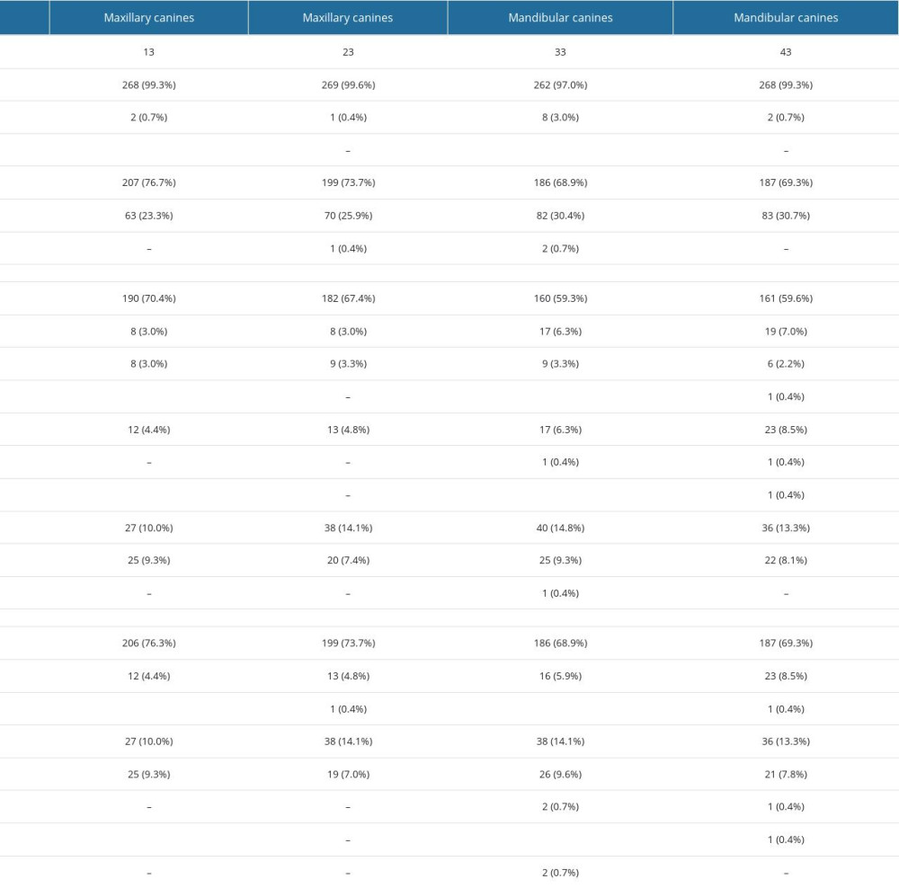 Table 2. Frequency and percentage distribution of bilateral symmetry (Z test for proportion).
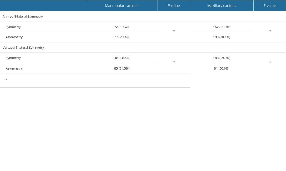 Table 3. Chi-square test for association between Ahmad Canal Configuration and gender in maxillary canine.
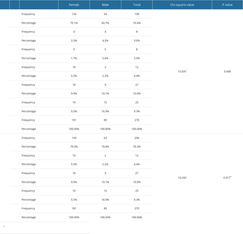 Table 4. Chi-square test for association between bilateral symmetry and gender in maxillary canine.
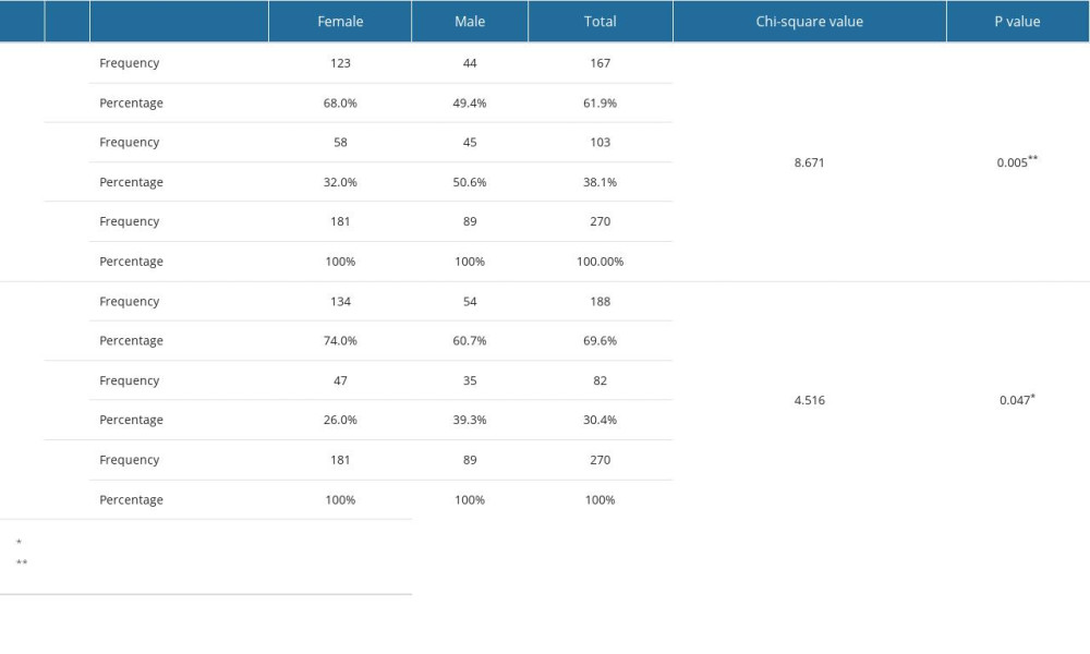 Table 5. Chi-square test for association between bilateral symmetry and gender in mandibular canines.
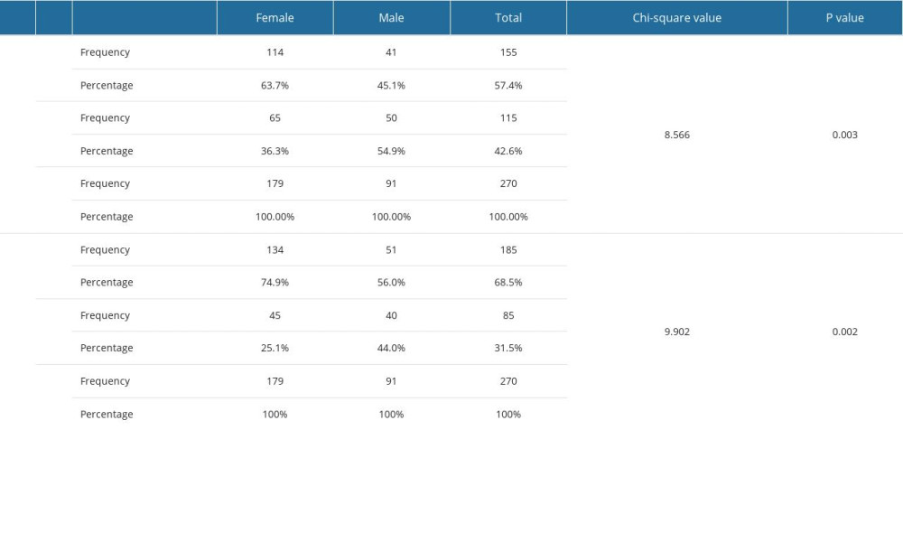 Table 6. Bilateral symmetry of the roots and canals among participants for maxillary canines.
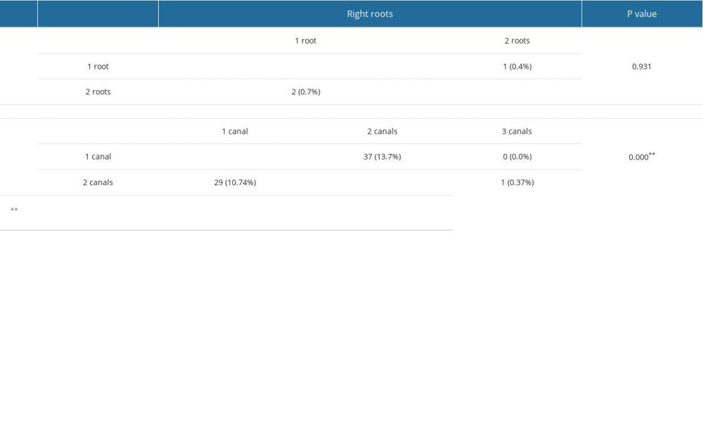 Table 7. Bilateral symmetry of the Ahmad and Vertucci among participants for maxillary canines.
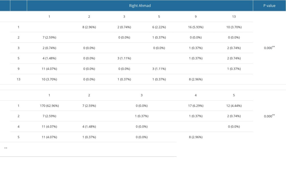 Table 8. Bilateral symmetry of roots and canals among participants for mandibular canines.
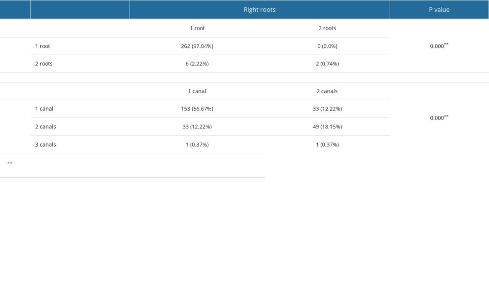 Table 9. Bilateral symmetry among participants for mandibular canines.
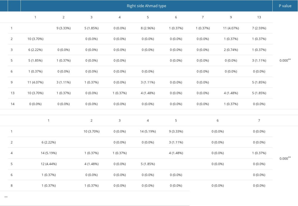 Table 10. Bilateral classification of Ahmad and Vertucci in maxillary and mandibular canines.
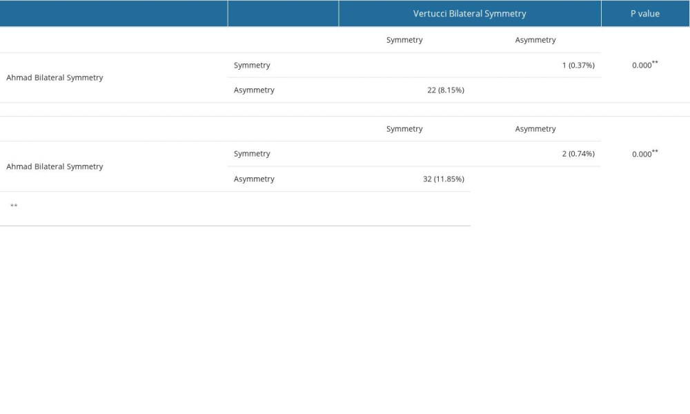
References
1. Mashyakhy M, Awawdeh M, Abu-Melha A, Anatomical evaluation of root and root canal configuration of permanent maxillary dentition in the population of the Kingdom of Saudi Arabia: Biomed Res Int, 2022; 2022; 3428229
2. Aminsobhani M, Sadegh M, Meraji N, Evaluation of the root and canal morphology of mandibular permanent anterior teeth in an Iranian population by cone-beam computed tomography: J Dent (Tehran), 2013; 10(4); 358-66
3. Talabani RMJ, Assessment of root canal morphology of mandibular permanent anterior teeth in an Iraqi subpopulation by cone-beam computed tomography: J Dent, 2021; 16(4); 1182-90
4. Martins JNR, Marques D, Mata A, Carames J, Root and root canal morphology of the permanent dentition in a Caucasian population: A cone-beam computed tomography study: Int Endod J, 2017; 50(11); 1013-26
5. Mirzaie M, Zaban PTJA, Cone-beam computed tomography study of root canals in a Hamadani population in Iran: J Dent Rserch, 2018; 4(2); 93-99
6. Vertucci FJ, Root canal anatomy of the human permanent teeth: Oral Surg Oral Med Oral Pathol Oral Radiol, 1984; 58(5); 589-99
7. Ahmed HMA, Versiani MA, De-Deus G, Dummer PMH, A new system for classifying root and root canal Morphology: Int Endod J, 2017; 50; 761-70
8. Karobari MI, Parveen A, Mirza MB, Root and root canal morphology classification systems: Inter J Dent, 2021; 2021; 6682189
9. Mashyakhy M, Abu-Melha AS, Analysis of bilateral symmetry of root canal anatomy in permanent dentition: An in vivo CBCT study in a Saudi Arabian population: J Contemp Dent Pract, 2021; 22(8); 867-75
10. Jain P, Balasubramanian S, Sundaramurthy J, Natanasabapathy V, A cone beam computed tomography of the root canal morphology of maxillary anterior teeth in an institutional-based study in Chennai urban population: An in vitro study: J Int Soc Prev Community Dent, 2017; 7(Suppl 2); S68-S74
11. Alshayban M, Abughosh T, Almalki W, Alrasheed M, Cone-beam computed tomographic evaluation of root canal morphology of mandibular anterior teeth in a Saudi subpopulation, retrospective in-vivo study: Saudi Dent J, 2022; 34(5); 390-96
12. Asiri AA, AlQahtani KW, Tarrosh MY, Root morphology and canal configuration of permanent canines among saudi population: Systematic review and comparison with worldwide studies: Inter J General Medicine, 2022; 15; 6849-60
13. Altunsoy M, Ok E, Nur BG, Aglarci OS, A cone-beam computed tomography study of the root canal morphology of anterior teeth in a Turkish population: Eur J Dent, 2014; 8(3); 302-6
14. Al-Dahman Y, Alqedairi A, Alfawaz H, Cone-beam computed tomographic evaluation of root canal morphology of mandibular canines in a Saudi subpopulation: AJSEJ, 2019; 9(2); 113
15. Almohaimede AA, Alqahtani AA, Alhatlani NM, Interpretation of root canal anatomy of maxillary and mandibular permanent canines in Saudi subpopulation: A cone-beam computed tomography (CBCT) study: Int J Dent, 2021; 2021; 5574512
16. Mashyakhy M, Prevalence of a second root and canal in mandibular and maxillary canines in a Saudi Arabian population: A cone-beam computed tomography study: J Contemp Dent Pract, 2019; 20(7); 773-77
17. Patel S, Horner KJ, The use of cone beam computed tomography in endodontics: Int Endo J, 2009; 42(9); 755-56
18. Singh S, Pawar M, Root and canal morphology of mandibular incisors and canines in South Asian Indian population by canal staining and tooth clearing technique: Endodontology, 2016; 28(2); 148-53
19. Zhao Y, Dong YT, Wang XYCone-beam computed tomography analysis of root canal configuration of 4 674 mandibular anterior teeth: Beijing Da Xue Xue Bao Yi Xue Ban, 2014; 46(1); 95-99 [in Chinese]
20. Zhengyan Y, Keke L, Fei W, Cone-beam computed tomography study of the root and canal morphology of mandibular permanent anterior teeth in a Chongqing population: Ther Clin Risk Manag, 2016; 12; 19-25
21. Pan JYY, Parolia A, Chuah SR, Root canal morphology of permanent teeth in a Malaysian subpopulation using cone-beam computed tomography: BMC Oral Helth, 2019; 19(1); 1-15
22. Soleymani A, Namaryan N, Moudi E, Gholinia A, Root canal morphology of mandibular canine in an Iranian population: A CBCT assessment: Iran Endod J, 2017; 12(1); 78-82
23. Pécora JD, Sousa Neto M, Saquy P, Internal anatomy, direction and number of roots and size of human mandibular canines: Braz Dent J, 1993; 4(1); 53-57
24. Rahimi S, Milani AS, Shahi S, Prevalence of two root canals in human mandibular anterior teeth in an Iranian population: Indian J Dent Res, 2013; 24(2); 234-36
25. Sert S, Bayirli GS, Evaluation of the root canal configurations of the mandibular and maxillary permanent teeth by gender in the Turkish population: J Endod, 2004; 30(6); 391-98
26. Lambrianidis T, Lyroudia K, Pandelidou O, Nicolaou A, Evaluation of periapical radiographs in the recognition of C-shaped mandibular second molars: Int Endo J, 2001; 34(6); 458-62
27. Martins JN, Gu Y, Marques D, Differences on the root and root canal morphologies between Asian and white ethnic groups analyzed by cone-beam computed tomography: J Endo, 2018; 44(7); 1096-104
28. Martins JNR, Marques D, Francisco H, Carames J, Gender influence on the number of roots and root canal system configuration in human permanent teeth of a Portuguese subpopulation: Quintessence Int, 2018; 49(2); 103-11
29. da Silva EJ, Queiroz de Castro RW, Nejaim Y, Evaluation of root canal configuration of maxillary and mandibular anterior teeth using cone beam computed tomography: An in-vivo study: Quintessence Int, 2016; 47(1); 19-24
30. Amardeep NS, Raghu S, Natanasabapathy V, Root canal morphology of permanent maxillary and mandibular canines in Indian population using cone beam computed tomography: Anat Res Int, 2014; 2014; 731859
31. Areqi A, Dallak A, Kariri M, Maxillary lateral incisor with two separated canals diagnosed with cbct technology: A six-month follow-up case report and minireview: Saudi Medi J Student, 2021; 2(1); 1-9
32. Michelotto AL, Gasparetto JC, Campos FR, Applying liquid chromatography-tandem mass spectrometry to assess endodontic sealer microleakage: Braz Oral Res, 2015; 29; 1-7
33. Ghasemi N, Rahimi S, Shahi S, A review on root anatomy and canal configuration of the maxillary second molars: Iran Endod J, 2017; 12(1); 1-9
Figures
Figure 1. Some shapes of root morphology and canal configuration of canine. A–D according to Ahmad’s classification are [(2) 43B (1) L(1)], [(1)43(1-2)], [(1)32(1-2-1)], and [(1)31(1-2-1-2)], respectively. A–D according to Virtucci’s classification are type I, V, III, VI, respectively.Figure 2. (A, B) CBCT of canine with a single root and single canal separated in the middle to 2 canals. According to Ahmad’s classification it is [(1)32(1-2-1)] and according to Virtucci’s classification it is type III. Tables
Table 1. Frequency and percentage distribution of roots and canals in maxillary and mandibular canines.Table 2. Frequency and percentage distribution of bilateral symmetry (Z test for proportion).Table 3. Chi-square test for association between Ahmad Canal Configuration and gender in maxillary canine.Table 4. Chi-square test for association between bilateral symmetry and gender in maxillary canine.Table 5. Chi-square test for association between bilateral symmetry and gender in mandibular canines.Table 6. Bilateral symmetry of the roots and canals among participants for maxillary canines.Table 7. Bilateral symmetry of the Ahmad and Vertucci among participants for maxillary canines.Table 8. Bilateral symmetry of roots and canals among participants for mandibular canines.Table 9. Bilateral symmetry among participants for mandibular canines.Table 10. Bilateral classification of Ahmad and Vertucci in maxillary and mandibular canines.Table 1. Frequency and percentage distribution of roots and canals in maxillary and mandibular canines.Table 2. Frequency and percentage distribution of bilateral symmetry (Z test for proportion).Table 3. Chi-square test for association between Ahmad Canal Configuration and gender in maxillary canine.Table 4. Chi-square test for association between bilateral symmetry and gender in maxillary canine.Table 5. Chi-square test for association between bilateral symmetry and gender in mandibular canines.Table 6. Bilateral symmetry of the roots and canals among participants for maxillary canines.Table 7. Bilateral symmetry of the Ahmad and Vertucci among participants for maxillary canines.Table 8. Bilateral symmetry of roots and canals among participants for mandibular canines.Table 9. Bilateral symmetry among participants for mandibular canines.Table 10. Bilateral classification of Ahmad and Vertucci in maxillary and mandibular canines. In Press
07 Mar 2024 : Clinical Research
Knowledge of and Attitudes Toward Clinical Trials: A Questionnaire-Based Study of 179 Male Third- and Fourt...Med Sci Monit In Press; DOI: 10.12659/MSM.943468
08 Mar 2024 : Animal Research
Modification of Experimental Model of Necrotizing Enterocolitis (NEC) in Rat Pups by Single Exposure to Hyp...Med Sci Monit In Press; DOI: 10.12659/MSM.943443
18 Apr 2024 : Clinical Research
Comparative Analysis of Open and Closed Sphincterotomy for the Treatment of Chronic Anal Fissure: Safety an...Med Sci Monit In Press; DOI: 10.12659/MSM.944127
08 Mar 2024 : Laboratory Research
Evaluation of Retentive Strength of 50 Endodontically-Treated Single-Rooted Mandibular Second Premolars Res...Med Sci Monit In Press; DOI: 10.12659/MSM.944110
Most Viewed Current Articles
17 Jan 2024 : Review article
Vaccination Guidelines for Pregnant Women: Addressing COVID-19 and the Omicron VariantDOI :10.12659/MSM.942799
Med Sci Monit 2024; 30:e942799
14 Dec 2022 : Clinical Research
Prevalence and Variability of Allergen-Specific Immunoglobulin E in Patients with Elevated Tryptase LevelsDOI :10.12659/MSM.937990
Med Sci Monit 2022; 28:e937990
16 May 2023 : Clinical Research
Electrophysiological Testing for an Auditory Processing Disorder and Reading Performance in 54 School Stude...DOI :10.12659/MSM.940387
Med Sci Monit 2023; 29:e940387
01 Jan 2022 : Editorial
Editorial: Current Status of Oral Antiviral Drug Treatments for SARS-CoV-2 Infection in Non-Hospitalized Pa...DOI :10.12659/MSM.935952
Med Sci Monit 2022; 28:e935952