07 August 2020: Animal Study
The Release of Norepinephrine in C57BL/6J Mice Treated with 6-Hydroxydopamine (6-OHDA) is Associated with Translocations in Enteric via the QseC Histidine Kinase Receptor
Jun Meng1ACDEF, Huamei Chen2BCF, Qin Lv2BC, Xiaodan Luo2BC, Kun Yang2AG*DOI: 10.12659/MSM.922986
Med Sci Monit 2020; 26:e922986


Abstract
BACKGROUND: We aimed to investigate the effects of norepinephrine (NE) released from endogenous stores on bacterial translocation of Escherichia coli in mice by administration of 6-hydroxydopamine (6-OHDA), which selectively destroys noradrenergic nerve terminals.
MATERIAL AND METHODS: E. coli strain BW25113 and its derivatives (BW25113ΔqseC and BW25113ΔqseC pQseC) were used in this study. The serum concentrations of endotoxin were analyzed. The strains BW25113, BW25113ΔqseC, and BW25113ΔqseC pQseC were detected respectively in tissue specimens harvested from mice treated with 6-OHDA.
RESULTS: Mice treated with BW25113ΔqseC showed reduced levels of bacterial translocation following administration of 6-OHDA compared with mice treated with BW25113. The defect of E. coli QseC receptor caused the norepinephrine-QseC signal chain to be interrupted, and the invasiveness and penetrating power of the bacteria on the intestinal mucosa was weakened, eventually leading to a significant decrease in the incidence of bacterial translocation.
CONCLUSIONS: NE modulates the interaction of enteric bacterial pathogens with their hosts via QseC. The blockade of the QseC receptor-mediated effects may be useful to attenuate bacterial translocation.
Keywords: Bacterial Translocation, Norepinephrine, Escherichia coli, Bacterial Toxins, Biological Transport, Escherichia coli Proteins, host-pathogen interactions, Intestines, Oxidopamine, Species Specificity
Background
The gastrointestinal tract of mammals is rich in catecholamine hormones, especially when exposed to stressful situations. Stress-related catecholamines such as norepinephrine (NE), epinephrine (EPI), and dopamine have been previously shown to decrease the immune effectiveness and increase the infection ability of enteric bacterial pathogens to their hosts [7,8]. QseC is a homolog of the adrenergic sensor kinase and acts as an important bacterial adrenergic receptor in this interkingdom interaction [9]. Previous researches have shown that
6-hydroxydopamine (6-OHDA) is a neurotoxic agent that selectively destroys sympathetic nerve terminals [12,14]. Since the sympathetic nerve terminals are destroyed, a large amount of NE in the postganglionic neurons of the sympathetic nervous system is immediately released into the circulating blood, resulting in a dramatic increase in the concentration of NE in the blood. In this study, an animal model of transient release of norepinephrine was established by intraperitoneal injection of 6-OHDA to investigate the role of
Material and Methods
STRAIN:
The experiments involving strains and primers are shown in Table 1. E. coli K-12 BW25113 was obtained from our laboratory and was confirmed by genome sequencing to contain the complete qseC gene. E. coli K-12 BW25113ΔqseC strain, which is a qseC gene deletion strain of BW25113 strain, was constructed by our laboratory. Its qseC gene was replaced by kanamycin resistance gene, and primer B1 and B2 were designed on the outside of the homologous region of E. coli chromosome qseC gene. E. coli K-12 BW25113 and BW25113ΔqseC colony PCR were used to identify the qseC gene deletion strain. E. coli K-12 BW25113ΔqseC pQseC, a qseC gene complementary strain of BW25113ΔqseC strain, was also constructed by our laboratory, and the qseC complementary vector pGFPuv-QseC was transformed into E. coli qseC negative mutant strain to obtain BW25113ΔqseC pQseC. P1 and P2 are sense and antisense primers respectively for the complementary vector pGFPuv-QseC, and the qseC gene complementary strain was identified by P1 and P2 E. coli K-12 BW25113ΔqseC pQseC colony PCR. Primers were synthesized by Invitrogen (Carlsbad, CA, USA).
ESTABLISHMENT OF TRACER BACTERIA:
pEGFP, a plasmid containing an ampicillin resistance gene and a gene encoding green fluorescent protein (GFP), was obtained commercially (Clontech, Tokyo, Japan). To establish the tracer bacteria, pEGFP was transformed into
EXPERIMENTAL ANIMALS:
The Imperial Cancer Research Fund (ICRF) specific pathogen-free (SPF) male C57BL/6J mice provided by Hunan Slack Jingda Experimental Animal Co., Ltd., animal license number: SCXK (Xiang) 2016-0002, weight 18~20 g, 42 days old, were used for experiments adaptive feeding: mice were housed in a peaceful, temperature and humidity-controlled room (ambient temperature, ~22°C; relative humidity, ~64%) with a 12 hour light/dark cycle, and a standard mouse diet and water was available ad libitum. Forty-two ICR mice with no ampicillin-resistant bacterial growth were randomly divided into 7 groups: blank+sham group (blank-S), BW25113+sham group (B-S), Δ
COLONIZATION OF TRACER BACTERIA IN THE INTESTINE:
All mice were free to drink a sterile aqueous solution containing 300 mg/L ampicillin for 3 consecutive days to inhibit the intrinsic flora in the intestine. The blank group was started on the fourth day, and the mice were given sterile physiological saline (1 mL/10 g, once a day) for 3 consecutive days. On the fourth day of the other groups, mice were fed with
PREPARATION OF AN ANIMAL MODEL OF TRANSIENT RELEASE OF NOREPINEPHRINE:
After the experimental strain was colonized in the intestine, an animal model was prepared. 6-OHDA (100 mg/kg body weight) was intraperitoneally injected, and 6-OHDA was temporarily prepared to 0.96 mmol/L with physiological saline before use, and sterilized by filtration through a 0.22 μm filter. The control group was intraperitoneally injected with an equal amount of physiological saline. All mice were fasted for 12 hours before the experiment, but they were allowed to drink. They were free to eat 6 hours after the experiment.
BACTERIAL CULTURE:
After 24 hours, the mice were intraperitoneally injected with pentobarbital sodium 60 mg/kg, and the mesenteric lymph nodes (MLN), spleen and liver specimens were obtained under strict aseptic conditions. The tissues were ground and homogenized with sterile physiological saline (0.5 mL/0.2 g tissue weight), and then the homogenate samples were all applied to an LB agar plate containing 100 mg/L ampicillin and cultured at 37°C for 18 hours. The bacterial translocation rate and the number of colonies were counted and converted to the total number of colonies per gram of tissue (CFU/g) based on tissue weight.
FLUORESCENCE MICROSCOPY:
We applied 5 μL of the bacterial culture solution to a glass slide, and a fluorescent image of the bacteria was observed under a fluorescence microscope (Zeiss Axiplan 2 microscope; Zeiss, Jena, Germany), and a fluorescent image was acquired using Axiovision 3.1 software.
PLASMA ENDOTOXIN CONTENT:
Portal vein blood samples were taken, and the endotoxin content of plasma was determined by
STATISTICAL ANALYSIS:
Using the SPSS 13.0 statistical software package, and date were expressed as mean±standard deviation (SD). The visceral bacterial content was expressed in median and range, using the Mann-Whitney U test. Bacterial translocation rates were expressed as relative numbers using chi-square test (χ2 test) and other results using one-way analysis of variance (ANOVA). The difference was statistically significant at
Results
POLYMERASE CHAIN REACTION (PCR) IDENTIFICATION OF 3 STRAINS:
Primers B1 and B2 were identified on the outside of the qseC gene, the sequence between them was 1488 bp in wild bacteria, and the sequence of qseC gene replaced by kanamycin resistance gene was 1659 bp. The genotype of the strain was identified by this primer. P1 and P2 are the primers of the complementary vector pGFPuv-QseC. The product obtained by PCR is the HindIII-Qsec-XbaI gene fragment (HindIII and XbaI are the cleavage sites when constructing the vector pGFPuv-QseC), and its size is 1363 bp, which was consistent with the theoretical value (Figure 1).
ANIMAL VISCERAL BACTERIAL CULTURE AND IDENTIFICATION:
The MLN, spleen, and liver specimen homogenates of each group of mice were cultured on the LB agar plate containing ampicillin. As a result, the grown colonies were found to have the following characteristics: uniform size and shape, yellow-white, round and moist, and were initially considered to be resistant to ampicillin (Figure 2A). In order to further determine that the positive colonies are from the intestine, the smears were observed under a fluorescence microscope and the results showed that the bacteria emitted intense green fluorescence (Figure 2B).
BACTERIAL CULTURE RESULTS OF MLN, SPLEEN, AND LIVER TISSUE:
Bacterial translocation was not detected in the blank-S group (Table 2). In addition, bacterial translocation occurred in the saline control group (B-S group, Δ-S group, and C-S group), but the visceral bacteria content was not high (Table 2). The bacterial translocation rate and visceral bacterial contents of the B-6OH group were significantly higher than those of the saline control group (P<0.01, Table 2). The bacterial translocation rate and bacterial content of MLN, spleen, and liver in the Δ-6OH group were significantly lower than those in the B-6OH group (P<0.01, Table 2). Furthermore, the bacterial contents of MLN, spleen, and liver of the C-6H group were significantly increased compared with the Δ-6H group (P<0.01, Table 2).
PLASMA ENDOTOXIN LEVELS:
The plasma endotoxin level in the B-6H group was significantly higher than that in the saline control group (B-S group, Δ-S group, and C-S group), and the difference was statistically significant (P<0.01, Table 3). The plasma endotoxin level in the Δ-6OH group was significantly lower than that in the B-6H group (P<0.01, Table 3). Similarly, the plasma endotoxin level in the C-6OH group was increased compared with the Δ-6OH group, and the difference was statistically significant (P<0.01, Table 3).
Discussion
The
In this study,
There were some limitations presented in our study. It is well known that intestinal pathogens such as enteric
Conclusions
In summary, this study provides evidence that the interkingdom signaling of QseC could be regulated by NE. Under various stress conditions, the body secretes a large amount of adrenaline and NE, and catecholamines. At this time, EPI (or NE) and
Figures
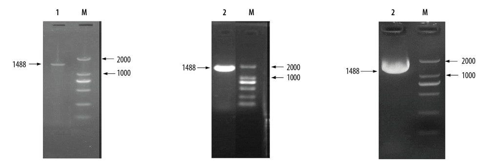 Figure 1. The genotype of the BW25113, BW25113ΔqseC, and BW25113ΔqseC pQseC was identified by polymerase chain reaction (PCR). M – DL2000 DNA Marker; 1 – BW25113; 2 – BW25113ΔqseC; 3 – BW25113ΔqseC pQseC.
 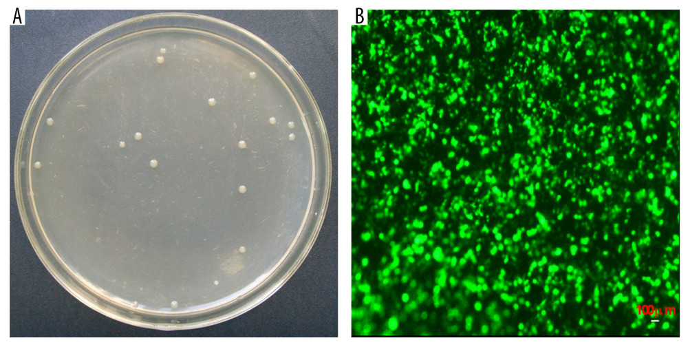 Figure 2. The ampicillin-resistant bacteria isolated from viscera. (A) The morphological characteristics of ampicillin-resistant bacteria. (B) The fluorescence microscopy image of the ampicillin-resistant bacteria. The photomicrograph was taken at 200× magnification.
 References
1. Ghalayini M, Launay A, Bridier-Nahmias A: Appl Environ Microbiol, 2018; 84(6); e02377-17
2. Mokszycki ME, Leatham-Jensen M, Steffensen JL: Appl Environ Microbiol, 2018; 84(24); e02166-18
3. Sorribas M, Jakob MO, Yilmaz B, FXR modulates the gut-vascular barrier by regulating the entry sites for bacterial translocation in experimental cirrhosis: J Hepatol, 2019; 71(6); 1126-40
4. Luo Y, Zeng B, Zeng L, Gut microbiota regulates mouse behaviors through glucocorticoid receptor pathway genes in the hippocampus: Transl Psychiatry, 2018; 8(1); 187
5. Filosa S, Di Meo F, Crispi S, Polyphenols-gut microbiota interplay and brain neuromodulation: Neural Regen Res, 2018; 13(12); 2055-59
6. Ikeda M, Shimizu K, Ogura H, Hydrogen-rich saline regulates intestinal barrier dysfunction, dysbiosis, and bacterial translocation in a murine model of sepsis: Shock, 2018; 50(6); 640-47
7. Carlson-Banning KM, Sperandio V: Curr Opin Microbiol, 2018; 41; 83-88
8. Sandrini S, Aldriwesh M, Alruways M, Freestone P, Microbial endocrinology: Host-bacteria communication within the gut microbiome: J Endocrinol, 2015; 225(2); R21-34
9. Clarke MB, Hughes DT, Zhu C, The QseC sensor kinase: A bacterial adrenergic receptor: Proc Natl Acad Sci USA, 2006; 103(27); 10420-25
10. Moreira CG, Weinshenker D, Sperandio V: Infect Immun, 2010; 78(3); 914-26
11. Bearson BL, Bearson SM: Microb Pathog, 2008; 44(4); 271-78
12. Pullinger GD, van Diemen PM, Carnell SC: Vet Res, 2010; 41(5); 68
13. Halang P, Toulouse C, Geißel B, Response of vibrio cholerae to the catecholamine hormones epinephrine and norepinephrine: J Bacteriol, 2015; 197(24); 3769-78
14. Feldman-Goriachnik R, Hanani M, The effects of sympathetic nerve damage on satellite glial cells in the mouse superior cervical ganglion: Auton Neurosci, 2019; 221; 102584
15. Parker CT, Russell R, Njoroge JW: J Bacteriol, 2017; 199(8); e00861-16
16. Lustri BC, Sperandio V, Moreira CG, Bacterial chat: Intestinal metabolites and signals in host-microbiota-pathogen interactions: Infect Immun, 2017; 85(12); e00476-17
17. Machado Ribeiro TR, Cardinali Lustri B, Elias WP, Moreira CG: J Bacteriol, 2019; 201(17); e00203-19
18. Machado Ribeiro TR, Cardinali Lustri B, Elias WP, Moreira CG: J Bacteriol, 2019; 201(17); e00203-19
19. Rooks MG, Veiga P, Reeves AZ, QseC inhibition as an antivirulence approach for colitis-associated bacteria: Proc Natl Acad Sci USA, 2017; 114(1); 142-47
20. He L, Dai K, Wen X: Front Microbiol, 2018; 9; 212
21. Njoroge J, Sperandio V: Infect Immun, 2012; 80(2); 688-703
22. Hernandez-Baltazar D, Zavala-Flores LM, Villanueva-Olivo A, The 6-hydroxydopamine model and parkinsonian pathophysiology: novel findings in an older model: Neurologia, 2017; 32(8); 533-39
23. Dong RF, Zhang B, Tai LW, The neuroprotective role of MiR-124-3p in a 6-hydroxydopamine-induced cell model of Parkinson’s disease via the regulation of ANAX5: J Cell Biochem, 2018; 119(1); 269-77
24. Real CC, Garcia PC, Britto LRG, Treadmill exercise prevents increase of neuroinflammation markers involved in the dopaminergic damage of the 6-OHDA Parkinson’s disease model: J Mol Neurosci, 2017; 63(1); 36-49
25. Feng XY, Yang J, Zhang X, Zhu J, Gastrointestinal non-motor dysfunction in Parkinson’s disease model rats with 6-hydroxydopamine: Physiol Res, 2019; 68(2); 295-303
26. Nagy E, Nagy G, Power CA, Anti-bacterial monoclonal antibodies: Adv Exp Med Biol, 2017; 1053; 119-53
27. Xu H, Xiong J, Xu J, Mosapride stabilizes intestinal microbiota to reduce bacterial translocation and endotoxemia in CCl(4)-induced cirrhotic rats: Dig Dis Sci, 2017; 62(10); 2801-11
28. Shao T, Zhao C, Li F, Intestinal HIF-1α deletion exacerbates alcoholic liver disease by inducing intestinal dysbiosis and barrier dysfunction: J Hepatol, 2018; 69(4); 886-95
Figures
Figure 1. The genotype of the BW25113, BW25113ΔqseC, and BW25113ΔqseC pQseC was identified by polymerase chain reaction (PCR). M – DL2000 DNA Marker; 1 – BW25113; 2 – BW25113ΔqseC; 3 – BW25113ΔqseC pQseC.Figure 2. The ampicillin-resistant bacteria isolated from viscera. (A) The morphological characteristics of ampicillin-resistant bacteria. (B) The fluorescence microscopy image of the ampicillin-resistant bacteria. The photomicrograph was taken at 200× magnification. Tables
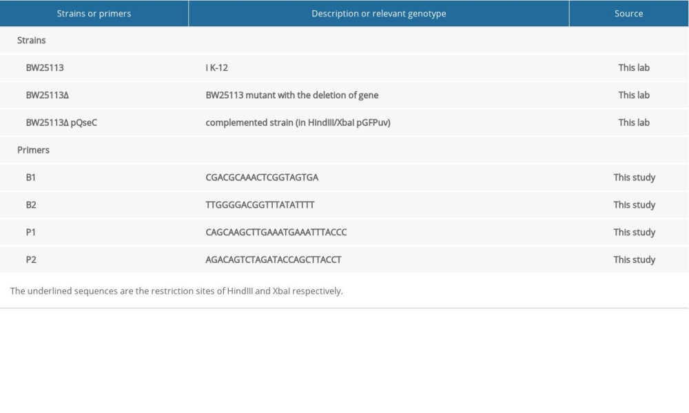 Table 1. Strains and primers used in this study.
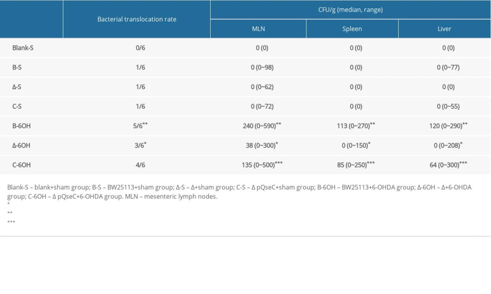 Table 2. Bacterial translocation after administration of 6-hydroxydopamine.
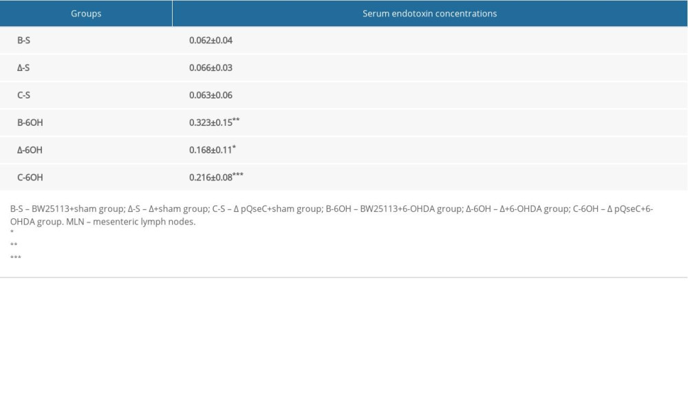 Table 3. The serum concentrations of endotoxin detected in the groups.Table 1. Strains and primers used in this study.Table 2. Bacterial translocation after administration of 6-hydroxydopamine.Table 3. The serum concentrations of endotoxin detected in the groups.
 In Press
Database Analysis
Epidemiology of Incidence and Mortality Due to Head and Neck Cancer in Poland in 2000 to 2022Med Sci Monit In Press; DOI: 10.12659/MSM.952477
Clinical Research
Comparison of Outcomes From Sequential Endoscopic Therapy in 60 Patients With Cirrhosis and Esophagogastric...Med Sci Monit In Press; DOI: 10.12659/MSM.952290
Clinical Research
Impact of a Staged Enteral Nutrition Nursing Pathway Based on Dynamic Assessment of Tolerance and Aspiratio...Med Sci Monit In Press; DOI: 10.12659/MSM.953263
Clinical Research
Risk of Vancouver-Type AG Greater Trochanteric Periprosthetic Fractures After Cementless Total Hip Arthropl...Med Sci Monit In Press; DOI: 10.12659/MSM.952936
Most Viewed Current Articles
17 Jan 2024 : Review article 14,176,688
Vaccination Guidelines for Pregnant Women: Addressing COVID-19 and the Omicron VariantDOI :10.12659/MSM.942799
Med Sci Monit 2024; 30:e942799
13 Nov 2021 : Clinical Research 3,764,843
Acceptance of COVID-19 Vaccination and Its Associated Factors Among Cancer Patients Attending the Oncology ...DOI :10.12659/MSM.932788
Med Sci Monit 2021; 27:e932788
14 Dec 2022 : Clinical Research 2,466,422
Prevalence and Variability of Allergen-Specific Immunoglobulin E in Patients with Elevated Tryptase LevelsDOI :10.12659/MSM.937990
Med Sci Monit 2022; 28:e937990
16 May 2023 : Clinical Research 708,979
Electrophysiological Testing for an Auditory Processing Disorder and Reading Performance in 54 School Stude...DOI :10.12659/MSM.940387
Med Sci Monit 2023; 29:e940387