31 July 2020: Lab/In Vitro Research
Curcumin Derivative C086 Combined with Cisplatin Inhibits Proliferation of Osteosarcoma Cells
Xi Jiang1ABCDF, Yulin Huang2ABCE*DOI: 10.12659/MSM.924507
Med Sci Monit 2020; 26:e924507
Abstract
BACKGROUND: Curcumin derivative C086 (cur C086) is a potential chemotherapeutic agent for patients with osteosarcoma. In this study, the effects of cur C086 combined with cisplatin on the biological processes of osteosarcoma cells were investigated.
MATERIAL AND METHODS: In this study, expression of BMIL1 was detected by real-time quantitative reverse transcription polymerase chain reaction and Western blotting in MG-63 cells treated with cur C086+cisplatin. Functions of cur C086+cisplatin on proliferation ability, apoptosis response, and metastatic potential of MG-63 cells were determined by MTT, flow cytometry, Hoechst 33258 staining and Transwell assays, respectively. In additionally, expression of P16, E-cadherin, epidermal growth factor (EGFR), and Notch1 was measured by Western blotting.
RESULTS: Expression of BMIL1 decreased significantly in MG-63 cells treated with cur C086 (20 µM)+cisplatin (1.28 nM). Treatment with cur C086+cisplatin considerably inhibited growth, migration, and invasion potential in MG-63 cells, whereas apoptosis was obviously upregulated. Moreover, cur C086+cisplatin suppressed BMIL1 expression or its potential downstream targets, P16, E-cadherin, EGFR, and Notch1.
CONCLUSIONS: The current results demonstrate that combined treatment with cur C086+cisplatin may be an effective form of chemotherapy for patients with osteosarcoma.
Keywords: Cisplatin, Curcumin, Osteosarcoma, Antigens, CD, Antineoplastic Agents, Phytogenic, Antineoplastic Combined Chemotherapy Protocols, Cadherins, Cyclin-Dependent Kinase Inhibitor p16, Drug Synergism, ErbB receptors, Inhibitory Concentration 50, Osteoblasts, Polycomb Repressive Complex 1, Receptor, Notch1
Background
Osteosarcoma is a malignant bone tumor more common in children or teenagers than in adults [1]. It develops from mesenchymal cells, and is also the most common primary bone tumor, characterized by rapid cell proliferation, strong metastatic potential, and resistance to chemotherapy [2]. At present, surgical excision, radiotherapy or chemotherapy are the main therapeutic strategies for osteosarcoma [3]. Recently, treatment of osteosarcoma has been improved by performing radical surgery. In addition, neoadjuvant chemotherapy increases 5-year overall survival from 30% to about 70% to 80% [4–6]. Although the survival rate has risen over the past decades, it is unsatisfactory and the prognosis for osteosarcoma patients with metastatic lesions is still poo. Despite the fact that chemotherapies and targeted drugs are useful against osteosarcoma, they can cause serious side effects [7,8]. Therefore, the molecular mechanism of osteosarcoma needs to be further studied to find better drugs with fewer adverse effects.
An increasing amount of evidence indicates that natural plant ingredients from traditional Chinese herbal medicines, such as ginsenoside, paclitaxel, and curcumin, have therapeutic advantages and effects on various tumors [9]. Curcumin reportedly is a powerful anticancer agent and can be isolated from ginger, which is encountered on a daily basis [10,11]. Multiple studies have revealed that curcumin can inhibit proliferation of the cells of various cancers, such as those of the breast, lung, and stomach, as well as osteosarcoma
The multifactorial pathogenesis of osteosarcoma involves genetic and epigenetic alterations of tumor suppressor genes, oncogenes or growth factors in osteosarcoma development [18].
C086 [4-(4-hydroxy-3-methoxy-phenyl-methyl)] is a new structural analog of curcumin. Being artificially synthesized, it has better properties than natural curcumin, such as solubility and antitumor effects. [26]. In this study, we intended to investigate the effect of the curcumin derivative C086 combined with cisplatin on proliferation and invasion of the MG-63 cells, as well as on expression of
Material and Methods
REAGENTS:
Cur C086, cisplatin, and 5-fluorouracil (5-FU) were purchased from Mclean Corp (Shanghai, China). For
CELL CULTURE:
MG-63 cells from ATCC (Manassas, Virginia, United States) were cultured in Dulbecco’s Modified Eagle Medium (Gibco; Thermo Fisher Scientific, Inc., United States) containing 10% fetal bovine serum, 100 μg/mL streptomycin and 100 U/mL penicillin. These cells were cultured in six-well plates under humidified conditions with 5% CO2 at 37°C. MG-63 cells were grown as a monolayer and cultured until 80% confluence was reached for use in further experiments.
QRT-PCR:
Total RNA was isolated with an RNA separation Kit (cat. no: Z3382, Promega, United States) and quantified with a spectrophotometer. Total RNA (400 ng) was used to synthesize cDNA using a PrimeScript reverse transcription polymerase chain reaction (RT-PCR) kit, according to reagent instructions (cat. no: RR055B, Takara, Japan). A SYBR Green I fluorescent dye (cat. no: RR420L, Takara, Japan) was used and quantitative PCR was performed on the ABI Prism PCR system. The experimental steps were as follows: 5 min at 95°C, followed by 36 cycles at 95°C for 35 s and 55°C for 45 s. All primers were from Invitrogen Company (Thermo Fisher Scientific, Inc., United States). Glyceraldehyde 3-phosphate dehydrogenase (GAPDH) was used as control. The primers sequences used were as follows:
Fold change in
CELL PROLIFERATION ASSAY:
Growth of MG-63 cells incubated with different concentrations of cur C086 for various durations was analyzed with MTT assays (cat. no: 88417, Sigma-Aldrich, United States). In brief, MG-63 cells were cultured in 96-well plates (Corning, NY, USA) at a density of 5×10³ cells/well. The cells were treated with cur C086 (0, 10, 20, 40 and 80 μM) for 48 h, or cur C086 (20 μM) for 0, 12, 24, 48, and 72 h. MTT dye was added to each well for at least 4 h. The reaction was stopped by adding DMSO and absorbance at 570 nm was measured at each time point using an enzyme immunoassay analyzer. All samples were analyzed in triplicate.
SENSITIVITY ANALYSIS OF CUR C086:
To assess the effect of cur C086 on the sensitivity of MG-63 cells to chemotherapeutic drugs, a single-drug intervention and an
SYNERGY OF CUR C086 AND CISPLATIN DETECTED BY MTT:
The collaborative experiment was divided into 7 groups: control, cisplatin (1.28 nM), 5-FU (2 μg/mL), cur C086 (20 μM), cisplatin (1.28 nM)+5-FU (2 μg/mL), cur C086 (20 μM)+5-FU (2 μg/mL), and cur C086 (20 μM)+cisplatin (1.28 nM) group. The cell culture system was the same as the previous cell proliferation experiment. The calculation formula for the coefficient of drug interaction (CDI) was: CDI=AB/(A×B).
APOPTOSIS ASSAYS:
Cisplatin (1.28 nM), cur C086 (20 μM), and cur C086 (20 μM)+cisplatin (1.28 nM) were used to treat MG-63 cells for 48 h. The MG-63 (1×10⁵) cells were collected and washed with PBS twice. Subsequently, the cells were stained with Hoechst 33258 reagents (cat. no: C1017, Beyotime Biotechnology, Shanghai, China) and observed under two-photon fluorescence microscopy (LaVision BioTec, Germany). At the same time, the treated cells (1.5×10⁵) were collected and washed with PBS twice. Annexin-V/PI (5 μL) (cat. no: 556547, BD Biosciences, United States) was added into the treated cells resuspended in PBS for 15 min in the dark. Apoptosis was detected through flow cytometry (BD Biosciences, United States). All samples were analyzed in triplicate.
MIGRATION AND INVASION ASSAY:
Nigration and invasion abilities assays were used with a 12-well Transwell chamber (8-mm pore size; Corning Incorporated) coated with Matrigel (200 mL per well, thickness: 3 mm; BD Biosciences) in triplicate. MG-63 cells were incubated with cisplatin (1.28 nM), cur C086 (4 μM), and cur C086 (20 μM)+cisplatin (1.28 nM) for 48 h. The treated cells (1×10⁵) were resuspended in a medium without serum, added into the upper chamber and cultured for 48 h, then in a medium with serum added to the lower chamber. Following incubation, non-migrating or non-invading cells in the upper chamber were erased with a cotton swab. Cells attached to the underside of the filter membrane were stained with crystal violet and 5 random fields were taken from the microscope (Olympus, Tokyo, Japan). Then the number of migration and invasion cells dyed purple in each field under microscopy was counted. All samples were investigated in triplicate.
WESTERN BLOT ANALYSIS:
Protein lysates of MG-63 cells treated with cisplatin (1.28 nM), cur C086 (20 μM) and cur C086 (20 μM)+cisplatin (1.28 nM) were lysed with a protein lysate buffer (Sigma-Aldrich; Merck KGaA) and the total protein concentration was quantified with a BCA Protein Assay kit (Pierce; Thermo Fisher Scientific, Inc.). Ten micrograms of each sample protein was resuspended in a loading buffer and electrophoresed on 10% sodium dodecyl sulfate polyacrylamide gel electrophoresis. Fractionated proteins were transferred onto polyvinylidene difluoride membranes (Invitrogen; Thermo Fisher Scientific, Inc.). The following primary antibodies were used: Anti-P16 (cat. no: sc-81866, Santa Cruz Biotechnology, United States), anti-E-cadherin (cat. no: sc-8426, Santa Cruz Biotechnology, United States), anti-
STATISTICAL ANALYSIS:
All experimental data analyses were performed in triplicate and all results were presented as the mean±SEM. Student’s
Results
:
First, we analyzed expression of BMIL1 from mRNA and protein levels in MG-63 cells exposed to cur C086 at different concentrations for 48 h (Figure 1A–1C). The inhibitory effects of cur C086 were also measured via an MTT assay (Figure 1D). The results demonstrated that BMIL1 expression was significantly downregulated by cur C086, and that cur C086 inhibited proliferation of MG-63 cells in a dose-dependent manner. The IC50 values of cur C086 and cisplatin weree 20 μM and 1.28 nM, respectively.
CONFIRMATION OF MOST SUITABLE DRUG CONCENTRATION OF CUR C086:
Expression of BMIL1 in MG-63 cells exposed to 20 μM cur C086 for different durations (Figure 2A, 2B) and effects of cur C086 on proliferation of MG-63 cells (Figure 2C) were measured. The results revealed that BMIL1 expression and MG-63 cell proliferation were affected in a time-dependent manner.
:
To determine whether BMIL1 expression was significantly affected by cur C086+cisplatin in MG-63 cells, quantitative RT-PCR and Western blotting were performed (Figure 3). The results proved that BMIL1 expression was obviously decreased following treatment with cur C086+cisplatin compared with the control, cur C086, and cisplatin groups.
CUR C086 INCREASED CHEMOSENSITIVITY OF MG-63 CELLS:
The sensitivity of MG-63 cells to cur C086 combined with chemotherapeutic drugs was studied in vitro (Figure 4). The results indicated that cell absorbance was significantly lower in the combined treatment groups after 48 h compared with the single-intervention groups. These results suggest that cur C086 intervention may enhance the chemosensitivity of MG-63 cells to cisplatin or 5-FU.
SYNERGISTIC EFFECT OF CUR C086 AND CISPLATIN ON MG-63 CELLS:
MG-63 cells were treated with cur C086 (20 μM) and cisplatin (1.28 nM) for 24 and 48 hours. MTT results showed that cur C086 and cisplatin increased the killing effect on MG-63 cells. Compared to the control group, the inhibition rates in the cur C086+cisplatin group were 42.29±3.5% and 46.32±4.1%, respectively. The CDI values of two different time groups (24 and 48 h) were 0.68 and 0.64, indicating that cur C086 and cisplatin were remarkably synergistic in inhibiting MG-63 cells.
CUR C086 INDUCED APOPTOSIS IN MG-63 CELLS:
To determine the effect of cur C086 on MG-63 cell apoptosis, cells were treated with cisplatin (1.28 nM), cur C086 (20 μM), and cur C086 (20 μM)+cisplatin (1.28 nM) for 48 h and analyzed using flow cytometry (Figure 5A, 5B) and Hoechst 33258 reagents (Figure 5C, 5D). Annexin V-FITC/PI double staining was used to detect cells in different phases of apoptosis. The results indicated that apoptosis of MG-63 cells increased considerably after cur C086+cisplatin treatment compared with the other groups.
CUR C086 SUPPRESSED MIGRATION AND INVASION OF MG-63 CELLS:
Cisplatin (1.28 nM), cur C086 (20 μM), and cur C086 (20 μM)+cisplatin were used to treat MG-63 cells for 48 h, and Transwell assays were conducted. The assays revealed that the migration abilities of MG-63 cells decreased significantly in the cur C086+cisplatin group compared to in the control cell group (Figure 6A, 6B). Similarly, the invasive abilities of MG-63 cells treated with cur C086+cisplatin obviously declined after 48 h of incubation (Figure 6C, 6D).
CUR C086 INHIBITED EXPRESSION OF P16, E-CADHERIN, EGFR, AND NOTCH1 IN MG-63 CELLS:
Expression of P16, E-cadherin, EGFR, and Notch1 in osteosarcoma cells treated with C086 and cisplatin was investigated. Experimental results suggested that P16 and E-cadherin proteins were markedly downregulated in the cur C086+cisplatin treatment group in comparison with the single-treatment groups (Figure 7A, 7B). Furthermore, EGFR and Notch1 inhibition was observed in MG-63 cells treated with cur C086+cisplatin (Figure 7C, 7D).
Discussion
Curcumin and curcumin derivatives exert significant inhibitory effects on the growth and metastasis of osteosarcoma cell lines, and they induce apoptosis in these cancerous cells [27]. Cisplatin is a platinum complex with broad-spectrum antitumor activity. At high doses, it inhibits synthesis of DNA, and at lower doses, it inhibits synthesis of RNA and protein in treatment of osteosarcoma [28,29]. In this study, the influence of curcumin C086 combined with cisplatin on osteosarcoma was evaluated.
Epithelial to mesenchymal transition (EMT) may increase the motility of cells and enable them to gain invasive properties, a process that is mediated by repression of E-cadherin [34,35]. EMT is a critical step in invasion and metastasis of cancers, and could be used as an indicator of cancer progression or prognosis, or a treatment target [36]. Invasion and metastasis of tumor cells are the main causes of death in patients with cancers including osteosarcoma. Interestingly, we discovered that cur C086 and cisplatin inhibited the migration and invasion potential of MG-63 cells and increased apoptosis of them.
A recent report showed that
EGFR expression has been detected in osteosarcoma, and because its activity is associated with tumor proliferation, invasion, and metastasis, EGFR been a significant target in cancer therapy [39,40]. EGFR is overexpressed in osteosarcoma and the response to EGFR-targeted agents is inversely correlated with EMT [41]. Notch signaling plays a role in development and progression of osteosarcoma [42]. Wang et al. [43] found that curcumin inhibits hypoxia-induced proliferation and invasion of MG-63 cells through downregulation of Notch1. We found that expression of EGFR and Notch1 was downregulated in MG-63 cells treated with cur C086 or cisplatin, and especially with combined treatment with cur C086 and cisplatin.
Conclusions
Our findings demonstrate that combined treatment with cur C086 and cisplatin inhibits expression of
Figures
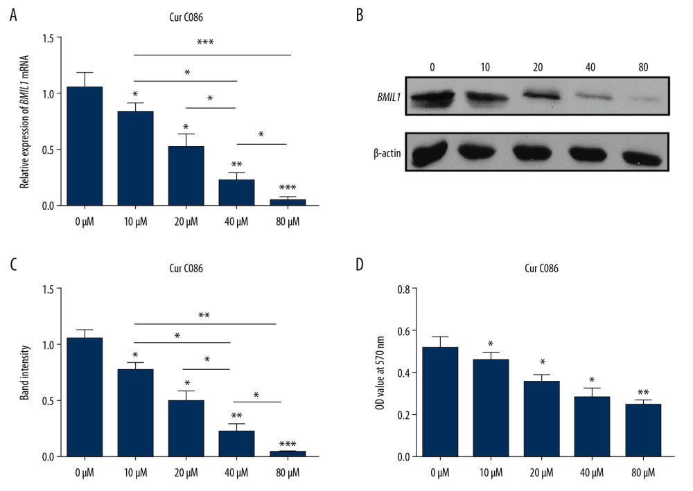 Figure 1. Expression of BMIL1 was inhibited by curcumin C086 in MG-63 cells. Levels of expression of BMIL1 mRNA (A) and protein (B, C) were downregulated by treatment with different concentrations of curcumin C086. (D) Curcumin C086 suppressed proliferation of MG-63 cells, as analyzed in MTT experiments. All data are presented as the mean±SEM. * P<0.05, ** P<0.01 and *** P<0.001.
 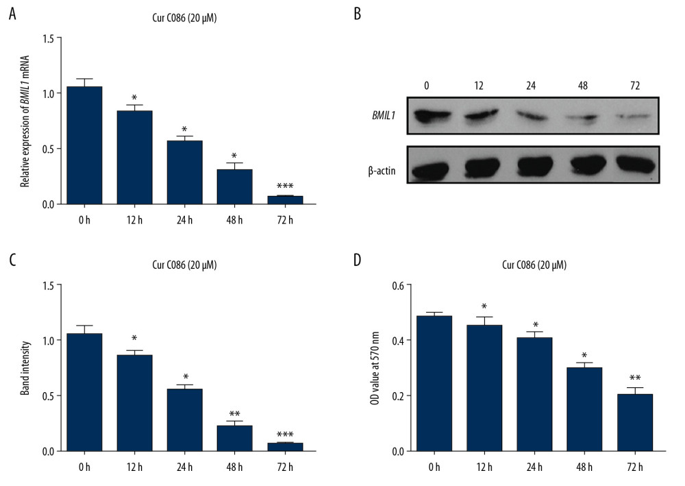 Figure 2. Expression of BMIL1 was downregulated in MG-63 cells incubated with curcumin C086 (20 μM) for 0, 12, 24, 48, and 72 h. Expression of BMIL1 mRNA (A) and protein (B) were repressed by curcumin C086. (C, D) Proliferation of MG-63 cells was inhibited by curcumin C086 (20 μM), as analyzed by MTT assay. Data are expressed as the mean±SEM. * P<0.05, ** P<0.01 and *** P<0.001.
 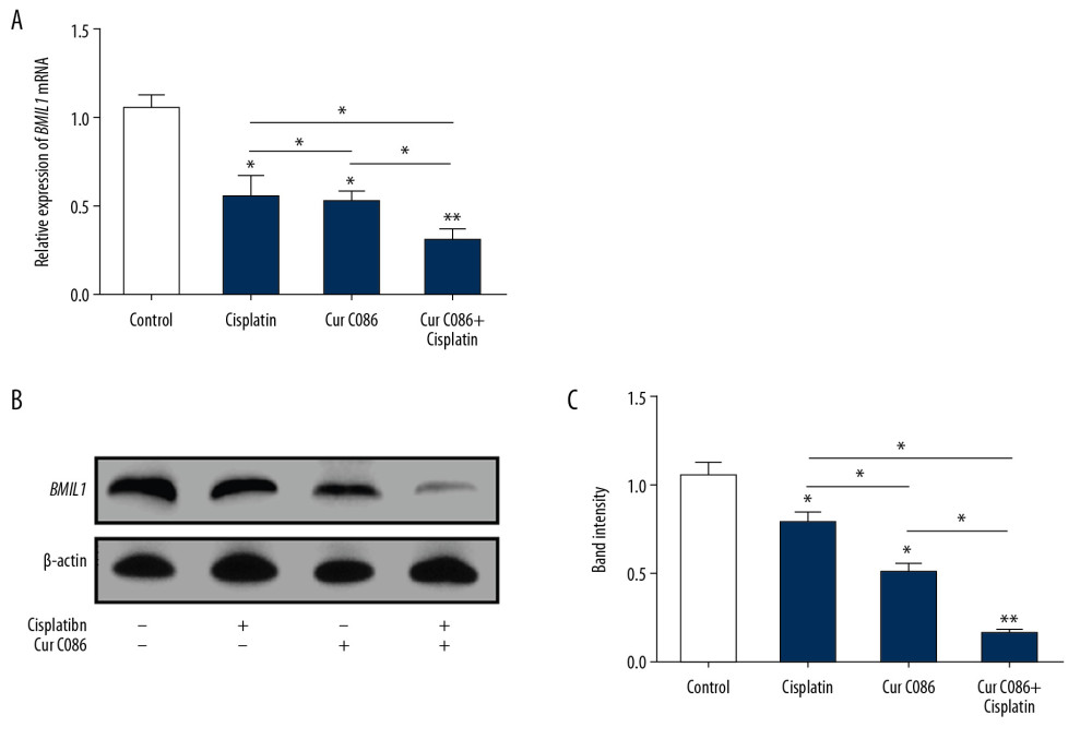 Figure 3. Expression of BMIL1 was analyzed in MG-63 cells treated with curcumin C086 and cisplatin. Expression of BMIL1 mRNA (A) and protein (B, C) was inhibited by curcumin C086 (20 μM)+cisplatin (1.28 nM). Data are expressed as the mean±SEM. * P<0.05 and ** P<0.01.
 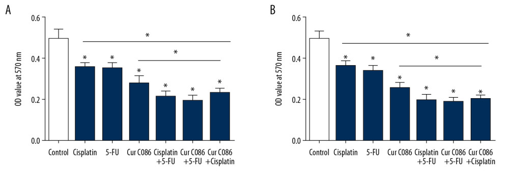 Figure 4. Curcumin C086 had synergic anticancer effects when combined with cisplatin and 5-FU. Proliferation of MG-63 cells was inhibited following treatment with curcumin C086 (20 μM) and cisplatin or 5-FU for (A) 24 or (B) 48 h. Data are expressed as the mean±SEM. * P<0.05.
 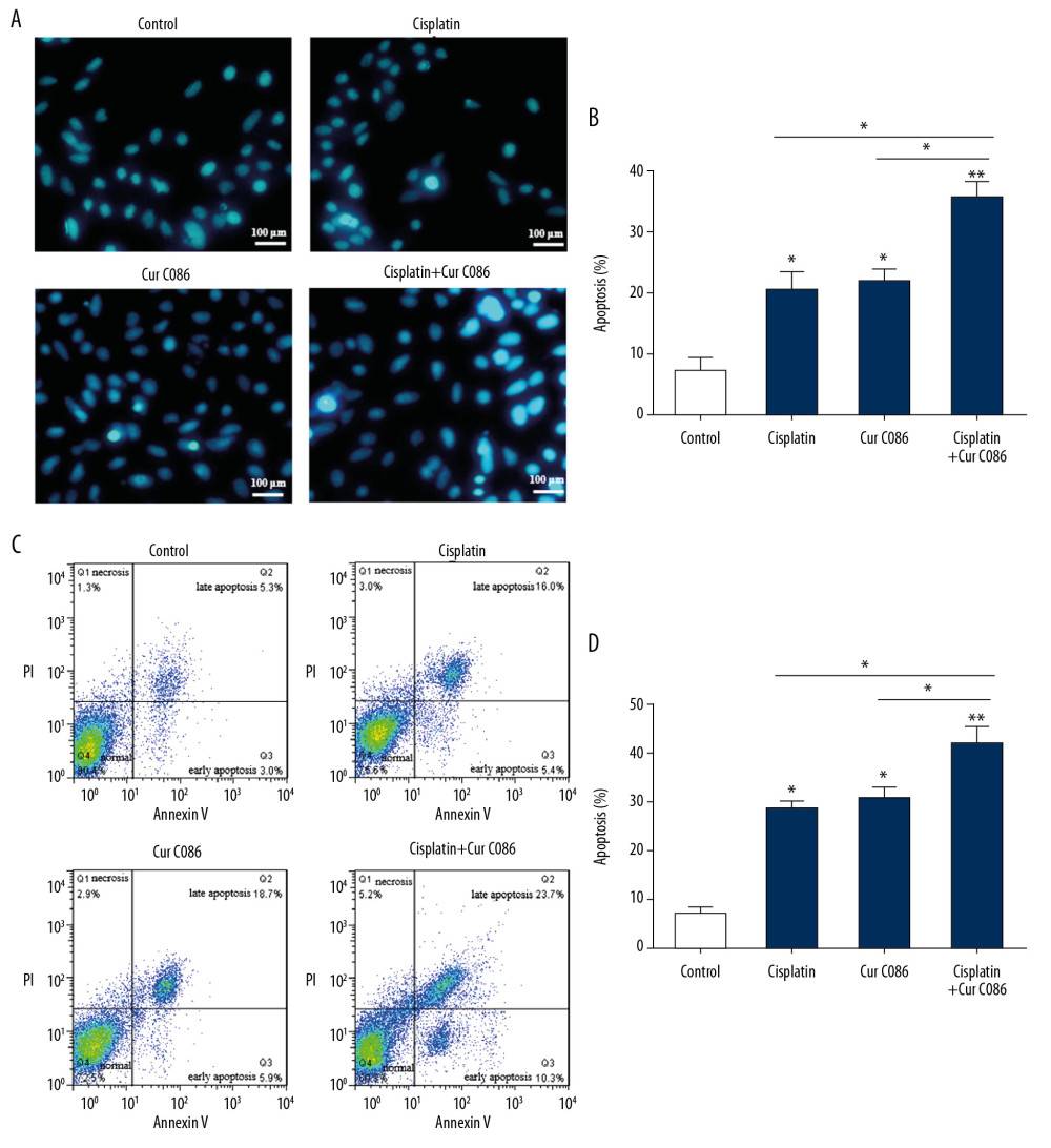 Figure 5. Treatment of MG-63 cells with curcumin C086+cisplatin significantly promoted apoptosis. Curcumin C086 (20 μM)+cisplatin increased apoptosis of MG-63 cells analyzed by flow cytometry (A, B) and Hoechst 33258 reagents (C, D). All images were acquired under an inverted microscope (200× magnification). Data are expressed as the mean±SEM. * P<0.05 and ** P<0.01.
 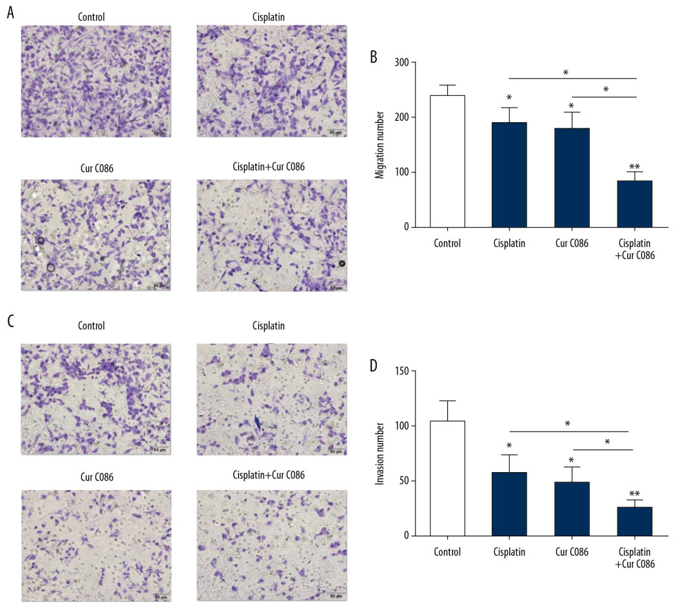 Figure 6. Migration and invasion abilities of MG-63 cells were inhibited by treatment with curcumin C086+cisplatin. (A, B) The effects of curcumin C086 (20 μM)+cisplatin on MG-63 cell migration potential were measured by Transwell assay. (C, D) The invasiveness of MG-63 cells incubated with curcumin C086 (20 μM)+cisplatin was evaluated through Transwell assay. Images were obtained under an inverted microscope with 200× magnification. Data are expressed as the mean±SEM. * P<0.05 and ** P<0.01.
 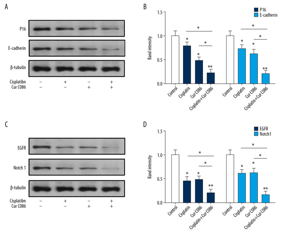 Figure 7. Levels of expression of P16, E-cadherin, Notch1 and EGFR in MG-63 cells were analyzed by Western blotting. (A, B) P16 and E-cadherin proteins were decreased in MG-63 cells treated with cisplatin, curcumin C086, and curcumin C086+cisplatin. (C, D) Meanwhile, Notch1 and EGFR proteins were downregulated in MG-63 cells treated with cisplatin, curcumin C086, and curcumin C086+cisplatin. Data are expressed as the mean±SEM. * P<0.05 and ** P<0.01.
 References
1. Zhou W, Hao M, Du X, Advances in targeted therapy for osteosarcoma: Discov Med, 2014; 17; 301-7
2. Liu YJ, Li W, Chang F, MicroRNA-505 is down-regulated in human osteosarcoma and regulates cell proliferation, migration and invasion: Oncol Rep, 2018; 39; 491-500
3. Cortini M, Avnet S, Baldini N, Mesenchymal stroma: Role in osteosarcoma progression: Cancer Lett, 2017; 405; 90-99
4. Provisor AJ, Ettinger LJ, Nachman JB, Treatment of nonmetastatic osteosarcoma of the extremity with preoperative and postoperative chemotherapy: A report from the Children’s Cancer Group: J Clin Oncol, 1997; 15; 76-84
5. Ottaviani G, Jaffe N, The epidemiology of osteosarcoma: Cancer Treat Res, 2009; 152; 3-13
6. Vassiliou LV, Lalabekyan B, Jay A, Head and neck sarcomas: A single institute series: Oral Oncol, 2017; 65; 16-22
7. Bishop MW, Janeway KA, Gorlick R, Future directions in the treatment of osteosarcoma: Curr Opin Pediatr, 2016; 28; 26-33
8. Otoukesh B, Boddouhi B, Moghtadaei M, Novel molecular insights and new therapeutic strategies in osteosarcoma: Cancer Cell Int, 2018; 18; 158
9. Chen , Jiang H, Cao Y, Drug target identification using network analysis: Taking active components in Sini decoction as an example: Sci Rep, 2016; 6; 24245
10. Golonko A, Lewandowska H, Świsłocka R, Curcumin as tyrosine kinase inhibitor in cancer treatment: Eur J Med Chem, 2019; 181; 111512
11. Zhao S, Pi C, Ye Y, Recent advances of analogues of curcumin for treatment of cancer: Eur J Med Chem, 2019; 180; 524-35
12. Xu Y, Zhang J, Han J, Curcumin inhibits tumor proliferation induced by neutrophil elastase through the upregulation of α1-antitrypsin in lung cancer: Mol Oncol, 2012; 6; 405-17
13. Lv ZD, Liu XP, Zhao WJ: Int J Clin Exp Pathol, 2014; 7; 2818-24
14. Wilken R, Veena MS, Wang MB, A review of anti-cancer properties and therapeutic activity in head and neck squamous cell carcinoma: Mol Cancer, 2011; 10; 12
15. Angulo P, Kaushik G, Subramaniam D, Natural compounds targeting major cell signaling pathways: A novel paradigm for osteosarcoma therapy: J Hematol Oncol, 2017; 10; 10
16. Sun Y, Liu L, Wang Y, Curcumin inhibits the proliferation and invasion of MG-63 cells through inactivation of the p-JAK2/p-STAT3 pathway: Onco Targets Ther, 2019; 12; 2011-21
17. Kelleher FC, O’Sullivan H, Monocytes, macrophages, and osteoclasts in osteosarcoma: J Adolesc Young Adult Oncol, 2017; 6; 396-405
18. Dang H, Wu W, Wang B, CXCL5 plays a promoting role in osteosarcoma cell migration and invasion in autocrine- and paracrine-dependent manners: Oncol Res, 2017; 25; 177-86
19. Cao L, Bombard J, Cintron K, BMI1 as a novel target for drug discovery in cancer: J Cell Biochem, 2011; 112; 2729-41
20. Kang MK, Kim RH, Kim SJ, Elevated BMI-1 expression is associated with dysplastic cell transformation during oral carcinogenesis and is required for cancer cell replication and survival: Br J Cancer, 2007; 96; 126-23
21. Liu J, Luo B, Zhao M, BMI-1 targeting suppresses osteosarcoma aggressiveness through the NF-κB signaling pathway: Mol Med Rep, 2017; 16; 7949-58
22. Xie X, Ye Z, Yang D, Effects of combined c-myc and Bmi-1 siRNAs on the growth and chemosensitivity of MG-63 osteosarcoma cells: Mol Med Rep, 2013; 8; 168-72
23. Wu Z, Min L, Chen D, Overexpression of BMI-1 promotes cell growth and resistance to cisplatin treatment in osteosarcoma: PLoS One, 2011; 6; e14648
24. Cheng C, Ding Q, Zhang Z, PTBP1 modulates osteosarcoma chemoresistance to cisplatin by regulating the expression of the copper transporter SLC31A1: J Cell Mol Med, 2020; 24(9); 5274-89
25. Fanelli M, Tavanti E, Patrizio MP: Front Oncol, 2020; 10; 331
26. Chen C, Liu Y, Chen Y, C086, a novel analog of curcumin, induces growth inhibition and down-regulation of NF-κB in colon cancer cells and xenograft tumors: Cancer Biol Ther, 2011; 12; 797-807
27. Maran A, Yaszemski MJ, Kohut A, Curcumin and osteosarcoma: Can invertible polymeric micelles help?: Materials (Basel), 2016; 9; E520
28. Zhang B, Zhang Y, Li R, Oncolytic adenovirus Ad11 enhances the chemotherapy effect of cisplatin on osteosarcoma cells by inhibiting autophagy: Am J Transl Res, 2020; 12; 105-17
29. Hou Z, Zhou Y, Li J: Sci Rep, 2015; 5; 13637
30. Benard A, Goossens-Beumer IJ, van Hoesel AQ, Prognostic value of polycomb proteins EZH2, BMI1 and SUZ12 and histone modification H3K27me3 in colorectal cancer: PLoS One, 2014; 9; e108265
31. Li Z, Wang Y, Yuan C, Oncogenic roles of BMI1 and its therapeutic inhibition by histone deacetylase inhibitor in tongue cancer: Lab Invest, 2014; 94; 1431-45
32. Mayr C, Wagner A, Loeffelberger M, The BMI1 inhibitor PTC-209 is a potential compound to halt cellular growth in biliary tract cancer cells: Oncotarget, 2016; 7; 745-58
33. Kreso A, van Galen P, Pedley NM, Self-renewal as a therapeutic target in human colorectal cancer: Nat Med, 2014; 20; 29-36
34. Kong D, Li Y, Wang Z, Cancer stem cells and epithelial-to-mesenchymal transition (EMT)-phenotypic cells: Are they cousins or twins?: Cancers (Basel), 2011; 3; 716-29
35. Brabletz T, Jung A, Reu S, Variable beta-catenin expression in colorectal cancers indicates tumor progression driven by the tumor environment: Proc Natl Acad Sci USA, 2001; 98; 10356-61
36. Liang Z, Wu R, Xie W, Curcumin reverses tobacco smoke-induced epithelial-mesenchymal transition by suppressing the MAPK pathway in the lungs of mice: Mol Med Rep, 2018; 17; 2019-25
37. Meng X, Wang Y, Zheng X, shRNA-mediated knockdown of Bmi-1 inhibit lung adenocarcinoma cell migration and metastasis: Lung Cancer, 2012; 77; 24-30
38. Yang MH, Hsu DS, Wang HW, BMI1 is essential in Twist1-induced epithelial-mesenchymal transition: Nat Cell Biol, 2010; 12; 982-92
39. Fard SS, Saliminejad K, Sotoudeh M, The correlation between EGFR and androgen receptor pathways: A novel potential prognostic marker in gastric cancer: Anticancer Agents Med Chem, 2019; 19; 2097-107
40. Benli Yavuz B, Koç M, Kozacıoğlu S, Prognostic importance of PTEN, EGFR, HER-2, and IGF-1R in patients treated with postoperative chemoradiation: Turk J Med Sci, 2019; 49; 1025-32
41. Wang T, Wang D, Zhang L, The TGFβ-miR-499a-SHKBP1 pathway induces resistance to EGFR inhibitors in osteosarcoma cancer stem cell-like cells: J Exp Clin Cancer Res, 2019; 38; 226
42. Yu L, Fan Z, Fang S, Cisplatin selects for stem-like cells in osteosarcoma by activating notch signaling: Oncotarget, 2016; 7; 33055-68
43. Wang Z, Zhang K, Zhu Y, Curcumin inhibits hypoxia-induced proliferation and invasion of MG-63 osteosarcoma cells via downregulating Notch1: Mol Med Rep, 2017; 15; 1747-52
Figures
 Figure 1. Expression of BMIL1 was inhibited by curcumin C086 in MG-63 cells. Levels of expression of BMIL1 mRNA (A) and protein (B, C) were downregulated by treatment with different concentrations of curcumin C086. (D) Curcumin C086 suppressed proliferation of MG-63 cells, as analyzed in MTT experiments. All data are presented as the mean±SEM. * P<0.05, ** P<0.01 and *** P<0.001.
 Figure 2. Expression of BMIL1 was downregulated in MG-63 cells incubated with curcumin C086 (20 μM) for 0, 12, 24, 48, and 72 h. Expression of BMIL1 mRNA (A) and protein (B) were repressed by curcumin C086. (C, D) Proliferation of MG-63 cells was inhibited by curcumin C086 (20 μM), as analyzed by MTT assay. Data are expressed as the mean±SEM. * P<0.05, ** P<0.01 and *** P<0.001.
 Figure 3. Expression of BMIL1 was analyzed in MG-63 cells treated with curcumin C086 and cisplatin. Expression of BMIL1 mRNA (A) and protein (B, C) was inhibited by curcumin C086 (20 μM)+cisplatin (1.28 nM). Data are expressed as the mean±SEM. * P<0.05 and ** P<0.01.
 Figure 4. Curcumin C086 had synergic anticancer effects when combined with cisplatin and 5-FU. Proliferation of MG-63 cells was inhibited following treatment with curcumin C086 (20 μM) and cisplatin or 5-FU for (A) 24 or (B) 48 h. Data are expressed as the mean±SEM. * P<0.05.
 Figure 5. Treatment of MG-63 cells with curcumin C086+cisplatin significantly promoted apoptosis. Curcumin C086 (20 μM)+cisplatin increased apoptosis of MG-63 cells analyzed by flow cytometry (A, B) and Hoechst 33258 reagents (C, D). All images were acquired under an inverted microscope (200× magnification). Data are expressed as the mean±SEM. * P<0.05 and ** P<0.01.
 Figure 6. Migration and invasion abilities of MG-63 cells were inhibited by treatment with curcumin C086+cisplatin. (A, B) The effects of curcumin C086 (20 μM)+cisplatin on MG-63 cell migration potential were measured by Transwell assay. (C, D) The invasiveness of MG-63 cells incubated with curcumin C086 (20 μM)+cisplatin was evaluated through Transwell assay. Images were obtained under an inverted microscope with 200× magnification. Data are expressed as the mean±SEM. * P<0.05 and ** P<0.01.
 Figure 7. Levels of expression of P16, E-cadherin, Notch1 and EGFR in MG-63 cells were analyzed by Western blotting. (A, B) P16 and E-cadherin proteins were decreased in MG-63 cells treated with cisplatin, curcumin C086, and curcumin C086+cisplatin. (C, D) Meanwhile, Notch1 and EGFR proteins were downregulated in MG-63 cells treated with cisplatin, curcumin C086, and curcumin C086+cisplatin. Data are expressed as the mean±SEM. * P<0.05 and ** P<0.01.
 In Press
15 Apr 2024 : Laboratory Research
The Role of Copper-Induced M2 Macrophage Polarization in Protecting Cartilage Matrix in OsteoarthritisMed Sci Monit In Press; DOI: 10.12659/MSM.943738
07 Mar 2024 : Clinical Research
Knowledge of and Attitudes Toward Clinical Trials: A Questionnaire-Based Study of 179 Male Third- and Fourt...Med Sci Monit In Press; DOI: 10.12659/MSM.943468
08 Mar 2024 : Animal Research
Modification of Experimental Model of Necrotizing Enterocolitis (NEC) in Rat Pups by Single Exposure to Hyp...Med Sci Monit In Press; DOI: 10.12659/MSM.943443
18 Apr 2024 : Clinical Research
Comparative Analysis of Open and Closed Sphincterotomy for the Treatment of Chronic Anal Fissure: Safety an...Med Sci Monit In Press; DOI: 10.12659/MSM.944127
Most Viewed Current Articles
17 Jan 2024 : Review article
Vaccination Guidelines for Pregnant Women: Addressing COVID-19 and the Omicron VariantDOI :10.12659/MSM.942799
Med Sci Monit 2024; 30:e942799
14 Dec 2022 : Clinical Research
Prevalence and Variability of Allergen-Specific Immunoglobulin E in Patients with Elevated Tryptase LevelsDOI :10.12659/MSM.937990
Med Sci Monit 2022; 28:e937990
16 May 2023 : Clinical Research
Electrophysiological Testing for an Auditory Processing Disorder and Reading Performance in 54 School Stude...DOI :10.12659/MSM.940387
Med Sci Monit 2023; 29:e940387
01 Jan 2022 : Editorial
Editorial: Current Status of Oral Antiviral Drug Treatments for SARS-CoV-2 Infection in Non-Hospitalized Pa...DOI :10.12659/MSM.935952
Med Sci Monit 2022; 28:e935952


