19 December 2023: Database Analysis
Exploring the Research Landscape of High Myopia: Trends, Contributors, and Key Areas of Focus
Kaiyao ChiDOI: 10.12659/MSM.941670
Med Sci Monit 2023; 29:e941670


Abstract
BACKGROUND: Myopia results when light rays focus before reaching the retina, causing blurred vision. High myopia (HM), defined by a refractive error of ≤-6 diopters (D) or an axial length of ≥26 mm, is an extreme form of this condition. The progression from HM to pathological myopia (PM) is marked by extensive ocular axis elongation. The rise in myopia has escalated concerns for HM due to its potential progression to pathological myopia. The covert progression of HM calls for thorough analysis of its current research landscape.
MATERIAL AND METHODS: HM-related publications from 2003-2022 were retrieved from the Web of Science database. Using VOSviewer and Citespace software, we conducted a bibliometric and visualized analysis to create document co-citation network maps. These maps detailed authors, institutions, countries, key terms, and significant literature.
RESULTS: From 9,079 articles, 8,241 were reviewed. An increasing trend in publications was observed, with Kyoko Ohno-Matsui identified as a top contributor. The Journal of Cataract and Refractive Surgery was the primary publication outlet. Chinese researchers and institutions were notably active. The document citation network identified five focal areas: refractive surgery, clinical manifestations/treatment, prevention/control, genetics, and open angle glaucoma.
CONCLUSIONS: Research emphasis in HM has shifted from refractive surgery for visual acuity enhancement to the diagnosis, classification, prevention, and control of HM complications. Proposals for early myopia intervention to prevent HM are gaining attention. Genetics and HM's link with open angle glaucoma, though smaller in focus, significantly enhance our understanding of HM.
Keywords: aplasia cutis congenita, high myopia, Cone-Rod Dysfunction
Background
Myopia is an optical condition in which incoming light rays converge anterior to the retina, yielding unclear vision. High myopia (HM), typified by a refractive error ≤-6 diopters (D) or axial length ≥26 mm, is a severe iteration of this condition. HM can further progress into pathological myopia (PM), a state characterized by excessive ocular axis elongation. Such elongation triggers mechanical stretching and thinning of the choroid and retinal pigment epithelium, ultimately inducing irreversible vision loss [1]. Over the past 2 decades, HM research has seen a surge in activity and has accrued significant advancements. Nonetheless, the expansive domain of study and the vast corpus of publications may pose challenges for newcomers in discerning key research progressions, core foci, and emerging frontiers within HM. As such, a comprehensive summation of the literature is crucial to elucidate the prevailing research foci in HM, thereby informing and directing future targeted research efforts.
Bibliometrics, a discipline leveraging mathematical statistics to spotlight key research themes and frontiers within a specific field [2], often draws data from metrics such as paper count, authorship, and keyword frequency. VOSviewer [3] and CiteSpace4[4] are notable bibliometric tools, both based on Java, facilitating researchers in decoding complex data sets through visualization techniques. Each tool has distinct strengths, and a dual application in data analysis is likely to yield a more comprehensive and accurate understanding. The goal of this study is to visually dissect the present landscape and evolutionary trends of basic and clinical research pertaining to HM through the lens of bibliometrics and knowledge mapping.
Material and Methods
DATA COLLECTION:
To determine prominent research themes and emerging trajectories in HM studies, a comprehensive retrieval of HM-related research published from January 1, 2003 to December 31, 2022 was conducted utilizing the Web of Science.
The search strategy encompassed the following terms: Topic=“high myopia” OR “pathologic myopia” OR “extreme myopia” OR “hypermyopia.” The data employed for the bibliometric analysis were extracted from Clarivate Analytics’s Web of Science (WoS) Core Collection, which comprises SCI-EXPANDED, SSCI, A&HCI, CPCI-S, CPCI-SSH, BKCI-S, BKCI-SSH, and ESCI. A total of 9079 pertinent literature entries were procured, of which 8241 were research original papers and reviews (other types were excluded) written in English. The process of inclusion and exclusion is delineated in Table 1. These search records were subsequently processed through VOSviewer and CiteSpace for an in-depth analysis. The extraction of studies occurred on April 16, 2023, and each downloaded study embodied elements such as authors, titles, abstracts, descriptors, and identifiers. In the process of retrieval, we found that HM research has seen a notable uptick in publications. To discern whether this proliferation is merely reflective of a global uptrend in scientific publishing or is uniquely associated with the HM field, we conducted another analysis. Our search strategy was delineated using the terms: Topic=“ophthalmology” OR “eye”, referencing the previously mentioned database.
ANALYZING TOOLS AND STATISTICAL METHODS:
VOSviewer is a specialized program designed to illustrate bibliometric networks. VOSviewer employs a three-stage process to formulate a map based on the co-occurrence matrix. Initially, a similarity matrix is derived from the co-occurrence matrix. Subsequently, the VOS mapping technique refines this matrix to craft a visual representation. Lastly, this map undergoes translation, rotation, and reflection [4]. Within VOSviewer’s co-occurrence graph, only terms recurring in titles and abstracts a minimum of 50 times are incorporated under the binary count [4]. The algorithm’s design ensures that prevalent terms are represented by larger visual bubbles, and terms exhibiting significant similarity are proximately positioned. Within the network map, discrete nodes represent different components such as authors, countries, institutions, and keywords. The magnitude of the node mirrors the quantity or frequency of the corresponding elements [5]. The interconnections between nodes signify associations, the thickness of the connecting lines represents the strength of the correlation between 2 elements, and the colors of nodes/lines reflect distinct clusters or years [6].
CiteSpace, akin to VOSviewer, is typically utilized for literature analysis and visualization. Beyond the functions shared with VOSviewer, CiteSpace also identifies keywords associated with strong citation bursts, investigates temporal co-occurrences of keywords, and predicts research frontiers while exploring co-evolutionary pathways of keywords [7]. CiteSpace-generated visualizations also employ nodes and links, analogously interpreted, as in VOSviewer. Nodes with a betweenness centrality exceeding 0.1 are typically highlighted with purple rings. A heightened centrality value denotes a node’s augmented collaboration with its counterparts. For clustering co-cited references and keywords, the Log-Likelihood Ratio algorithm was selected. Each cluster amalgamates numerous interrelated terms. Fewer cluster labels imply a greater inclusion of references or keywords within that cluster. The silhouette metric is commonly used for cluster evaluation. A silhouette value over 0.7 signifies pronounced cluster member homogeneity, underscoring the credibility of the clustering outcome. Conversely, a silhouette value over 0.5 generally indicates the clustering is satisfactory [3].
The operation and analysis of VOSviewer are more concise and easier to understand, so our study is mainly based on VOSviewer and uses CiteSpace to augment the VOSviewer analysis. The procured data were imported into VOSviewer (version 1.6.19) and CiteSpace VI (version 6.2.R2) for further scrutiny. By running the software, we drew knowledge maps and conducted cooperative network analysis. Cluster analysis and burst keywords analysis were used to study the evolving research themes and core foci within the domain of HM, effectively encapsulating the present state, burgeoning trends, and recent developments in HM research.
Results
PUBLICATION YEARS AND JOURNALS:
Between 2003 and 2022, HM research yielded 8241 scholarly articles. As illustrated in Figure 1, the annual output of HM-associated research between 2000 and 2021 exhibits an oscillatory upward trajectory. Publication numbers witnessed a decline in 2005, 2014, 2017, and 2022, while the intervening years experienced a rising trajectory in publication count. A zenith of 834 articles was attained in 2021, and it is expected that this record will be broken in the coming years. To elucidate trends in the publication output, a polynomial curve fitting was applied to the data, correlating the year with the number of annual publications. As portrayed in Figure 1, the R-square correlation coefficient of the polynomial equation reached 0.976. Over the designated timeframe, we amassed 405 712 articles pertinent to ophthalmology. Subsequent analysis yielded a polynomial curve-like equation, with an impressive r-squared correlation coefficient of 0.9901.
HM-related articles have been published in approximately 200 distinct journals. Leading the ranks in terms of article count was the Journal of Cataract and Refractive Surgery with 498 entries, a specialized periodical focusing on diagnostic and therapeutic modalities pertaining to anterior segment surgery. Investigative Ophthalmology Visual Science claimed the second position with 450 articles, its content primarily being original research covering clinical and laboratory ophthalmology and vision research at large. The most influential journal, judging by an IF of 14.277 and an H-index of 217, was Ophthalmology, a publication venue for clinical and basic scientific research and other manuscripts relevant to visual perception. Table 2 enumerates the 10 journals that have contributed most significantly to the publication of HM studies, potentially serving as valuable reference sources for budding researchers.
RESULTS OF COOPERATION:
Scientific collaboration involves scholars from similar or disparate fields working synergistically on a shared research task with the intention of uncovering novel scientific knowledge [4]. Within VOSviewer, our objects of interest for analysis include all authors, along with their affiliated institutions and countries/regions, contributing to a publication borne out of such scientific cooperation. We can, firstly, acknowledge and commend the individuals, institutions, and countries that have significantly advanced HM research. Secondly, this provides pivotal guidance for lead investigators when selecting international research collaborators and aids young researchers in determining their educational pursuits.
CO-AUTHORSHIP: Publications from 2003 to 2022 were selected for the analysis, with selection criteria stipulating that an author must have at least 10 publications and citations. Following the unification of variant spellings of an author’s name, the VOSviewer software was deployed, revealing 403 nodes within the author distribution map. A subset of these nodes, totaling 32, were unconnected to others, so they were concealed in the network for a less cluttered visualization. The resultant co-authorship network is depicted in Figure 2, where the size of a circle is indicative of the author’s publication output. The proximity between 2 circles signifies the extent of collaboration between the respective authors, whereas the color denotes authors belonging to the same cluster. Figure 2 shows that numerous authors tend to collaborate with a consistent group of colleagues, giving rise to several prominent author clusters. Within each cluster, typically 2 or more authors emerge as core contributors. As exemplified by Figure 2, the most prolific author in the HM realm is Ohno-matsui Kyoko, with a cumulative total of 155 published studies. This analysis could offer a personalized snapshot of scholarly contributions to other researchers in the field.
The authors with the top 10 publication counts are listed in Table 3. Together, they have generated a total of 859 publications over the past 2 decades, accounting for 10.42% of the global total. It is important to emphasize that this analysis includes all authors without regard to their contribution ranking (first author, corresponding author, or co-author).
CO-INSTITUTE: The selection of articles for analysis from 2003 to 2022 was based on having at least 10 publications and citations for an institution. After reconciliation of different orthographic variations of the same institute’s name, use of VOSviewer software revealed a distribution map with 383 institutional nodes. Two disconnected nodes were excluded for a less cluttered visualization, as exhibited in Figure 3. The 10 foremost institutions contributing to HM research literature are listed in Table 4. Sun Yat-Sen University emerges as the leading contributor in terms of HM-related SCI publications, closely followed by Fudan University, Capital Medical University, and National University of Singapore, each with more than 200 published articles.
CO-COUNTRY: For the analysis of inter-country collaboration, selection criteria included a minimum of 10 citations for a country. Utilizing VOSviewer software, 55 country nodes were identified in the distribution map, as shown in Figure 4. The 10 countries with the most substantial contribution to HM literature are listed in Table 4, with the People’s Republic of China and the USA topping the list in HM-related SCI publications.
CO-OCCURRING KEYWORDS ANALYSIS:
Co-occurring keywords analysis aims to discern the frequency distribution of pivotal or topical words that encapsulate the core content of the literature under review, thereby revealing the evolution and emerging trends within the field. As such, co-occurring keywords reflect research focal points within the HM realm.
Using VOSviewer and CiteSpace for analysis, all studies published from 2003 to 2022 were examined. In Figure 5, nodes represent keywords, with the size and color of each node corresponding to the co-occurrence frequency of the keywords and distinct clustering groups, respectively.
In VOSviewer, the minimum keyword frequency was set to 50. After amalgamation of synonymous keywords, 6 distinct clusters were identified: red (68 items), green (42 items), blue (37 items), yellow (22 items), purple (21 items), and cyan (10 items). The red cluster predominantly comprises terms associated with refractive surgery. The clusters most intimately linked to HM are depicted in green, with local magnifications illustrated in Figure 5A. The blue and yellow clusters predominantly relate to adolescent myopia and its prevention, while the purple signifies advancements in the genetic research of HM. The cyan cluster pertains to open-angle glaucoma associated with HM. Node size reflects the frequency of a given keyword’s occurrence; larger nodes indicate more frequent co-occurrences. VOSviewer delineates keywords with different colors, with the color of nodes and labels denoting the cluster to which they belong. Higher co-occurrence frequencies between 2 keywords result in closer proximity within the network.
Within CiteSpace, the g-index was chosen as the selection criterion for modifying graph size, with a 2-year time slice for analysis. The co-occurring keywords network map (Figure 5C) comprises 260 unique nodes and 727 lines, with a modularity Q of 0.6146 and a weighted mean silhouette S of 0.8567. These metrics evaluate the efficacy of clustering, with Q >0.3 signifying a highly valuable network, and silhouette >0.5 indicating a valid clustering outcome.
Figure 5B, 5D highlights the temporal shift in keywords, while Table 5 provides the clustering results from VOSviewer and CiteSpace. The 30 most frequently occurring HM keywords are listed in Table 6.
Beyond the frequently cited keywords previously noted, CiteSpace also offers a feature to identify keywords with robust citation bursts. This can be accessed by selecting the ‘Burstness’ option once the clustering in Figure 5 is displayed. Keywords exhibiting citation surges serve as markers of nascent trends within the field of high myopia. While such terms may not register a substantial overall frequency, they offer insights into the episodic focal points of research within a specified timeframe. This is shown in Figure 6, organized chronologically.
DOCUMENT CO-CITATION ANALYSIS:
Analyses were conducted on studies published between 2003 and 2022, selected based on a citation threshold of a minimum of 25 for a cited reference. This criterion yielded a co-citation distribution map, presented in Figure 7. TOP 10 references on HM with the most frequent occurrence were listed in Table 7.
Analogous to keywords with robust citation bursts, references exhibiting strong citation bursts can guide us to pivotal articles that might be cited less frequently but hold significant relevance. Articles exhibiting citation surges illustrate a marked amplification of research interest in the HM arena. Figure 8 lists the 30 most impactful references spanning the years 2003 through 2022.
Discussion
GENERAL INFORMATION:
Our study spanned 8241 HM-related articles and 405 712 broader ophthalmology articles from the WoS, covering 2003 to 2022. During this interval, we discerned a consistent increase in both HM-specific and broader ophthalmology publications, aptly captured by the functions y=167.7e^{0.0761(x-2002)} and y=9323.3e^{0.0669(x-2002)}, respectively. Because our analysis ended in May 2023, it encompasses all articles until this juncture. Interpreting these functions reveals that HM constitutes approximately 1.8% (167.7/9323.3) of all ophthalmological research, a proportion that will likely rapidly increase given the disparity in exponential growth rates (0.0761 >0.0669). This trend suggests that HM’s prominence in research is rising. According to this formula, it is plausible that HM-focused publications could exceed 6000 annually by mid-century. The reason for this growth, we believe, is the rapid development of economy, education, and other fields in emerging economies with large population bases such as China and India.
Over the last 20 years, countries from the East and West, predominantly China and the USA, have cemented their roles as leading contributors to HM literature, covering its many aspects. The USA has seen steady growth, with its publications doubling over this timeframe. In contrast, China began the century at a more measured pace but had an exponential surge in the subsequent decade, amplifying its contributions by nearly 30-fold compared to 2003. We postulate that this rapid increase is due to China’s swift economic progress. This economic ascent has not only elevated the living standards and lifestyles of its populace but has also empowered China to channel considerable resources into scientific exploration. Notably, while Singapore is not among the top 10 countries in publication volume, it has 3 of the top 10 publishing institutions, compared to China’s 4 and the USA’s zero. We surmise that this phenomenon is a byproduct of Singapore’s modest population, its populace’s diverse educational background, and robust financial backing for research endeavors.
The
Authors from countries including China, Singapore, Japan, Switzerland, Spain, and Iran dominate the roster of the top 10 contributors in HM literature. The primary research foci of these scholars in HM are detailed in Table 3, serving as a roadmap for emerging researchers. Dr. Kyoko Ohno-Matsui led the way with 155 articles over the past 20 years. Her most cited work, “Myopia,” published in Lancet, has received 1057 citations, ranking second among HM studies in terms of citation bursts (Figure 8, Line 12) [8]. Her comprehensive review has profound implications for understanding the pathophysiological mechanisms of myopia, informing preventive measures, and shaping relevant public health strategies.
Amongst these research articles, Holden’s seminal 2016 Ophthalmology piece,
EVOLUTION OF RESEARCH TRENDS:
Figure 6 shows that in the initial decade of our study period, HM research largely continued to focus on refractive surgery and its associated complications, themes inherited from the previous century. Keywords related to photorefractive keratotomy, in situ keratomileusis, and excimer laser were prevalent. However, as refractive surgery matured, its prominence as a research focus gradually waned in the subsequent decade. Ophthalmologists recognized that, although refractive surgery improved uncorrected visual acuity in HM patients, the age-related progression to pathologic myopia (PM) was largely unmitigated. Notably, neovascularization, a leading cause of visual impairment in PM patients, became a research hotspot by the end of the first decade, particularly in the context of verteporfin photodynamic therapy. Although not captured in Figure 6, the advent of anti-VEGF therapy superseded verteporfin photodynamic therapy as the new vanguard for treating neovascularization in the second decade. Beyond neovascularization, other retinal changes linked with PM garnered increased attention, including retinopathy, low vision, and classification. Recent years have seen a shift in focus toward preventing and controlling myopia and enhancing visual quality, with emergent themes such as myopia control, childhood, and quality.
RESEARCH HOTSPOTS:
We used co-occurrence cluster analysis to generate a network graph delineating the co-occurrence relationships within the keywords of HM studies. Five potential research directions were identified, spanning 8 clusters in CiteSpace and 6 clusters in VOSviewer. These research hotspots demonstrate that HM research extends beyond ophthalmology, with anticipated advancements in allied disciplines such as genetics, neuroscience, pharmacology, biochemistry, and cell biology.
The cornea is the primary refractive element of the eye, making corneal refractive surgery an essential tool in HM correction. The evolution of this surgery is characterized by 6 key turning points:
Following the cornea, the lens is the second most critical refractive medium in the eye and holds a central role in treating HM patients. Studies reviewing early 21st century outcomes of cataract surgery in HM patients reported a higher incidence of intraoperative and postoperative complications related to phacoemulsification, such as posterior capsule rupture, retinal detachment, progressive myopic traction maculopathy, capsular contraction syndrome, intraocular lens dislocation, and transient intraocular pressure elevation. Despite this, the surgery has generally improved vision in HM patients [20,21]. Unlike corneal refractive surgery, lens refractive surgery preserves the cornea’s physiological structure, thus virtually eliminating the change in postoperative high-order aberrations and ensuring optimal visual quality [22,23]. The implantable collamer lens (ICL), due to its advantages such as no corneal thickness limitation, broad refractive error correction range, reversibility, and ocular accommodation preservation, has been increasingly studied and applied. Compared with SMILE, early postoperative visual quality in HM patients is superior following ICL implantation, exhibiting a higher safety index, lower high-order aberration, coma aberration, and spherical aberration, albeit with an increased risk of halos and endothelial cell density loss [24,25].
Given these developments, the challenge facing clinicians is the refinement of surgical procedures to minimize complications and to identify the most cost-effective surgical approach for HM patients. Artificial intelligence has been harnessed in many ways to aid preoperative evaluation and to curtail complications [26].
Optical coherence tomography (OCT) serves as a cornerstone tool for the early detection, prognostication, and therapeutic evaluation of PM. The progression of corneal refractive surgery is delineated by 4 fundamental milestones:
Following OCT, OCT angiography (OCTA) has supplanted fluorescein angiography by offering non-invasive, swift capture of high-quality retinal/choroidal blood flow images. As a result, the detection of myopic choroidal neovascularization [33] and myopic glaucoma-like optic neuropathy [34] has become very efficient. Additionally, artificial intelligence-enhanced OCT image analysis offers considerable potential for PM diagnosis and detection of associated fundus lesions [35].
PM predominantly manifests 3 specific fundus lesions: atrophy, neovascularization, and traction. While established therapies exist for neovascularization and traction, atrophy currently lacks efficacious treatment options [36]. The criterion standard for managing PM-associated choroidal neovascularization is the intravitreal administration of anti-VEGF agents such as ranibizumab, following a 1+ pro re nata protocol [37]. Verteporfin photodynamic therapy, once a staple in clinical practice, has gradually been superseded due to its relative inefficacy, but remains a viable alternative when VEGF therapy is unavailable. Initial traction typically manifests as retinoschisis, whereas severe traction can result in macular holes or even retinal detachment. The origin of traction is associated with the anterior traction exerted by the epiretinal membrane or posterior vitreous cortex, compounded by posterior traction factors of the posterior scleral staphyloma [38]. Consequently, standard surgical interventions to mitigate traction involve pars plana vitrectomy with epiretinal membrane excision and inner limiting membrane (ILM) peeling. Nonetheless, the question of whether complete ILM peeling or fovea-sparing ILM peeling provides the greatest benefit for PM patients remains contentious. Numerous investigations have demonstrated that preserving the ILM of fovea can mitigate the occurrence of postoperative macular holes and augment postoperative visual acuity compared to exhaustive ILM peeling [39–41]. Conversely, a probe of 33 ocular instances discerned no substantial disparity in postoperative visual acuity or macular hole emergence between these 2 methodologies [42]. Furthermore, a feasible alternative to ILM peeling has been posited. Deviating from ILM peeling, scholars performed scleral imbrication to withdraw the posterior scleral staphyloma post pars plana vitrectomy in PM patients, contributing to a reduction in retinal traction [43,44].
Over recent years, the incidence of myopia has experienced a remarkable rise in East and Southeast Asia, exceeding global trends [45]. Thus, regions such as China have significant investment in myopia research. This investigation uncovered an abundance of clusters and keywords pertinent to myopia prevention and control. However, the primary influence of HM on axial elongation, coupled with the global challenge of rectifying axial length, drives much of the research focus toward early myopia prevention and control, rather than HM. The copious data on early myopia prevention and control within HM studies can be traced back to 2 crucial factors. Firstly, the quest for viable HM prevention and control strategies persists, and secondly, appropriate tactics for populations predisposed to HM are under active investigation. Because the progression from low myopia to HM can be effectively controlled prior to the emergence of unrestrained PM, scrupulous ocular care prior to adulthood becomes imperative [46]. This text succinctly covers measures for myopia prevention and reduction of its progression, including public health interventions, topical application of low-dose atropine eye drops, and optical techniques deploying multifocal glasses and contact lenses with aspheric or discrete dual-focus designs, and orthokeratology.
Pioneering research by Jones [47] and Rose [48,49] showed that the duration of outdoor activity, albeit via an undefined mechanism, is the only confirmed intervention to reduce the onset of myopia in adolescents. Numerous theories have been proposed to decipher this connection, including increased light intensity, shifts in chromatic light composition, alterations in dioptric landscapes, decreased near work, and reduced accommodative demand [50]. A meta-analysis from 2012 suggested that each additional hour of weekly outdoor activity lowers the risk of myopia onset by 2% [51]. This was supported by a subsequent meta-analysis in 2017, although it failed to show a reduction in myopia progression among pre-existing myopic schoolchildren [52]. Notably, recent studies indicate that 30 minutes of outdoor activity every workday for a year has minimal effect on myopia. While the initial year of outdoor activity was linked with a significant reduction in myopia incidence, this decline gradually attenuated over the next 2 years following the cessation of outdoor activity. By the third year, the myopia rate paralleled that of children who refrained from outdoor activity [53].
The first randomized controlled trial investigating atropine’s efficacy in myopia control, conducted by Yen, established the effectiveness of 1% atropine eye drops. Nonetheless, due to associated adverse effects, such as photophobia and near blurred vision, this approach did not initially receive significant interest [54]. A 2017 meta-analysis found that increasing atropine concentrations did not significantly improve myopia control but rather intensified adverse drug reactions [55]. Subsequently, 0.01% atropine eye drops gained prominence in research circles. In recent years, however, these drops have seen declining usage. A meta-analysis from the previous year demonstrated that 0.05% atropine was as efficacious as higher doses (1% and 0.5%), exhibited fewer adverse effects in children, and all 3 were significantly superior to 0.01% atropine in controlling myopia [56]. Echoing trends seen in outdoor activities, a rebound phenomenon seems to occur in atropine-induced myopia controls. Studies have found that upon discontinuation of atropine in children after 1 year, there was a gradual regression of axial length and spherical equivalent over 3 years, with the extent of regression correlating with the concentration of atropine previously used [57]. Moreover, responses to myopia progression interventions may differ between Asian and White children [58]. Further subgroup analysis is necessary to elucidate the relationship between ethnicity and optimal atropine dosage.
Induced peripheral myopic defocus is an additional myopia control tactic. The feedback mechanisms during emmetropization appear to not only originate from the central retinal refractive state, but peripheral refractive errors are also implicated in exacerbating the progression of central refractive errors in primate models [59]. Importantly, myopia correction with conventional glasses often engenders peripheral hyperopic defocus, and the degree of this defocus tends to increase with the glasses’ diopter [60,61]. As a result, several studies have monitored and compared children wearing progressive multifocal lenses and single-vision lenses, finding the former to be more effective in controlling myopia [62,63]. Current optical designs encompass dual/multi-focus spectacles [64–66]/contact lenses [67–69] and orthokeratology contact lenses [70–72]. However, each design possesses inherent limitations, and some show no significant difference in control efficacy compared to conventional single-vision lenses [73,74]. Therefore, future investigations should strive to develop a refractive power distribution that maximally slows myopia progression without compromising functional vision.
Prior to 2009, investigations of genes associated with HM relied heavily on familial linkage analysis or exploration of candidate gene variants. However, these approaches often fell short in identifying common myopia genes within the general population. Genome-wide association studies (GWAS) are a new way to assess genotype-phenotype associations. This methodology, utilizing high-density genetic markers such as single-nucleotide polymorphisms or copy number variations, facilitated genetic analysis on large-scale population DNA samples [75]. The inaugural case-control GWAS, considering HM as a dichotomous outcome, was carried out on a Japanese cohort in 2009 [76]. Subsequent studies incorporated various parameters, including refractive error [77], spherical equivalent [78], axial length [79], corneal curvature [80], and age at myopia diagnosis [81], to identify potential genetic loci. Despite these advancements, numerous large-scale GWAS have indicated that individual genes exert a modest influence on myopia risk [82]. Thus, the interplay of additional factors, encompassing multiple genes, environmental triggers, and epigenetic modifications, is likely instrumental in the genesis and progression of myopia. Exceptions include several HM syndromes such as Marfan syndrome, Stickler syndrome, and Donnai-Barrow syndrome, primarily inherited via Mendelian patterns with a single causative gene exhibiting high penetrance [83]. Polygenic risk scores quantify the cumulative risk conferred by multiple risk alleles [84]. As GWAS continue to evolve and delve deeper, the application of polygenic risk scores incorporating common, low-frequency, and even rare variants may prove invaluable for risk stratification of HM patients, supplementing familial and environmental risk factors. This development may substantially enhance screening, prevention, therapeutic strategies, and clinical research on HM [85]. Although whole-genome sequencing is considered the criterion standard for GWAS [86], its adoption in HM population studies has been hampered by the requisite high-throughput computational infrastructure and associated costs. Consequently, GWAS have largely employed whole-exome sequencing, requiring only a fraction of the effort and resources needed for whole-genome sequencing. This approach has led to the identification of an increasing number of potential loci [87,88]. As sequencing technology matures and becomes more cost-effective, we anticipate the emergence of large-scale GWAS employing whole-genome sequencing, offering continued identification and supplementation of rare variants.
The correlation between HM and open-angle glaucoma (OAG) is clear. An extensive meta-analysis comprising 514 265 individuals, predominantly of Asian descent, delineated a dose-response interplay between myopia and OAG risk, suggestive of an exponential function. This relationship has a noticeable risk escalation commencing at −6 D and intensifying significantly from −8 D [89]. Intriguingly, intraocular pressure measurements in HM-associated OAG subjects commonly fall within normal parameters, while altered optic disc and peripapillary zone morphology in HM patients confounds the assessment of the cup-to-disc ratio, thereby complicating diagnosis and therapeutic intervention [90].
The structural scrutiny of HM eyes is rendered complex by the distorted scleral canal geometry, optic disc tilt and rotation, and widespread parapapillary atrophy (PPA), encompassing both microstructural and microvascular elements. HM typically induces damage analogous to glaucoma [91], such as posterior bowing or constriction of the lamina cribrosa and/or displacement of laminar sheets within the lamina cribrosa. Such alterations could obstruct axonal transport of retinal ganglion cells by exerting shear stress. Optic disc tilt and torsion imply skewed optic nerve insertion into the eyeball, potentially elevating stress exposure for a subset of retinal ganglion cell axons [92]. Additionally, HM subjects presenting with normal-tension glaucoma have smaller discs and a higher disc-tilt ratio, with disc torsion direction correlating with the scope of retinal nerve fiber layer defects [93]. The β-zone of PPA (βPPA) is the locus of retinal pigment epithelium loss, and is likely associated with the site of prospective visual field defect progression [94]. Scholars further classify βPPA into βPPA+BM and βPPA-BM, based on the presence or absence of the Bruch membrane. Observations indicate that βPPA+BM occurs in myopic children and adolescents [95], while βPPA-BM is linked to axial elongation [96]. This seemingly paradoxical conclusion may be attributed to the deformation of the eyeball due to age and axial growth, resulting in localized displacement and rupture of Bruch’s membrane. It is widely accepted that the prelamina cribrosa tissue and lamina cribrosa derive their nourishment from the short posterior ciliary arteries. Specifically, deep retinal microvessels and the choroid encircling the optic disc are primarily engaged as branches of these arteries [97]. However, axial length elongation can inflict damage, instigating not only vascular structural changes, such as diminished microvascular density, but also functional alterations in vascular autoregulation, as evidenced by impaired vasoreactivity [98].
Morphological transformations around the optic disc furnish critical indications for the incidence and location of HM-associated OAG. Deeper investigation into the precise associations between these morphological hints could shed light on a more specific etiology and pathogenesis. From another angle, considering that HM patients with partial visual field defects may not exhibit characteristic OAG progression and fail to meet the ISNT rule [99,100], it becomes imperative to provide nutritional support and anti-glaucoma medication for prophylactic purposes. Moreover, the absence of standardized ocular data for these patients underscores the importance of routine intraocular pressure monitoring and establishment of baseline data as the cornerstone for HM-associated OAG diagnosis and treatment.
LIMITATIONS:
This investigation, as the first attempt to assess HM research focal points through a bibliometric lens, has several limitations. Primarily, the software employed in this study has limitations in its current state, as it lacks the capability to autonomously eradicate duplicate entries across disparate databases for simultaneous analysis. Consequently, this study was limited to the inclusion of only the WOS core database, rendering the literature dataset somewhat circumscribed. Secondly, the process of merging synonyms (keywords, author names) was undertaken manually by the researchers during the analytical process, thereby introducing potential bias into the outcomes due to inadvertent omissions.
Conclusions
Over the past 20 years, the number of publications and scholarly exploration of HM have surged. This momentum is poised to gain even greater traction, propelled by the burgeoning economies of populous nations, chiefly China and India. This upswing in research volume has, in turn, elevated the impact factors of specialized journals. Consequently, the bar for the quality of future HM studies is set ever higher. In the last 2 decades, the focal points of HM research have evolved from an emphasis on refractive surgery, which ameliorates uncorrected visual acuity, toward the diagnosis, categorization, prevention, and management of HM complications that forestall visual deterioration. The prospective role of artificial intelligence in shaping refractive surgery protocols and in identifying and diagnosing PM-associated retinal damage holds considerable promise. Proactive measures addressing the onset and progression of myopia have been suggested as potential strategies against HM. Future endeavors will probe the optimal duration and consistency of outdoor exposures, the ideal concentration for varying populations of agents like atropine, and the intricacies of lens refractive distribution. While the genetics of HM and its interplay with open-angle glaucoma currently occupy a more niche segment of research, they are set to substantially enrich our understanding of HM’s etiology, pathogenesis, and clinical presentation. Large-scale GWAS, grounded in WGS, will progressively illuminate the landscape of rare genetic variants. Prioritizing the standardization of OAG prevention, detection, diagnosis, and treatment in HM patients remains an avenue of paramount significance.
Figures
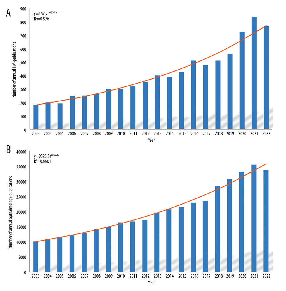 Figure 1. (A) The annual number of published HM studies, 2003–2022. (B) The annual number of published ophthalmic studies, 2003–2022. Produced by Office (version 2021, Redmond, USA).
 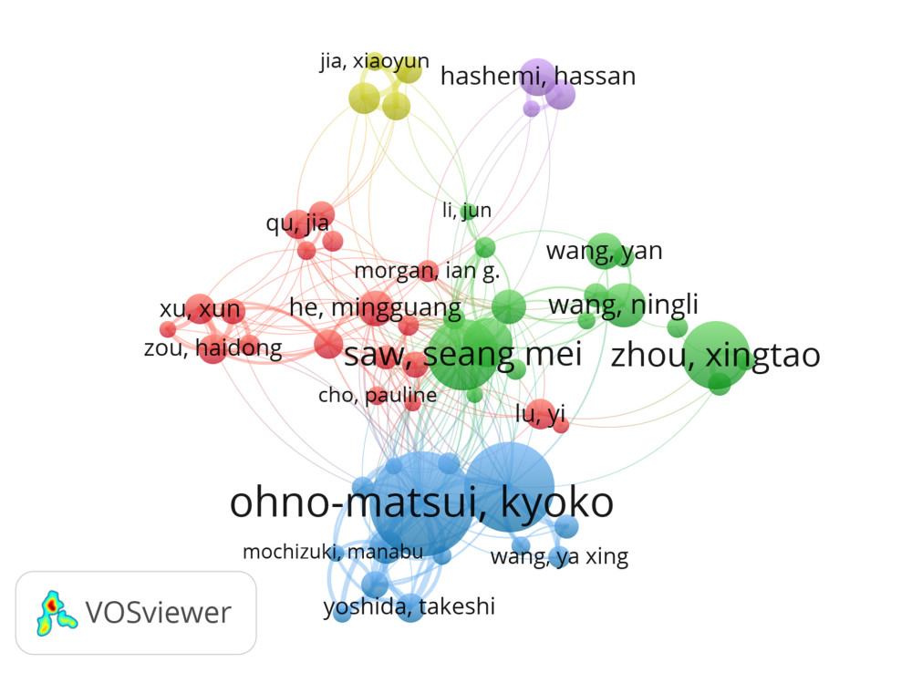 Figure 2. Cooperation map of authors of studies on HM. The bubble’s size corresponds to a researcher’s influence. A denser connecting line between the bubbles indicates more collaboration between the authors. Scholars sharing the same color focus on similar research themes. Threshold: A minimum of 25 documents/citations per author and a clustering resolution of 0.3. Produced using VOSviewer (version 1.6.19, Leiden University, Leiden, Netherlands).
 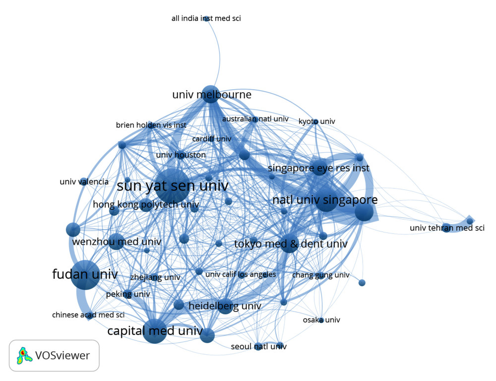 Figure 3. Cooperation map of institutions in the studies of HM. The bubble’s sizes are indicative of the institution’s influence. A more pronounced line connecting the bubbles signifies intensified collaboration between the institutions. Threshold: A minimum of 50 documents/citations per institution and a clustering resolution of 0.1. Produced using VOSviewer (version 1.6.19, Leiden University, Leiden, Netherlands).
 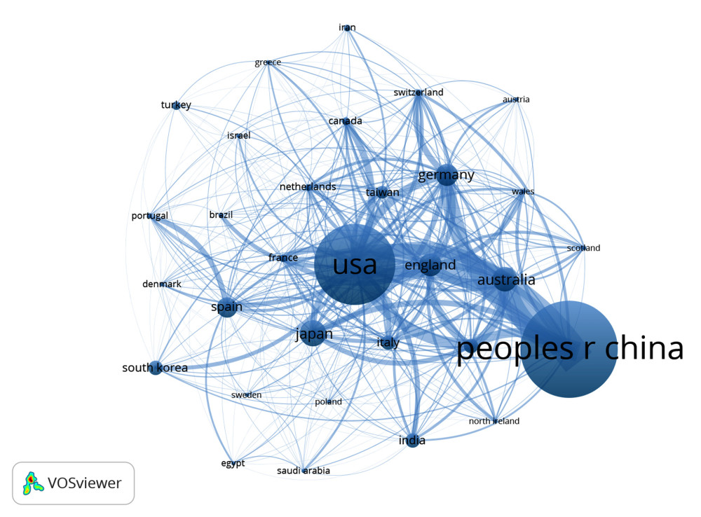 Figure 4. Cooperation map of countries in the studies of HM. The size of the bubble correlates with the country’s influence. A more robust connecting line between the bubbles denotes deeper collaboration between the countries. Threshold: A minimum of 50 documents/citations per country and a clustering resolution of 0.6. Produced using VOSviewer (version 1.6.19, Leiden University, Leiden, Netherlands).
 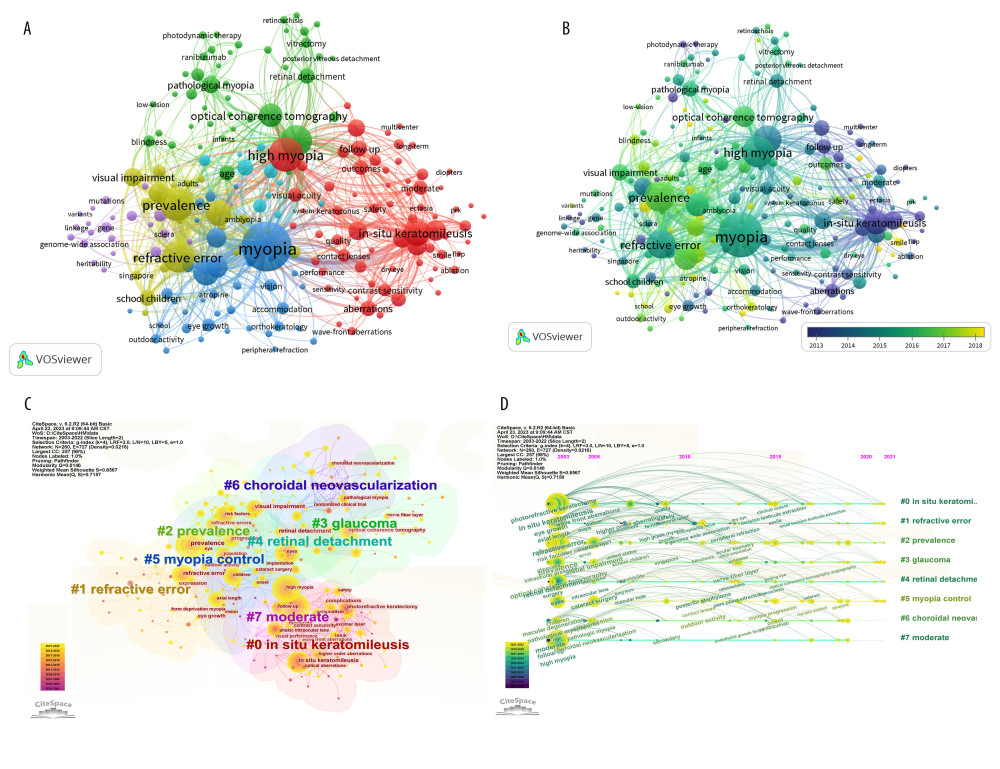 Figure 5. (A, C) The co-occurrence network and the clusters of keywords on HM. Keywords sharing a common color are part of the same cluster. A denser line between keywords suggests a higher probability of their concurrent appearance in a single article. In Figure 5C, keywords in yellow indicate proximity to the article’s publication year, whereas those that are purple have greater temporal distance from it. Threshold: A minimum of 25 occurrences for a keyword and a clustering resolution of 1.3. (B, D) Co-cited reference timeline map of publications on HM. In Figure 5B, the color gradient from purple to yellow signifies the average year of keyword occurrence, transitioning from distant to recent. In Figure 5D, keywords in yellow are nearer to the article’s publication year, while those in purple suggest a greater temporal distance from it. (A, B) Produced using CiteSpace (version 6.2.R2, Drexel University, Philadelphia, USA), (C, D) produced using VOSviewer (version 1.6.19, Leiden University, Leiden, Netherlands).
 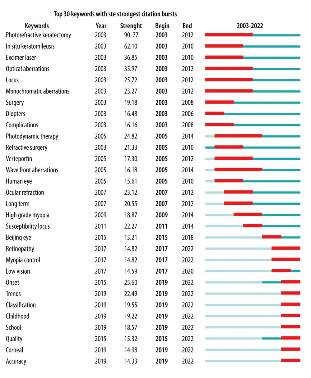 Figure 6. The top keywords with the strongest citation bursts. The red line within the blue line demarcates the time span during which the keyword had its peak research focus over the past 2 decades. Produced using CiteSpace (version 6.2.R2, Drexel University, Philadelphia, USA).
 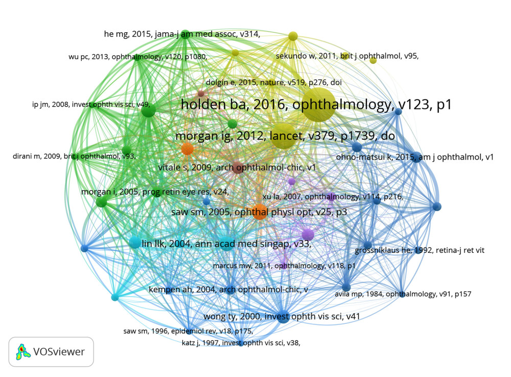 Figure 7. The network of co-cited references. The bubble’s magnitude reflects the article’s influence. A denser line interconnecting the bubbles signifies a stronger interrelation between the articles. Works sharing a common color have similar research themes. Parameters set include: A minimum of 25 citations for a referenced article and a clustering resolution of 1.3. Produced using VOSviewer (version 1.6.19, Leiden University, Leiden, Netherlands).
 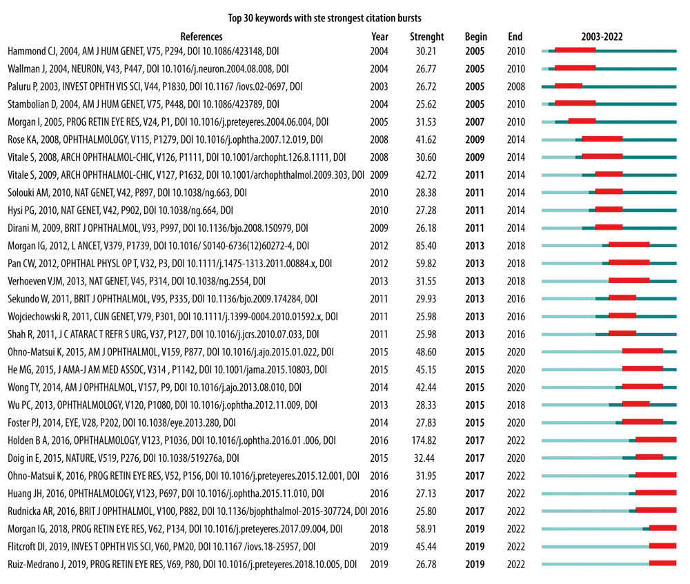 Figure 8. The top references with the strongest citation bursts. The red line within the blue line indicates the timeframe over the last 2 decades during which this reference received its highest citation frequency. Produced using CiteSpace (version 6.2.R2, Drexel University, Philadelphia, USA).
 Tables
Table 1. Retrieval strategies for HM articles.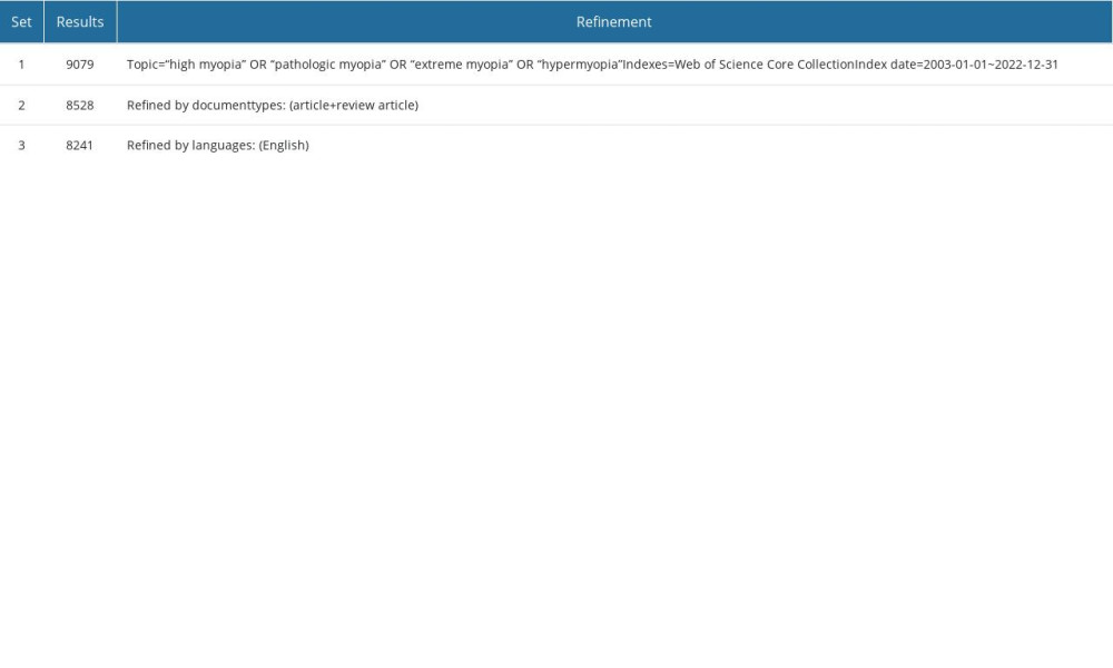 Table 2. Top 10 influential source journals for HM studies from 2003 to 2022.
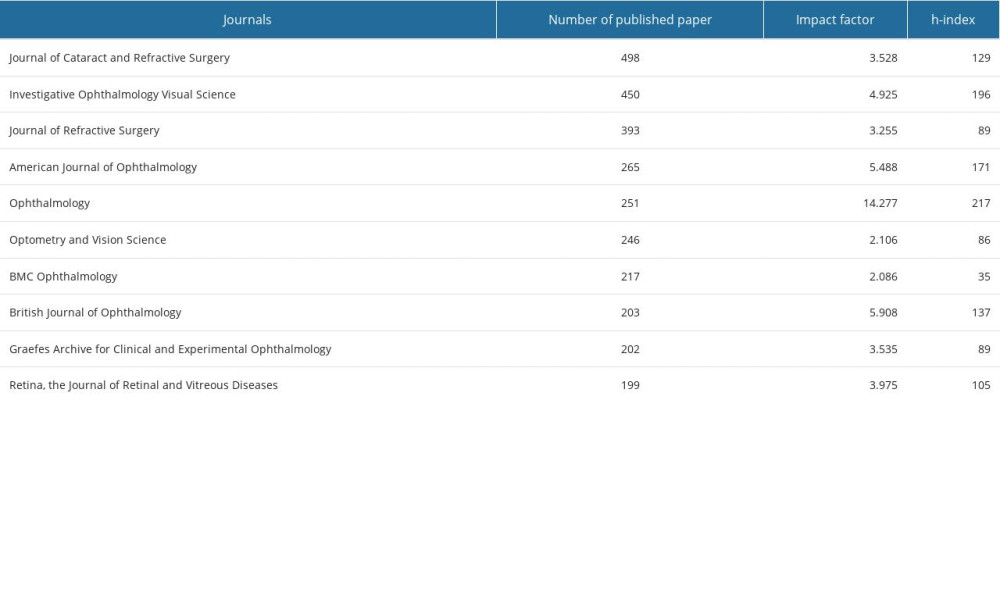 Table 3. Top 10 most influential authors for HM studies from 2003 to 2022.
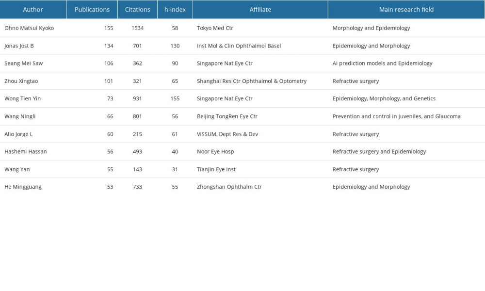 Table 4. The top 10 countries/regions and institutions contributed to publications of HM from 2003 to 2022.
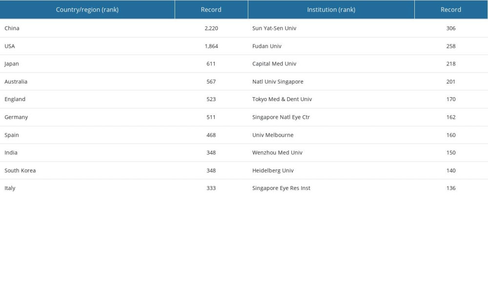 Table 5. Comparison of VOSviewer and CiteSpace on clustering analysis of HM keywords.
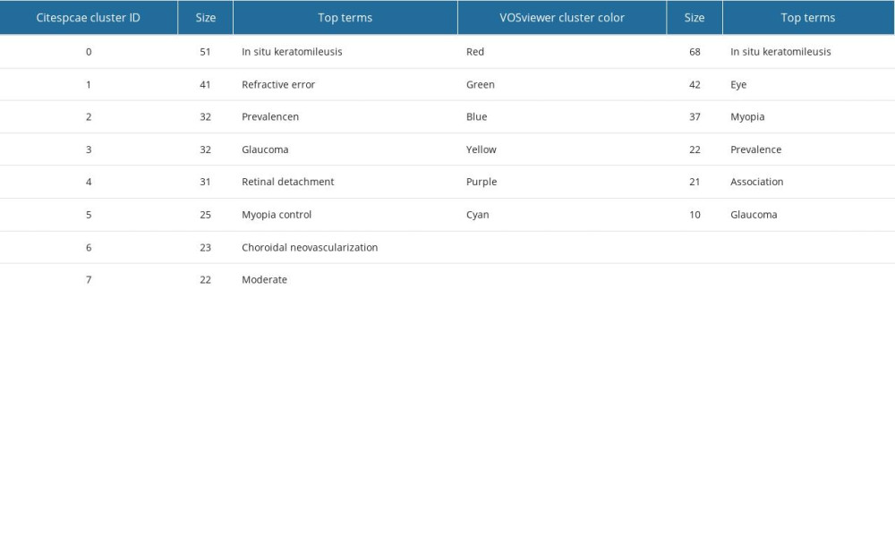 Table 6. TOP 30 keywords on HM with the most frequent occurrence.
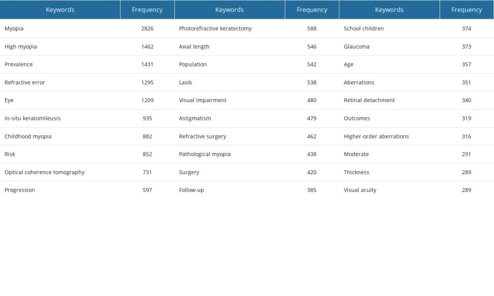 Table 7. TOP 10 references on HM with the most frequent occurrence.
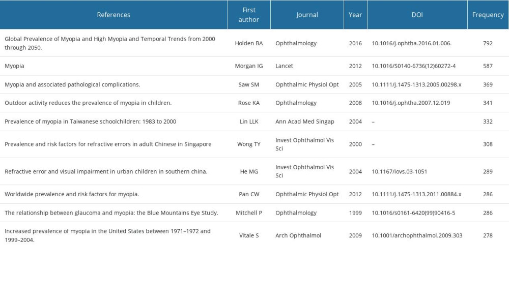
References
1. Flitcroft D, He M, Jonas J, IMI – defining and classifying myopia: A proposed set of standards for clinical and epidemiologic studies: Invest Ophthalmol Vis Sci, 2019; 60(3); M20-M30
2. Ellegaard O, Wallin J, The bibliometric analysis of scholarly production: How great is the impact?: Scientometrics, 2015; 105(3); 1809-31
3. van Eck N, Waltman L, Software survey: VOSviewer, a computer program for bibliometric mapping: Scientometrics, 2010; 84(2); 523-38
4. Chen C, Chen Y, Searching for clinical evidence in CiteSpace: AMIA Annual Symposium proceedings AMIA Symposium, 2005; 2005; 121-15
5. Liang C, Luo A, Zhong Z, Knowledge mapping of medication literacy study: A visualized analysis using CiteSpace: SAGE Open Med, 2018; 6; 2050312118800199
6. Liang Y, Li Y, Zhao J, Study of acupuncture for low back pain in recent 20 years: A bibliometric analysis via CiteSpace: J Pain Res, 2017; 10; 951-64
7. Chen C, Searching for intellectual turning points: Progressive knowledge domain visualization: Proc Natl Acad Sci USA, 2004(Suppl 1); 5303-10
8. Morgan I, Ohno-Matsui K, Saw S, Myopia: Lancet (London, England), 2012; 379(9827); 1739-48
9. Tuteja SY, Ramappa M, Radial keratotomy: N Engl J Med, 2019; 380(4); e4
10. Sánchez P, Moutsouris K, Pandolfi A, Biomechanical and optical behavior of human corneas before and after photorefractive keratectomy: J Cataract Refract Surg, 2014; 40(6); 905-17
11. Vestergaard A, Hjortdal J, Ivarsen A, Long-term outcomes of photorefractive keratectomy for low to high myopia: 13 to 19 years of follow-up: J Refract Surgery, 2013; 29(5); 312-19
12. Updegraff SA, Kritzinger MS, Laser in situ keratomileusis technique: Curr Opin Ophthalmol, 2000; 11(4); 267-72
13. Teus M, de Benito-Llopis L, Sánchez-Pina J, LASEK versus LASIK for the correction of moderate myopia: Optom Vis Sci, 2007; 84(7); 605-10
14. Lim T, Yang S, Kim M, Tchah H, Comparison of the IntraLase femtosecond laser and mechanical microkeratome for laser in situ keratomileusis: Am J Ophthalmol, 2006; 141(5); 833-39
15. Liu M, Chen Y, Wang D, Clinical outcomes after SMILE and femtosecond laser-assisted LASIK for myopia and myopic astigmatism: A prospective randomized comparative study: Cornea, 2016; 35(2); 210-16
16. Li M, Zhao J, Shen Y, Comparison of dry eye and corneal sensitivity between small incision lenticule extraction and femtosecond LASIK for myopia: PLoS One, 2013; 8(10); e77797
17. Wei S, Wang Y, Comparison of corneal sensitivity between FS-LASIK and femtosecond lenticule extraction (ReLEx flex) or small-incision lenticule extraction (ReLEx smile) for myopic eyes: Graefes Arch Clin Exp Ophthalmol, 2013; 251(6); 1645-54
18. Miraftab M, Hashemi H, Aghamirsalim M, Matched comparison of corneal higher order aberrations induced by SMILE to femtosecond assisted LASIK and to PRK in correcting moderate and high myopia: 3.00mm vs. 6.00mm: BMC Ophthalmol, 2021; 21(1); 216
19. Qin B, Li M, Chen X, Early visual outcomes and optical quality after femtosecond laser small-incision lenticule extraction for myopia and myopic astigmatism correction of over −10 dioptres: Acta Ophthalmol, 2018; 96(3); e341-e46
20. Lee H, Yong Kang D, Reinstein D, Comparing corneal higher-order aberrations in corneal wavefront-guided transepithelial photorefractive keratectomy versus small-incision lenticule extraction: J Cataract Refract Surg, 2018; 44(6); 725-33
21. Yao Y, Lu Q, Wei L, Efficacy and complications of cataract surgery in high myopia: J Cataract Refract Surg, 2021; 47(11); 1473-80
22. Wan T, Yin H, Wu Z, Yang Y, Comparative study of implantable collamer lens implantation in treating four degrees of myopia: Six-month observation of visual results, higher-order aberrations, and amplitude of accommodation: Curr Eye Res, 2020; 45(7); 839-46
23. Hashemian S, Farrokhi H, Foroutan A, Ocular higher-order aberrations changes after implantable collamer lens implantation for high myopic astigmatism: J Curr Ophthalmol, 2018; 30(2); 136-41
24. Chen D, Zhao X, Chou Y, Luo Y, Comparison of visual outcomes and optical quality of femtosecond laser-assisted SMILE and visian implantable collamer lens (ICL V4c) implantation for moderate to high myopia: A meta-analysis: J Refract Surg, 2022; 38(6); 332-38
25. Cao K, Zhang J, Wang J, Implantable collamer lens versus small incision lenticule extraction for high myopia correction: A systematic review and meta-analysis: BMC Ophthalmol, 2021; 21(1); 450
26. Redd T, Campbell J, Chiang M, Artificial intelligence for refractive surgery screening: finding the balance between myopia and hyperopia: JAMA Ophthalmol, 2020; 138(5); 526-27
27. Swanson E, Izatt J, Hee M, In vivo retinal imaging by optical coherence tomography: Optics letters, 1993; 18(21); 1864-66
28. Drexler W, Morgner U, Ghanta R, Ultrahigh-resolution ophthalmic optical coherence tomography: Nat Med, 2001; 7(4); 502-7
29. Jia Y, Bailey S, Wilson D, Quantitative optical coherence tomography angiography of choroidal neovascularization in age-related macular degeneration: Ophthalmology, 2014; 121(7); 1435-44
30. Lim L, Cheung G, Lee S, Comparison of spectral domain and swept-source optical coherence tomography in pathological myopia: Eye (Lond), 2014; 28(4); 488-91
31. Saito R, Shinohara K, Tanaka N, Association between dome-shaped macula and posterior staphyloma in highly myopic eyes investigated by ultra-widefield optical coherence tomography: Retina, 2021; 41(3); 646-52
32. Xiao W, Zhu Z, Odouard C, Wide-field en face swept-source optical coherence tomography features of extrafoveal retinoschisis in highly myopic eyes: Invest Ophthalmol Visual Sci, 2017; 58(2); 1037-44
33. Querques G, Corvi F, Querques L, Optical coherence tomography angiography of choroidal neovascularization secondary to pathologic myopia: Dev Ophthalmol, 2016; 56; 101-6
34. Suwan Y, Fard M, Geyman L, Association of myopia with peripapillary perfused capillary density in patients with glaucoma: An optical coherence tomography angiography study: JAMA ophthalmology, 2018; 136(5); 507-13
35. Li Y, Foo L, Wong C, Pathologic myopia: Advances in imaging and the potential role of artificial intelligence: Br J Ophthalmol, 2023; 107(5); 600-6
36. Ruiz-Medrano J, Montero J, Flores-Moreno I, Myopic maculopathy: Current status and proposal for a new classification and grading system (ATN): Prog Retin Eye Res, 2019; 69; 80-115
37. Cheung C, Arnold J, Holz F, Myopic choroidal neovascularization: Review, guidance, and consensus statement on management: Ophthalmology, 2017; 124(11); 1690-711
38. Shinohara K, Tanaka N, Jonas J, Ultrawide-field OCT to investigate relationships between myopic macular retinoschisis and posterior staphyloma: Ophthalmology, 2018; 125(10); 1575-86
39. Shimada N, Sugamoto Y, Ogawa M, Fovea-sparing internal limiting membrane peeling for myopic traction maculopathy: Am J Ophthalmol, 2012; 154(4); 693-701
40. Iwasaki M, Miyamoto H, Okushiba U, Imaizumi H, Fovea-sparing internal limiting membrane peeling versus complete internal limiting membrane peeling for myopic traction maculopathy: Jpn J Ophthalmol, 2020; 64(1); 13-21
41. Tian T, Jin H, Zhang Q, Long-term surgical outcomes of multiple parfoveolar curvilinear internal limiting membrane peeling for myopic foveoschisis: Eye (Lond), 2018; 32(11); 1783-89
42. Wang L, Wang Y, Li Y, Comparison of effectiveness between complete internal limiting membrane peeling and internal limiting membrane peeling with preservation of the central fovea in combination with 25G vitrectomy for the treatment of high myopic foveoschisis: Medicine, 2019; 98(9); e14710
43. Baba T, Tanaka S, Nizawa T, Scleral imbrication combined with pars plana vitrectomy without internal limiting membrane peeling for myopic schisis: Retina, 2016; 36(10); 1927-34
44. Fujikawa M, Kawamura H, Kakinoki M, Scleral imbrication combined with vitrectomy and gas tamponade for refractory macular hole retinal detachment associated with high myopia: Retina, 2014; 34(12); 2451-57
45. Holden B, Fricke T, Wilson D, Global prevalence of myopia and high myopia and temporal trends from 2000 through 2050: Ophthalmology, 2016; 123(5); 1036-42
46. Morgan I, French A, Ashby R, The epidemics of myopia: Aetiology and prevention: Prog Rtin Eye Res, 2018; 62; 134-49
47. Jones L, Sinnott L, Mutti D, Parental history of myopia, sports and outdoor activities, and future myopia: Invest Ophthalmol Vis Sci, 2007; 48(8); 3524-32
48. Rose K, Morgan I, Ip J, Outdoor activity reduces the prevalence of myopia in children: Ophthalmology, 2008; 115(8); 1279-85
49. Rose K, Morgan I, Smith W, Myopia, lifestyle, and schooling in students of Chinese ethnicity in Singapore and Sydney: Arch Ophthalmol, 2008; 126(4); 527-30
50. Jonas J, Ang M, Cho P, IMI Prevention of myopia and its progression: Invest Ophthalmol Vis Sci, 2021; 62(5); 6
51. Sherwin J, Reacher M, Keogh R, The association between time spent outdoors and myopia in children and adolescents: A systematic review and meta-analysis: Ophthalmology, 2012; 119(10); 2141-51
52. Xiong S, Sankaridurg P, Naduvilath T, Time spent in outdoor activities in relation to myopia prevention and control: A meta-analysis and systematic review: Acta Ophthalmol, 2017; 95(6); 551-66
53. Guo Y, Liu L, Lv Y, Outdoor jogging and myopia progression in school children from rural Beijing: The Beijing children eye study: Transl Vis Sci Technol, 2019; 8(3); 2
54. Yen M, Liu J, Kao S, Shiao C, Comparison of the effect of atropine and cyclopentolate on myopia: Ann Ophthalmol, 1989; 21(5); 180-82
55. Gong Q, Janowski M, Luo M, Efficacy and adverse effects of atropine in childhood myopia: A meta-analysis: JAMA Ophthalmol, 2017; 135(6); 624-30
56. Ha A, Kim S, Shim S, Efficacy and safety of 8 atropine concentrations for myopia control in children: A network meta-analysis: Ophthalmology, 2022; 129(3); 322-33
57. Yam J, Li F, Zhang X, Two-year clinical trial of the low-concentration atropine for myopia progression (LAMP) study: Phase 2 report: Ophthalmology, 2020; 127(7); 910-19
58. Huang J, Wen D, Wang Q, Efficacy comparison of 16 interventions for myopia control in children: A network meta-analysis: Ophthalmology, 2016; 123(4); 697-708
59. Smith E, Hung L, Huang J, Relative peripheral hyperopic defocus alters central refractive development in infant monkeys: Vision Res, 2009; 49(19); 2386-92
60. Lin Z, Martinez A, Chen X, Peripheral defocus with single-vision spectacle lenses in myopic children: Optom Vis Sci, 2010; 87(1); 4-9
61. Mutti D, Sinnott L, Mitchell G, Relative peripheral refractive error and the risk of onset and progression of myopia in children: Invest Ophthalmol Vis Sci, 2011; 52(1); 199-205
62. Cheng D, Woo G, Drobe B, Schmid K, Effect of bifocal and prismatic bifocal spectacles on myopia progression in children: Three-year results of a randomized clinical trial: JAMA Ophthalmol, 2014; 132(3); 258-64
63. Berntsen D, Barr C, Mutti D, Zadnik K, Peripheral defocus and myopia progression in myopic children randomly assigned to wear single vision and progressive addition lenses: Invest Ophthalmol Vis Sci, 2013; 54(8); 5761-70
64. Lu Y, Lin Z, Wen L, The adaptation and acceptance of defocus incorporated multiple segment lens for Chinese children: Am J Ophthalmol, 2020; 211; 207-16
65. Lam C, Tang W, Tse D, Defocus Incorporated Soft Contact (DISC) lens slows myopia progression in Hong Kong Chinese schoolchildren: A 2-year randomised clinical trial: Br J Ophthalmol, 2014; 98(1); 40-45
66. Correction of Myopia Evaluation Trial 2 Study Group for the Pediatric Eye Disease Investigator Group., Progressive-addition lenses versus single-vision lenses for slowing progression of myopia in children with high accommodative lag and near esophoria: Inv Ophthalmol Vis Sci, 2011; 52(5); 2749-57
67. Chamberlain P, Peixoto-de-Matos S, Logan N, A 3-year randomized clinical trial of MiSight lenses for myopia control: Optom Vis Sci, 2019; 96(8); 556-67
68. Walline J, Walker M, Mutti D, Effect of high add power, medium add power, or single-vision contact lenses on myopia progression in children: The BLINK randomized clinical trial: JAMA, 2020; 324(6); 571-80
69. Sankaridurg P, Bakaraju R, Naduvilath T, Myopia control with novel central and peripheral plus contact lenses and extended depth of focus contact lenses: 2 year results from a randomised clinical trial: Ophthalmic Physiol Optics, 2019; 39(4); 294-307
70. Si J, Tang K, Bi H, Orthokeratology for myopia control: A meta-analysis: Optom Vis Sci, 2015; 92(3); 252-57
71. Cho P, Cheung S, Protective role of orthokeratology in reducing risk of rapid axial elongation: A reanalysis of data from the ROMIO and TO-SEE studies: Invest Ophthalmol Vis Sci, 2017; 58(3); 1411-16
72. Hiraoka T, Sekine Y, Okamoto F, Safety and efficacy following 10-years of overnight orthokeratology for myopia control: Ophthalmic Physiol Opt, 2018; 38(3); 281-89
73. Kanda H, Oshika T, Hiraoka T, Effect of spectacle lenses designed to reduce relative peripheral hyperopia on myopia progression in Japanese children: A 2-year multicenter randomized controlled trial: Jpn J Ophthalmol, 2018; 62(5); 537-43
74. Sankaridurg P, Donovan L, Varnas S, Spectacle lenses designed to reduce progression of myopia: 12-month results: Optom Vis Sci, 2010; 87(9); 631-41
75. Klein R, Zeiss C, Chew E, Complement factor H polymorphism in age-related macular degeneration: Science, 2005; 308(5720); 385-89
76. Nakanishi H, Yamada R, Gotoh N, A genome-wide association analysis identified a novel susceptible locus for pathological myopia at 11q24.1: PLoS Genet, 2009; 5(9); e1000660
77. Stambolian D, Wojciechowski R, Oexle K, Meta-analysis of genome-wide association studies in five cohorts reveals common variants in RBFOX1, a regulator of tissue-specific splicing, associated with refractive error: Hum Mol Genet, 2013; 22(13); 2754-64
78. Verhoeven V, Hysi P, Wojciechowski R, Genome-wide meta-analyses of multiancestry cohorts identify multiple new susceptibility loci for refractive error and myopia: Nat Genet, 2013; 45(3); 314-18
79. Cheng C, Schache M, Ikram M, Nine loci for ocular axial length identified through genome-wide association studies, including shared loci with refractive error: Am J Hum Genet, 2013; 93(2); 264-77
80. Chen P, Miyake M, Fan Q, CMPK1 and RBP3 are associated with corneal curvature in Asian populations: Hum Mol Genet, 2014; 23(22); 6129-36
81. Abecasis G, Auton A, Brooks L, An integrated map of genetic variation from 1,092 human genomes: Nature, 2012; 491(7422); 56-65
82. Ku C, Loy E, Pawitan Y, Chia K, The pursuit of genome-wide association studies: Where are we now?: J Hum Genet, 2010; 55(4); 195-206
83. Li J, Zhang Q, Insight into the molecular genetics of myopia: Mol Vis, 2017; 23; 1048-80
84. Torkamani A, Wineinger N, Topol E, The personal and clinical utility of polygenic risk scores: Nat Rev Genet, 2018; 19(9); 581-90
85. Chatterjee N, Shi J, García-Closas M, Developing and evaluating polygenic risk prediction models for stratified disease prevention: Nat Rev Genet, 2016; 17(7); 392-406
86. Lek M, Karczewski K, Minikel E, Analysis of protein-coding genetic variation in 60,706 humans: Nature, 2016; 536(7616); 285-91
87. Guggenheim J, Clark R, Cui J, Whole exome sequence analysis in 51 624 participants identifies novel genes and variants associated with refractive error and myopia: Hum Mol Genet, 2022; 31(11); 1909-19
88. Haarman A, Thiadens A, van Tienhoven M, Whole exome sequencing of known eye genes reveals genetic causes for high myopia: Hum Mol Genet, 2022; 31(19); 3290-98
89. Ha A, Kim C, Shim S, Degree of myopia and glaucoma risk: A dose-response meta-analysis: Am J Ophthalmol, 2022; 236; 107-19
90. Chang R, Singh K, Myopia and glaucoma: Diagnostic and therapeutic challenges: Curr Opin Ophthalmol, 2013; 24(2); 96-101
91. Miki A, Ikuno Y, Asai T, Defects of the lamina cribrosa in high myopia and glaucoma: PloS One, 2015; 10(9); e0137909
92. Park H, Lee K, Park C, Optic disc torsion direction predicts the location of glaucomatous damage in normal-tension glaucoma patients with myopia: Ophthalmology, 2012; 119(9); 1844-51
93. Lan Y, Chang S, Sun F, Hsieh J, Different disc characteristics associated with high myopia and the location of glaucomatous damage in primary open-angle glaucoma and normal-tension glaucoma: J Glaucoma, 2019; 28(6); 519-28
94. Teng C, De Moraes C, Prata T, The region of largest β-zone parapapillary atrophy area predicts the location of most rapid visual field progression: Ophthalmology, 2011; 118(12); 2409-13
95. Sung M, Heo H, Piao H, Parapapillary atrophy and changes in the optic nerve head and posterior pole in high myopia: Sci Rep, 2020; 10(1); 4607
96. Yamada H, Akagi T, Nakanishi H, Microstructure of peripapillary atrophy and subsequent visual field progression in treated primary open-angle glaucoma: Ophthalmology, 2016; 123(3); 542-51
97. Onda E, Cioffi G, Bacon D, Van Buskirk E, Microvasculature of the human optic nerve: Am J Ophthalmol, 1995; 120(1); 92-102
98. Fan X, Xu H, Zhai R, Peripapillary vascular reactivity in primary open-angle glaucoma with high myopia by using optical coherence tomography angiography: Front Med, 2022; 9; 850483
99. Qiu K, Wang G, Lu X, Application of the ISNT rules on retinal nerve fibre layer thickness and neuroretinal rim area in healthy myopic eyes: Acta Ophthalmol, 2018; 96(2); 161-67
100. Bedggood P, Mukherjee S, Nguyen B, Geometry of the retinal nerve fibers from emmetropia through to high myopia at both the temporal raphe and optic nerve: Invest Ophthalmol Vis Sci, 2019; 60(14); 4896-903
Figures
Figure 1. (A) The annual number of published HM studies, 2003–2022. (B) The annual number of published ophthalmic studies, 2003–2022. Produced by Office (version 2021, Redmond, USA).Figure 2. Cooperation map of authors of studies on HM. The bubble’s size corresponds to a researcher’s influence. A denser connecting line between the bubbles indicates more collaboration between the authors. Scholars sharing the same color focus on similar research themes. Threshold: A minimum of 25 documents/citations per author and a clustering resolution of 0.3. Produced using VOSviewer (version 1.6.19, Leiden University, Leiden, Netherlands).Figure 3. Cooperation map of institutions in the studies of HM. The bubble’s sizes are indicative of the institution’s influence. A more pronounced line connecting the bubbles signifies intensified collaboration between the institutions. Threshold: A minimum of 50 documents/citations per institution and a clustering resolution of 0.1. Produced using VOSviewer (version 1.6.19, Leiden University, Leiden, Netherlands).Figure 4. Cooperation map of countries in the studies of HM. The size of the bubble correlates with the country’s influence. A more robust connecting line between the bubbles denotes deeper collaboration between the countries. Threshold: A minimum of 50 documents/citations per country and a clustering resolution of 0.6. Produced using VOSviewer (version 1.6.19, Leiden University, Leiden, Netherlands).Figure 5. (A, C) The co-occurrence network and the clusters of keywords on HM. Keywords sharing a common color are part of the same cluster. A denser line between keywords suggests a higher probability of their concurrent appearance in a single article. In Figure 5C, keywords in yellow indicate proximity to the article’s publication year, whereas those that are purple have greater temporal distance from it. Threshold: A minimum of 25 occurrences for a keyword and a clustering resolution of 1.3. (B, D) Co-cited reference timeline map of publications on HM. In Figure 5B, the color gradient from purple to yellow signifies the average year of keyword occurrence, transitioning from distant to recent. In Figure 5D, keywords in yellow are nearer to the article’s publication year, while those in purple suggest a greater temporal distance from it. (A, B) Produced using CiteSpace (version 6.2.R2, Drexel University, Philadelphia, USA), (C, D) produced using VOSviewer (version 1.6.19, Leiden University, Leiden, Netherlands).Figure 6. The top keywords with the strongest citation bursts. The red line within the blue line demarcates the time span during which the keyword had its peak research focus over the past 2 decades. Produced using CiteSpace (version 6.2.R2, Drexel University, Philadelphia, USA).Figure 7. The network of co-cited references. The bubble’s magnitude reflects the article’s influence. A denser line interconnecting the bubbles signifies a stronger interrelation between the articles. Works sharing a common color have similar research themes. Parameters set include: A minimum of 25 citations for a referenced article and a clustering resolution of 1.3. Produced using VOSviewer (version 1.6.19, Leiden University, Leiden, Netherlands).Figure 8. The top references with the strongest citation bursts. The red line within the blue line indicates the timeframe over the last 2 decades during which this reference received its highest citation frequency. Produced using CiteSpace (version 6.2.R2, Drexel University, Philadelphia, USA). Tables
Table 1. Retrieval strategies for HM articles.Table 2. Top 10 influential source journals for HM studies from 2003 to 2022.Table 3. Top 10 most influential authors for HM studies from 2003 to 2022.Table 4. The top 10 countries/regions and institutions contributed to publications of HM from 2003 to 2022.Table 5. Comparison of VOSviewer and CiteSpace on clustering analysis of HM keywords.Table 6. TOP 30 keywords on HM with the most frequent occurrence.Table 7. TOP 10 references on HM with the most frequent occurrence.Table 1. Retrieval strategies for HM articles.Table 2. Top 10 influential source journals for HM studies from 2003 to 2022.Table 3. Top 10 most influential authors for HM studies from 2003 to 2022.Table 4. The top 10 countries/regions and institutions contributed to publications of HM from 2003 to 2022.Table 5. Comparison of VOSviewer and CiteSpace on clustering analysis of HM keywords.Table 6. TOP 30 keywords on HM with the most frequent occurrence.Table 7. TOP 10 references on HM with the most frequent occurrence. In Press
08 Mar 2024 : Animal Research
Modification of Experimental Model of Necrotizing Enterocolitis (NEC) in Rat Pups by Single Exposure to Hyp...Med Sci Monit In Press; DOI: 10.12659/MSM.943443
18 Apr 2024 : Clinical Research
Comparative Analysis of Open and Closed Sphincterotomy for the Treatment of Chronic Anal Fissure: Safety an...Med Sci Monit In Press; DOI: 10.12659/MSM.944127
08 Mar 2024 : Laboratory Research
Evaluation of Retentive Strength of 50 Endodontically-Treated Single-Rooted Mandibular Second Premolars Res...Med Sci Monit In Press; DOI: 10.12659/MSM.944110
11 Mar 2024 : Clinical Research
Comparison of Effects of Sugammadex and Neostigmine on Postoperative Neuromuscular Blockade Recovery in Pat...Med Sci Monit In Press; DOI: 10.12659/MSM.942773
Most Viewed Current Articles
17 Jan 2024 : Review article
Vaccination Guidelines for Pregnant Women: Addressing COVID-19 and the Omicron VariantDOI :10.12659/MSM.942799
Med Sci Monit 2024; 30:e942799
14 Dec 2022 : Clinical Research
Prevalence and Variability of Allergen-Specific Immunoglobulin E in Patients with Elevated Tryptase LevelsDOI :10.12659/MSM.937990
Med Sci Monit 2022; 28:e937990
16 May 2023 : Clinical Research
Electrophysiological Testing for an Auditory Processing Disorder and Reading Performance in 54 School Stude...DOI :10.12659/MSM.940387
Med Sci Monit 2023; 29:e940387
01 Jan 2022 : Editorial
Editorial: Current Status of Oral Antiviral Drug Treatments for SARS-CoV-2 Infection in Non-Hospitalized Pa...DOI :10.12659/MSM.935952
Med Sci Monit 2022; 28:e935952