13 September 2020: Animal Study
AEB-071 Ameliorates Muscle Weakness by Altering Helper T Lymphocytes in an Experimental Autoimmune Myasthenia Gravis Rat Model
Feng Jing1AE, Wei Huang1BC, Qian Ma1BC, Sheng-jie Xu1BC, Chang-jin Wu2BC, Yu-xiu Guan2BC, Bing Chen1A*DOI: 10.12659/MSM.924393
Med Sci Monit 2020; 26:e924393


Abstract
BACKGROUND: Myasthenia gravis (MG) is an autoimmune neurological disorder of neuromuscular junctions. In this study we established experimental autoimmune myasthenia gravis (EAMG) rat models to investigate the effects of AEB-071 (AEB), which is a specific inhibitor of protein kinase C that prevents T lymphocyte activation.
MATERIAL AND METHODS: We utilized animals divided into 4 groups: (1) control rats, (2) EAMG, (3) AEB-071+EAMG, and (4) AZP+EAMG. Drug treatment was continued for 10 days. Ten weeks after immunization we measured body weights, assessed mortality rates, and used Lennon scores to evaluate EAMG grades. We also assessed the proportions of Treg, Th1, Th2, Th17, and lymphocytes using flow cytometry.
RESULTS: In the absence of drug treatment, we found a significant decline in body weights in the EAMG group in comparison to control rats, and EAMG group rats also had higher Lennon scores (P<0.05). Interestingly, we found that AEB-071 restored the body weight of EAMG rats and the decreased mortality rate compared to AZP treatment. Although a decrease in the number of Treg cells was observed, the proportion of Th lymphocytes was significantly increased in the EAMG group, and AEB-071 treatment decreased the proportion of Th lymphocytes.
CONCLUSIONS: We concluded that AEB-071 treatment imparts beneficial effects in EAMG rat models by reducing mortality rate and restoring Th lymphocyte balance, and thus may be an attractive candidate for use in MG treatment.
Keywords: Autoimmune Diseases, Myasthenia Gravis, Neurology, Lymphocyte Activation, Muscle Weakness, Myasthenia Gravis, Autoimmune, Experimental, Pyrroles, Quinazolines, Rats, Inbred Lew, T-Lymphocytes, Helper-Inducer
Background
MG is an autoimmune neurological disorder of neuromuscular junctions. It is a T-cell-dependent and B-cell-mediated disorder caused by antibodies targeting the neuromuscular junction, primarily the acetylcholine receptor (AChR) [1]. Despite extensive research in the field, the exact pathogenesis of MG remains unclear [2]. MG leads to cellular immune abnormalities mediated by CD4+ T lymphocytes [3]. Native CD4+ helper T lymphocytes (Th1, Th2, and Th17 subsets) [4] are elevated in MG [5] and are correlated with higher AChR antibody titers [6]. In addition, Treg lymphocytes inhibit activation of B cells and T cells to maintain immune homeostasis [7]. A loss of active Treg cells increases susceptibility to autoimmune diseases, as observed in MG [8,9]. Cumulatively, imbalances of Th17, Treg, and Th1/Th2 lymphocytes mediate MG progression [10].
Protein kinase C (PKC) is a serine/threonine kinase that regulates a wide range of biological processes, including T cell activation and the downstream signaling of CD28 and T cell receptors [11]. PKC inhibition has been proposed as a therapeutic strategy to treat autoimmune diseases [12]. AEB-071 (AEB, Sotrastaurin) has previously been shown to inhibit the activity of PKC isoforms [13] and thus holds promise for the treatment of T-cell-associated disease. AEB-071 also has the potential to prevent allograft rejection and reduce the inflammatory response [14]. AEB-071 has been shown to have curative effects on psoriasis and autoimmune diabetes [15,16]. However, the role of AEB-071 in MG treatment remains unknown. Azathioprine (AZP) is an oral immunosuppressive drug commonly used in clinical practice. AZP is a pro-drug that is converted
Material and Methods
ANIMAL MODELS:
Female Lewis rats (6–8 weeks of age) were used to establish the EAMG model according to a previously described protocol [19]. Rats were purchased from the Vital River Laboratory Beijing (SCXK, Beijing, 2012-0001), and average weights were recorded. Animals were maintained in the Experimental Animal Center of the 8th Medical Center of Chinese PLA General Hospital, with 3 animals per cage. Feeding and handling in SPF-class facilities was performed at 23±1°C and 50–70% humidity with a light cycle of 12 h and free access to water and food. All experiments were approved by the Institutional Animal Care and Use Committee of PLA General Hospital and were conducted following its guidelines. The license number was SCXK (Beijing) 2012-0023.
REAGENTS AND INSTRUMENTS:
The R97-116 peptide derived from the rat (AChRα) subunit was used as an immunogen. The peptide (DGDFAIVKFTKVLLDYTGHI) was obtained from Bankpeptide Technology Co. (Hefei, China) (MW: 2252.57 Da, purity >95%). Mycobacterium tuberculosis H37RA powder was purchased from DIFCO (Franklin Lakes, NJ, USA). Freund’s adjuvants were obtained from Sigma (St. Louis, MO, USA.). Rat AChR-Ab ELISA test kits (QS41981) were purchased from Qisong Biotechnology Co. (Beijing, China). Sotrastaurin (AEB-071) was produced by GLPBIO (Montclair, CA, USA). Antibodies (anti-rat CD4-FITC, CD25-PE, and Foxp3-APC), fixation buffer, true-nuclear transcription factor buffer sets, intracellular staining permeabilization buffer, and leukocyte cell activation cocktail were purchased from Bio Legend, Inc. Erythrocyte Lysis Buffer and rat interleukin (IL)-17A-APC were purchased from eBioscience, Inc. (San Diego, CA, USA) Azathioprine (AZP) and 0.9% saline (NS) were provided by the Pharmacy Department of the 8th Medical Center of Chinese PLA General Hospital. RPMI-1640 was prepared in-house. The optical density of the test sample and the standard was determined with a Bio-Kinetics microplate reader at a wavelength of 450 nm. Flow cytometry was performed on a BD LSRII flow cytometer (BD Biosciences, Franklin Lakes, NJ, USA).
CHARACTERIZATION OF EAMG MODELS:
EAMG models were established according to previously described protocols [20]. R97-116, CFA, and 0.9% NS solutions (200 μL, ratio: 1: 1.5: 1.5) were added to H37RA (1 mg), which was subcutaneously injected into the footpad, back, and groin of rats. Control rats received 0.9% NS only. Booster immunizations were performed at 30, 45, and 60 days after injection.
GROUPING AND DRUG TREATMENT:
Rats were randomly divided into 4 groups (n=10 per group): the Control group, in which control rats were treated with 0.9% NS orally; the EAMG group, in which EAMG rats were treated with 0.9% NS orally; the AEB-071+EAMG group, which received 30 mg/kg AEB-071 per day by gavage 15 days after EAMG immunization in accordance with previous studies [14,16]; and the AZP+EAMG group, which received the AZP (25 mg/kg) per day orally 15 days after EAMG immunization. The concentration of AEB071 was based on previous experiments [21]. The AZP manual recommends a concentration of 1–3 mg/kg, which was calculated based on the conversion ratio from human to animal dosage. A gavage needle was used for intragastric administration. Drug trials lasted for 10 days.
CLINICAL EVALUATIONS:
Clinical evaluation was performed using Lennon scales according to the EAMG Clinical Rating Scale [22]. This was defined as 0: no weakness; Grade 1: mild weakness, easy fatigue, especially after repeated grasping; Grade 2: obvious weakness symptoms, tremor, posture change, weak grip; Grade 3: serious muscle weakness, unable to grasp, moribund state; Grade 4: death. Rats showing severe muscle weakness or Lennon scores ≥Grade 3 were humanely sacrificed. After initial immunization, we blindly observed the clinical symptoms of disease, and body weights were assessed on a weekly basis to investigate health status.
ELISA FOR SERUM ANTI-ACHR ANTIBODY ASSAY:
The AChR antibody in the serum was used to assess the successful establishment of the EAMG animal model. A commercial rat AChR antibody ELISA kit was used to determine serum antibody titers. The ELISA kit was provided with a set of standards for establishing a curve for determination of absolute AChR antibody concentration in serum. Blood was obtained from the ocular veins of sacrificed rats and centrifuged for serum collection. Serum was diluted into assay plates (50 μL/well, 60 min) and incubated with enzyme-conjugated antibodies for 30 min at room temperature. Chromogenic reagent was then added for 20 min followed by stop solution (1M H2SO4) to terminate the reactions (50 μL). The optical density of the test sample and the standard was determined with a microplate reader at a wavelength of 450 nm.
FLOW CYTOMETRY ANALYSIS:
Treg/Th lymphocyte ratio and the total number of CD4+ T cells in the blood samples were measured. T helper cells were collected after drug intervention, washed in cytological washing solution (PBS+1% FBS), and centrifuged. Cells were labeled with anti-CD4-FITC and anti-CD3-APC antibodies in PBS+1% FBS for 20 min at room temperature and resuspended in permeabilization buffer. Cells were subsequently labeled with fluorescently-conjugated anti-IFN-γ PE for Th1 detection, anti-IL-4 PE for Th2 detection, and IL-17A PE for Th17 detection for 30 min at room temperature. Cells were washed in permeabilization buffer, resuspended in PBS+1% FBS, and analyzed by flow cytometry. For Treg lymphocyte detection, cells treated as above were labeled with anti-CD4-FITC and anti-CD25-APC antibodies, washed, and labeled in FOXP3 staining solution for 60 min in the dark. Cells were then washed, resuspended in PBS+1% FBS, and analyzed by flow cytometry.
STATISTICAL ANALYSIS:
SPSS statistical software (SPSS, Chicago, IL, USA) and GraphPad Prism 7.0 (GraphPad, San Diego, CA, USA) were used for the statistical analysis. Data are shown as the mean ±standard deviation. All experiments were carried out at least 3 times. The data of the 2 groups were assessed using the
Results
ASSESSMENT OF CLINICAL SYMPTOMS, BODY WEIGHT AND LENNON SCORE:
Two weeks after R97-116 inoculation, the EAMG group showed muscle weakness and significant weight loss. The animals exhibited dull coats and listlessness. Focal inflammatory response was present at the injection sites. EAMG rats also showed hypo-activity, including reduced crawling and grip, weak bite and vocalization, and weakness aggravated by activity. EAMG rats also showed decreased appetite, reduced food intake, and weight loss. Ten weeks after immunization, the EAMG group had higher Lennon scores than the control group, as shown in Figure 1A. The body weights of EAMG rats (179.68±8.59) were significantly lower compared to the control group (238.63±7.46) (P<0.05) (Figure 1B). MG is a disease with unknown etiology and diverse pathogenesis, which eventually results in dysfunction of neuromuscular junction transmission due to production of autoantibodies to AChR. To confirm EAMG, serum anti-AChR was assessed using ELISA (Figure 1C). ELISA results showed that the level of serum anti-AChR in the EAMG group was higher (P<0.05). These results show the successful establishment of the EAMG model.
AEB-071 IMPROVED THE CLINICAL SYMPTOMS OF EAMG:
In animal models of AEB071 intervention, the experimental period is typically short (typically less than 30 days). In our models, we observed that the symptoms of weakness and local inflammation in rats were significantly improved after about 10 days of intervention. Thus, the experiment was terminated after 10 days. Rats in the AEB-071+EAMG and AZP+EAMG groups showed fewer MG symptoms than the EAMG group. The body weights of rats in the AEB-071+EAMG, AZP+EAMG, EAMG, and control groups were 212.62±7.63, 189.95±9.86, 172.53±13.57, and 247.52±10.66, respectively (Figure 2A), indicating the restoration of body weight in response to AEB-071 treatment. The Lennon scores of the AEB-071+EAMG, AZP+EAMG, and EAMG groups were (1.56±0.43, 3.06±1.67, and 3.52±0.93), respectively. There was a significant decrease in local inflammatory response with the AZP and AEB-071 treatment, associated with slowed onset of disease and alleviation of symptoms of EAMG (Figure 2B). Lower Lennon scores and higher body weights suggested the curative effect of AEB-071 and AZP. Also, ELISA results showed that AEB-071 treatment decreased the level of serum anti-AChR in EAMG rats (Figure 2C). Furthermore, the AEB-071+EAMG group had lower Lennon scores and higher body weights than the AZP+EAMG group (P<0.05). In addition, the mortality rates of the AEB-071+EAMG, AZP+EAMG, and EAMG groups were 20%, 40%, and 70%, respectively, at 10 days after the intervention. The mortality rates were significantly lower in the AEB-071 group compared to the EAMG group (without drug treatment) or AZP+EAMG group. These results suggest that AEB-071 has a better therapeutic effect compared to AZP.
AEB-071 ALTERED HELPER T LYMPHOCYTE COUNTS IN BLOOD OF EAMG RATS:
On day 10 after treatment, we observed that the proportion of Treg cells was decreased, and the ratios of Th1, Th2, and Th17 cells were increased in the EAMG group compared to the control group (P<0.05). Interestingly, treatment with AEB-071 and AZP decreased the ratios of Th1, Th2, and Th17 lymphocytes in the EAMG group (P<0.05). Furthermore, the AEB-071+EAMG group had Treg numbers similar to those in the EAMG group, and the number of Treg cells in the AZP+EAMG group was lower (Figure 3). These results indicated that AEB-071 intervention significantly reversed the T cell subtype imbalance induced by EAMG in rats, and the effect was better than that of AZP.
Discussion
The equilibrium of Treg and Th cells is critical for immune homeostasis, and an alteration in the equilibrium leads to autoimmune diseases such as MG. CD4+ helper T cells are important T cell subtypes that exhibit distinct cytokine secretion patterns. For example, Th1 lymphocytes secrete IFN-γ, which is increased in EAMG models and enhances the severity of MG [23]. Also, Th2 cells have been shown to secrete IL-4 and IL-6, induce B cell growth and differentiation, and enhance production of Tregs. Th17 lymphocytes have been found to secrete IL-17, a pro-inflammatory cytokine [24], the levels of which are enhanced in MG and are correlated with anti-AChR titers [6,25]. Imbalances in helper T cells and Treg lymphocytes induce MG [26]. In this study we observed an increased proportion of Th1, Th2, and Th17 lymphocytes in EAMG and decreased Treg lymphocyte numbers, in agreement with previous reports [3]. Dysregulated numbers of CD4+ helper T cells have been linked to a loss of immune balance [26,27] as reported in multiple sclerosis, systemic lupus erythematosus, and MG [26–28]. Restoring this balance offers a viable way to treat MG [29]. The prevalence of T cells in peripheral tissues can be highly variable, and both memory T cells and Tregs can be resident in specific tissues, with these tissue-resident lymphocytes playing important roles in maintaining immune homeostasis and in autoimmunity [30]. The tissue distribution and inhibitory effect on these lymphocyte populations in neuromuscular tissues may be critical for the efficacy of AEB071 and for the activity observed in this study.
PKC signaling regulates an array of cellular processes, including T cell activation [31], and is crucial for Th2 and Th17 lymphocyte differentiation [32,33]. The general consensus is that PKCθ positively regulates Th1, Th2, and Th17 lymphocyte functions [34] and is highly expressed in immunological and inflammatory disorders [35]. However, PKCθ negatively regulates Treg function [36], suggesting that it can promote T cell imbalances. The restriction of positive regulation by PKCθ through an NFκB-activating complex to effector T cells may explain the lack of effect on Treg numbers by AEB071 in the present study [37].
AEB-071 is a selective PKC inhibitor with strong activity against PKCθ, α, and β isoforms [38]. AEB-071 can prevent allograft rejection and reduce inflammatory responses [14] and has been assessed as an immunosuppressant during renal and liver transplantation in clinical trials [39]. In this study, we investigated the effects of AEB-071 on MG disease progression. EAMG rats showed an increase in weight and reduced muscle weakness in response to AEB-071 treatment. Attenuation of focal inflammation was also observed. These results indicated that AEB-071 inhibits autoimmune disease and inflammatory responses. In the AEB-071 group, Th1, Th2, and Th17 lymphocyte ratios were decreased, while Treg levels showed no significant decrease. These results indicated that AEB-071 restores Th lymphocyte balance, with clear therapeutic potential for MG treatment. In the AZP group, MG symptoms and focal inflammation were also reduced, highlighting its therapeutic effects. However, AZP has a different mechanism of action than AEB-071. AZP is a non-specific immunosuppressant that interferes with DNA synthesis, leading to a decline in Th and Treg cell proportions in the peripheral blood. Despite the improved symptoms, rats in the AZP group showed increased weight loss, increased Lennon scores, and higher mortality rates compared to the AEB-071 group, indicating a lower efficacy of AZP compared to AEB-071.
PKC has been shown to activate B lymphocytes [40], while its inhibition results in decreased B cell proliferation. AEB-071 also has shown promising curative effects in B cell lymphoma [41]. Therefore, we speculated that in addition to regulating cellular immunity, AEB-071 inhibits humoral immunity and antigen presentation through its ability to inhibit B cells via PKC inhibition. This may underlie its effectiveness in the treatment of MG disease. In addition to its role in the regulation of lymphocyte activation and proliferation, PKC may also regulate the differentiation of embryonic muscles [42]. We observed minimal levels of muscle atrophy in AEB-071-treated rats, although conclusions regarding long-term atrophy effects cannot be drawn from the present results. PKC has been shown to participate in embryonic myoblast differentiation but not fetal myoblast differentiation [42]. Based on our data, we speculate that use of AEB-071 will not lead to significant muscle atrophy in the short term; however, its impact during prolonged treatment needs further investigation. While we did not evaluate adverse effects that may be associated with longer-term use of AEB071, which can include bone marrow suppression, liver damage, and carcinogenesis, we generally observed a lack of known possible short-term adverse effects, including gastrointestinal symptoms. We also observed the general health of mice under treatment by assessment of posture and behavior and measured body weight, showing that general health was significantly improved in EAMG rats treated with AEC071 compared to the control rats and untreated EAMG rats, and treated animals did not display signs of general adverse effects.
Our literature search found no reports on the application of AEB-071 or other protein kinase C inhibitors in treatment of myasthenia gravis (MG). Therefore, the effect of these drugs on MG has been previously unknown. The purpose of this study was to explore the short-term therapeutic efficacy and toxicity of AEB-071 in a rat model of MG. While the scope of the present study does not address the underlying mechanism, we clearly demonstrated effective short-term amelioration of symptoms in a relevant model of MG with AEB071 treatment. As a first step in establishing the phenotypic and clinically relevant effects of the drug, these results justify further in-depth mechanistic studies, including those regarding PKC inhibition using these models in future studies.
There were some limitations to this study. First, we only observed the short-term curative effects of AEB-071 (the observation period was only 10 days), meaning no rats were completely cured and the Lennon scores remained high after drug intervention. Improved curative effects may be detected with extended observation periods. The long-term safety and efficacy of this compound requires further investigation. Also, this was a preliminary study on the use of AEB-071 to treat MG. We only focused on the curative effects and drug safety and assessed T lymphocyte frequency to explore potential mechanisms. Changes in AChR antibodies and muscle and thymus histopathology were not assessed. We therefore could not discount alternative therapeutic mechanisms. Future studies in this area are required. Finally, the sample size was relatively small. While the numbers were sufficient for confidence in the results we have reported, larger numbers will be needed to evaluate rarer adverse effects, long-term effects, and the mechanism, which will be the subjects of further research. The scope of this study was limited to the cellular level. Further research on interleukin levels and effector proteins are warranted to clarify the mechanism of action.
Conclusions
In summary, our results indicate that AEB-071 shows minimal adverse effects and superior beneficial effects in EAMG rat models. Therefore, AEB-071 may be a candidate as an effective treatment for MG. Compared to AZP, AEB-071 showed significantly lower mortality and Lennon scores and improved body weight, indicating its superiority to AZP in the treatment of EAMG. Further investigations regarding the mechanism may provide new avenues for MG treatment.
Figures
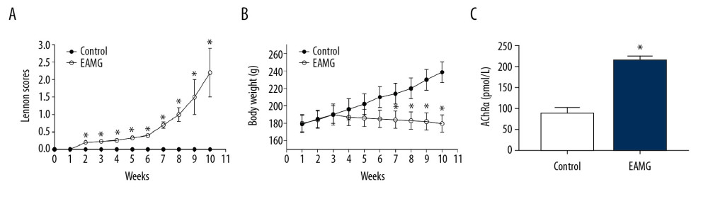 Figure 1. Assessment of the EAMG rat model. (A) Lennon scores were increased in EAMG models compared to control animals. (B) Body weight was reduced in EAMG models compared to the control group. (C) Levels of serum anti-AChR were increased in EAMG models compared to control animals as determined by ELISA analysis at 10 weeks after establishment of the EAMG model. The results are expressed as mean±SD (n=5 rats/group). * p<0.05 compared to control group.
 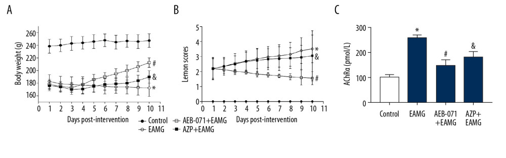 Figure 2. AEB-071 improved the clinical symptom of EAMG rats. (A) AEB-071 treatment reversed the body weight decline in EAMG rats. (B) AEB-071 treatment reduced the Lennon score in EAMG rats. (C) AEB-071 treatment reduced serum anti-AChR level in EAMG rats as measured by ELISA analysis after 10 days of drug intervention. The results are expressed as mean±SD (n=5 rats/group). * P<0.05 compared to control group; # P<0.05 compared to EAMG group; & P<0.05 compared to AEB-071+EAMG group.
 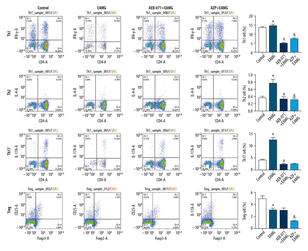 Figure 3. Proportion of Th1, Th2, Th17, and Treg lymphocytes as measured by flow cytometry. The percentages of IFN-y+CD4-Th1 cells, TL-4+CD4-Th2 cells, TL-17A+CD4-Th17 cells, and CD25 + Foxp3-Treg cells among the 4 groups were assessed after 10 days of drug intervention. The results are expressed as mean±SD (n = 5 rats/group). * P<0.05 compared to control group; # p<0.05 compared to EAMG group, & P<0.05 compared to AEB-071+EAMG group.
 References
1. Behin A, Le Panse R, New pathways and therapeutic targets in autoimmune myasthenia gravis: J Neuromuscul Dis, 2018; 5(3); 265-77
2. Meng QF, Zhang Z, Wang YJ, Astilbin ameliorates experimental autoimmune myasthenia gravis by decreased Th17 cytokines and up-regulated T regulatory cells: J Neuroimmunol, 2016; 298; 138-45
3. Jing F, Yang F, Cui Z, Rapamycin alleviates inflammation and muscle weakness, while altering the Treg/Th17 balance in a rat model of myasthenia gravis: Biosci Rep, 2017; 37(4); BSR20170767
4. Ziegler SF, Division of labour by CD4(+) T helper cells: Nat Rev Immunol, 2016; 16(7); 403
5. Berrih-Aknin S, Le Panse R, Myasthenia gravis: A comprehensive review of immune dysregulation and etiological mechanisms: J Autoimmun, 2014; 52; 90-100
6. Wang Z, Wang W, Chen Y, Wei D, T helper type 17 cells expand in patients with myasthenia-associated thymoma: Scand J Immunol, 2012; 76(1); 54-61
7. Meng X, Yang J, Dong M, Regulatory T cells in cardiovascular diseases: Nat Rev Cardiol, 2016; 13(3); 167-79
8. Masuda M, Matsumoto M, Tanaka S, Clinical implication of peripheral CD4+CD25+ regulatory T cells and Th17 cells in myasthenia gravis patients: J Neuroimmunol, 2010; 225(1–2); 123-31
9. Thiruppathi M, Rowin J, Ganesh B, Impaired regulatory function in circulating CD4(+)CD25(high)CD127(low/−) T cells in patients with myasthenia gravis: Clin Immunol, 2012; 145(3); 209-23
10. Wang Z, Yan Y, Immunopathogenesis in myasthenia gravis and neuromyelitis optica: Front Immunol, 2017; 8; 1785
11. Gruber T, Pfeifhofer-Obermair C, Baier G, PKCtheta is necessary for efficient activation of NFkappaB, NFAT, and AP-1 during positive selection of thymocytes: Immunol Lett, 2010; 132(1–2); 6-11
12. Miller AT, Dahlberg C, Sandberg ML, Inhibition of the inositol kinase Itpkb augments calcium signaling in lymphocytes and reveals a novel strategy to treat autoimmune disease: PLoS One, 2015; 10(6); e0131071
13. Evenou JP, Wagner J, Zenke G, The potent protein kinase C-selective inhibitor AEB071 (sotrastaurin) represents a new class of immunosuppressive agents affecting early T-cell activation: J Pharmacol Exp Ther, 2009; 330(3); 792-801
14. Fang YH, Joo DJ, Lim JB, AEB-071 versus tacrolimus monotherapy to prevent acute cardiac allograft rejection in the rat: A preliminary report: Transplant Proc, 2010; 42(3); 976-79
15. Manicassamy S, Sotrastaurin, a protein kinase C inhibitor for the prevention of transplant rejection and treatment of psoriasis: Curr Opin Investig Drugs, 2009; 10(11); 1225-35
16. Merani S, Edgar RL, Emamaulllee J, AEB-071 has minimal impact on onset of autoimmune diabetes in NOD mice: Autoimmunity, 2009; 42(3); 242-48
17. Evans WE, Pharmacogenetics of thiopurine S-methyltransferase and thiopurine therapy: Ther Drug Monit, 2004; 26(2); 186-91
18. Armstrong VW, Oellerich M, New developments in the immunosuppressive drug monitoring of cyclosporine, tacrolimus, and azathioprine: Clin Biochem, 2001; 34(1); 9-16
19. Losen M, Martizez-Martizez P, Molenaar PC, Standardization of the experimental autoimmune myasthenia gravis (EAMG) model by immunization of rats with Torpedo californica acetylcholine receptors – Recommendations for methods and experimental designs: Exp Neurol, 2015; 270; 18-28
20. Baggi F, Annoni A, Ubiali F, Breakdown of tolerance to a self-peptide of acetylcholine receptor alpha-subunit induces experimental myasthenia gravis in rats: J Immunol, 2004; 172(4); 2697-703
21. Fang YH, Joo JD, Lim BJ, The effects of AEB071 (sotrastaurin) with tacrolimus on rat heterotopic cardiac allograft rejection and survival: J Surg Res, 2011; 171(1); e133-37
22. Lennon VA, Lindstrom LM, Seybold ME, Experimental autoimmune myasthenia: A model of myasthenia gravis in rats and guinea pigs: J Exp Med, 1975; 141(6); 1365-75
23. Wang HB, Shi FD, Li H, Role for interferon-gamma in rat strains with different susceptibility to experimental autoimmune myasthenia gravis: Clin Immunol, 2000; 95(2); 156-62
24. Yang J, Sundrud MS, Skepner J, Yamagata T, Targeting Th17 cells in autoimmune diseases: Trends Pharmacol Sci, 2014; 35(10); 493-500
25. Xie Y, Li H, Jiang B, Elevated plasma interleukin-17A in a subgroup of myasthenia gravis patients: Cytokine, 2016; 78; 44-46
26. Mu L, Sun B, Kong Q, Disequilibrium of T helper type 1, 2 and 17 cells and regulatory T cells during the development of experimental autoimmune myasthenia gravis: Immunology, 2009; 128(1 Suppl); e826-36
27. Talaat RM, Mohamed SF, Bassyouni IH, Raouf AA, Th1/Th2/Th17/Treg cytokine imbalance in systemic lupus erythematosus (SLE) patients: Correlation with disease activity: Cytokine, 2015; 72(2); 146-53
28. Raphael I, Nalawade S, Eagar TN, T cell subsets and their signature cytokines in autoimmune and inflammatory diseases: Cytokine, 2015; 74(1); 5-17
29. Xie X, ATRA alters humoral responses associated with amelioration of EAMG symptoms by balancing Tfh/Tfr helper cell profiles: Clin Immunol, 2013; 148(2); 162-76
30. Kumar BV, Mu L, Yao X, Human T cell development, localization, and function throughout life: Immunity, 2018; 48(2); 202-13
31. Wang XD, Gong Y, Chen Z-L, TCR-induced sumoylation of the kinase PKC-theta controls T cell synapse organization and T cell activation: Nat Immunol, 2015; 16(11); 1195-203
32. Wachowicz K, Hermann-Kleiter N, Meisel M, Protein kinase C theta regulates the phenotype of murine CD4+ Th17 cells: PLoS One, 2014; 9(5); e96401
33. Salek-Ardakani S, So T, Halteman BS, Differential regulation of Th2 and Th1 lung inflammatory responses by protein kinase C theta: J Immunol, 2004; 173(10); 6440-47
34. Salek-Ardakani S, So T, Halteman BS, Protein kinase Ctheta controls Th1 cells in experimental autoimmune encephalomyelitis: J Immunol, 2005; 175(11); 7635-41
35. Zhang EY, Kong K-F, Altman A, The yin and yang of protein kinase C-theta (PKCtheta): A novel drug target for selective immunosuppression: Adv Pharmacol, 2013; 66; 267-312
36. Zanin-Zhorov A, Ding Y, Kumari S, Protein kinase C-theta mediates negative feedback on regulatory T cell function: Science, 2010; 328(5976); 372-76
37. Zanin-Zhorov A, Dustin ML, Blazar BR, PKC-θ function at the immunological synapse: Prospects for therapeutic targeting: Trends Immunol, 2011; 32(8); 358-63
38. Hage-Sleiman R, Hamze AB, Reslan L, The novel PKCtheta from benchtop to clinic: J Immunol Res, 2015; 2015 348798
39. Kovarik JM, Slade A, Overview of sotrastaurin clinical pharmacokinetics: Ther Drug Monit, 2010; 32(5); 540-43
40. Zuidscherwoude M, Dunlock V-ME, van den Bogaart G, Tetraspanin microdomains control localized protein kinase C signaling in B cells: Sci Signal, 2017; 10(478); eaag2755
41. Robertson MJ, Kahl BS, Vose JM, Phase II study of enzastaurin, a protein kinase C beta inhibitor, in patients with relapsed or refractory diffuse large B-cell lymphoma: J Clin Oncol, 2007; 25(13); 1741-46
42. Zappelli F, Willems D, Osada S, The inhibition of differentiation caused by TGFbeta in fetal myoblasts is dependent upon selective expression of PKCtheta: A possible molecular basis for myoblast diversification during limb histogenesis: Dev Biol, 1996; 180(1); 156-64
Figures
Figure 1. Assessment of the EAMG rat model. (A) Lennon scores were increased in EAMG models compared to control animals. (B) Body weight was reduced in EAMG models compared to the control group. (C) Levels of serum anti-AChR were increased in EAMG models compared to control animals as determined by ELISA analysis at 10 weeks after establishment of the EAMG model. The results are expressed as mean±SD (n=5 rats/group). * p<0.05 compared to control group.Figure 2. AEB-071 improved the clinical symptom of EAMG rats. (A) AEB-071 treatment reversed the body weight decline in EAMG rats. (B) AEB-071 treatment reduced the Lennon score in EAMG rats. (C) AEB-071 treatment reduced serum anti-AChR level in EAMG rats as measured by ELISA analysis after 10 days of drug intervention. The results are expressed as mean±SD (n=5 rats/group). * P<0.05 compared to control group; # P<0.05 compared to EAMG group; & P<0.05 compared to AEB-071+EAMG group.Figure 3. Proportion of Th1, Th2, Th17, and Treg lymphocytes as measured by flow cytometry. The percentages of IFN-y+CD4-Th1 cells, TL-4+CD4-Th2 cells, TL-17A+CD4-Th17 cells, and CD25 + Foxp3-Treg cells among the 4 groups were assessed after 10 days of drug intervention. The results are expressed as mean±SD (n = 5 rats/group). * P<0.05 compared to control group; # p<0.05 compared to EAMG group, & P<0.05 compared to AEB-071+EAMG group. In Press
Clinical Research
Body Weight and Insulin Resistance Indicators Among ChildrenMed Sci Monit In Press; DOI: 10.12659/MSM.951434
Clinical Research
Comparison of Radiographic Cervical Sagittal Alignment Parameters in Patients With Nonspecific Neck Pain, D...Med Sci Monit In Press; DOI: 10.12659/MSM.952950
Clinical Research
Combined Fibrinogen and Urinary α1-Microglobulin as Predictors of Respiratory Tract Infection in Children w...Med Sci Monit In Press; DOI: 10.12659/MSM.951066
Database Analysis
Evaluation of Salivary Total Oxidant Status (TOS) and Total Antioxidant Status (TAS) in Orthodontic Patient...Med Sci Monit In Press; DOI: 10.12659/MSM.952052
Most Viewed Current Articles
17 Jan 2024 : Review article 14,175,576
Vaccination Guidelines for Pregnant Women: Addressing COVID-19 and the Omicron VariantDOI :10.12659/MSM.942799
Med Sci Monit 2024; 30:e942799
13 Nov 2021 : Clinical Research 3,756,620
Acceptance of COVID-19 Vaccination and Its Associated Factors Among Cancer Patients Attending the Oncology ...DOI :10.12659/MSM.932788
Med Sci Monit 2021; 27:e932788
14 Dec 2022 : Clinical Research 2,465,966
Prevalence and Variability of Allergen-Specific Immunoglobulin E in Patients with Elevated Tryptase LevelsDOI :10.12659/MSM.937990
Med Sci Monit 2022; 28:e937990
16 May 2023 : Clinical Research 708,651
Electrophysiological Testing for an Auditory Processing Disorder and Reading Performance in 54 School Stude...DOI :10.12659/MSM.940387
Med Sci Monit 2023; 29:e940387