10 August 2020: Clinical Research
Abnormal Fractional Amplitude of Low-Frequency Fluctuation Changes in Patients with Monocular Blindness: A Functional Magnetic Resonance Imaging (MRI) Study
Jian-Wen Fang1ACDEF, Ya-Jie Yu1ACD, Li-Ying Tang23CD, Si-Yi Chen1C, Meng-Yao Zhang1F, Tie Sun1AF, Shi-Nan Wu1B, Kang Yu1B, Biao Li1B, Yi Shao1ACDG*DOI: 10.12659/MSM.926224
Med Sci Monit 2020; 26:e926224


Abstract
BACKGROUND: We used fractional amplitude of low-frequency fluctuation (fALFF) technology to investigate spontaneous cerebral activity in patients with monocular blindness (MB) and in healthy controls (HCs).
MATERIAL AND METHODS: Thirty MB patient and 15 HCs were included in this study. All subjects were scanned by resting-state functional magnetic resonance imaging (rs-fMRI). The independent sample t test and chi-squared test were applied to analyze demographics of MB patients and HCs. The 2-sample t test and receiver operating characteristic (ROC) curves were applied to identify the difference in average fALFF values between MB patients and HCs. Pearson’s correlation analysis was applied to explore the relationship between the average fALFF values of brain areas and clinical behavior in the MB group.
RESULTS: MB patients had lower fALFF values in the left anterior cingulate and higher fALFF values in the left precuneus and right and left inferior parietal lobes than in HCs. Moreover, the mean fALFF values of MB patients in the left anterior cingulate had negative correlations with the anxiety scale score (r=–0.825, P<0.001) and the depression scale score (r=–0.871, P<0.001).
CONCLUSIONS: Our study found that MB patients had abnormal spontaneous activities in the visual and vision-related regions. The finding of abnormal neuronal activity helps to reveal the underlying neuropathologic mechanisms of vision loss.
Keywords: Anxiety, Blindness, Depression, Magnetic Resonance Imaging, Brain Mapping, Case-Control Studies
Background
Worldwide, there are estimated to be 285 million people with impaired vision; of these, 39 million are considered blind [1]. Blindness is related to several different diseases. If not be treated in a timely manner, it may result in permanently impaired vision and adversely affect patients’ quality of life. Blindness has become a serious global social problem. The common causes of blindness include trachoma [2], glaucoma [3], cataracts [4], diabetic retinopathy [5] (DR), and age-related macular degeneration [6] (AMD). Monocular blindness (MB) is a severe ocular condition that affects patients of all age and can cause vision loss in 1 eye and visual interference in stereovision, field coverage, and exteroception of shape and color, thereby affecting the performance of visuomotor tasks [7]. Ocular trauma is the leading cause of MB worldwide [8]. MB is likely to develop into binocular blindness. The ideal treatment of irreversible blindness has been unclear. Determining the potential pathology leading to blindness is crucial to discover a cure for diseases leading to blindness [9].
The visual system is associated with transmission of visual signals, and destruction of the visual pathway can result in blindness. Therefore, it is important to explore whether the visual system of MB patients has substantial changes. Functional magnetic resonance imaging (fMRI), as an imaging technology, is commonly used to detect diseases of the brain and abnormal metabolism of the nerves [10]. The stimulation of neural activity can be a task-based neural response or spontaneous brain activity fluctuations in an unconscious state (“resting state”). fMRI has been used to assess neural activities in blind patients. Dormal et al. [11] used fMRI analysis to assess the effect of sound and external stimuli on the brain of patients with early blindness. Chan et al. [12] used blood-oxygenation-level-dependent (BOLD) fMRI to compare congenital versus acquired blindness to understand brain visual cortex activity and response during sensory replacement tasks and rest. Previous studies have shown that blindness can induce changes to the occipital cortex. The occipital cortex of early blind (EB) patients were thicker than those of normal people [13]. Furthermore, resting-state fMRI (rs-fMRI) showed the change of functional connectivity between visual cortex and cognitive control networks in EB patients [14]. The abovementioned studies were focused on brain network changes in blindness. However, the effect of MB on brain activities remains unknown.
Low-frequency oscillations (LFOs, typically in the 0.01–0.08 Hz frequency band) are linked to brain neuronal activity. rs-fMRI is considered a useful tool to study brain function to investigate brain development, aging, and diseases [15]. The brain exerts synchronous low-frequency fluctuations in particular areas when the brain is quiet without any form of activity such as cognitive or language actions [16]. The use of rs-fMRI allows researchers to investigate functional connectivity in the brain based on low-frequency fluctuations in the BOLD signal [16]. Amplitude of low-frequency fluctuation (ALFF) is an rs-fMRI indicator of partial change resulting from cerebral activity [17]. ALFF is an effective tool; it is easy to calculate and reliably analyzes rs-fMRI data [18]. ALFF has been used to study neurological conditions, such as optic neuritis [19], Parkinson’s disease [20], epilepsy [21], bipolar disorder [22], and schizophrenia [23]. ALFF also has been used widely in research on ocular diseases, such as MB [24], high myopia [25], glaucoma [26], optic neuritis [27], and diabetic retinopathy [28].
Fractional ALFF (fALFF) is a relatively new method to detect ALFF for rs-fMRI. The fALFF approach effectively restrains non-specific signals in rs-fMR, which increases the sensitivity and specificity of studying neural activities [17]. In our research, rs-fMRI was used through the fALFF approach to assess regional spontaneous cerebral activity in MB patients and in sighted subjects.
Material and Methods
SUBJECTS:
This study included 30 MB patients (18 caused by ocular trauma, 12 caused by keratitis), and 15 healthy controls (HCs). All MB patients were enrolled from the Department of Ophthalmology, the First Affiliated Hospital of Nanchang University. The diagnostic criteria for MB patients were: 1) one eye with no light perception and 2) healthy other eye without any diseases such as glaucoma or cataracts. The exclusion criteria were patients with: 1) a history of eye surgery, 2) severe mental health conditions such as schizophrenia, bipolar disorder, or other complex nervous system diseases, or 3) those that had undergone a long therapeutic procedure for blindness.
The HCs were matched by sex and age with MB patients. The procedures of this research were approved by the First Affiliated Hospital of Nanchang University Ethics Committee. All participants were acquainted with the purposes, contents, and risks of the research, and signed the informed consent form.
MRI PARAMETERS:
The MRI scans were performed using the same 3-Tesla MR scanner (Trio; Siemens, Munich, Germany). A three-dimensional metamorphic gradient echo pulse sequence was used to get the functional data. The following scanning parameters were used: repeat time=2,000 ms, echo time=40 ms, flip angle=90°, slice thickness/gap=4.0/1 mm, field of view=240×240 mm, and plane resolution=64×64. Overall, 30 axial slices covering the brain were recorded, and 240 functional images were finally obtained. The entire scanning process was completed in 8 min.
FMRI DATA ANALYSIS:
MRIcro software (
FALFF ANALYSIS:
The fALFF value is calculated on the trend data by REST software. REST has a built-in fast Fourier transform function that converts time series data into the frequency domain and calculates the power spectrum. Using the ratio of each frequency in the low-frequency range (0.01–0.08 Hz) to the power in the whole frequency range (0–0.25 Hz), fALFF was obtained. Then, a band-pass filtering of 0.01–0.08 Hz was used to ensure that the influence of low-frequency drift and high-frequency physiological noise such as from heartbeats and the respiratory rhythm is small.
STATISTICAL ANALYSIS:
SPSS 17.0 (SPSS, IBM Corp, USA) was used to determine differences in clinical features by independent sample
Results
DEMOGRAPHICS AND VISUAL MEASUREMENTS:
There were no significant differences in age (P=0.721), weight (P=0.765), or intraocular pressure of 2 eyes (P=0.748, P=0.783, respectively) between MB patients and HCs. The differences in best-corrected VA – right (P=0.008) and best-corrected VA – left (P=0.006) between the 2 groups were significant. More information is shown in Table 1.
FALFF DIFFERENCES:
MB patients had lower fALFF values in the left anterior cingulate and higher fALFF values in the left precuneus and right inferior parietal lobe and left inferior parietal lobes than in the HC group (Figure 1, Table 2). The result of the 2-sample t test revealed that the difference in mean fALFF values between MB patients and HCs was significant (P<0.001) (Table 3).
RECEIVER OPERATING CHARACTERISTIC CURVE:
The mean fALFF values in these regions were evaluated by ROC curves. The AUCs of fALFF values were as follows: left anterior cingulate (0.969) (Figure 2A), left precuneus (0.887), right inferior parietal lobe (0.936), and left inferior parietal lobe (0.911) (Figure 2B).
CORRELATION ANALYSIS:
The mean fALFF values of MB patients in the left anterior cingulate had negative correlations with the score of anxiety scale (r=−0.825, P<0.001) and depression scale (r=−0.871, P<0.001) (Figure 3).
Discussion
ANALYSIS OF THE INCREASED FALFF VALUES IN MB PATIENTS:
As a portion of the superior parietal lobule forward of the occipital lobe (cuneus), the precuneus has a significant role in the visuospatial imagery [29], episodic memory retrieval [30], and consciousness [31]. A study found that blind patients had less local gray matter in the precuneus than sighted individuals [32]. However, MB patients in our study had increased fALFF values in the left precuneus. The compensation mechanism is crucial for monocular vision loss. We speculated that the high activity of the anterior wedge may reflect obstacles of the visual space perception, as well as compensated vision loss and visual field defects in MB patients.
The parietal lobe supports higher cognitive functions, including mathematical cognition, semantics, and thought processing [33]. The function of the inferior parietal lobule (IPL) is associated with oculomotor and attentional mechanisms and the adaptive recalibration of eye-hand coordination [34]. The angular (ANG) and supramarginal (SMG) gyri of the left IPL make significant functional contributions to visual word recognition [35]. Normally, ANG plays an important role in semantic processing, whereas SMG is essential in phonological processing during reading. A previous fMRI study found that EB patients had more spatial variability in a bilateral parietal network than normal sighted individuals when at rest or listening to a vocal drama [36]. In our research, MB patients had increased fALFF values in the right inferior parietal lobe and left inferior parietal lobe, which may indicate the functional improvement to compensate for impaired visual acuity.
ANALYSIS OF THE DECREASED FALFF VALUES IN MB PATIENTS:
The anterior cingulate cortex (ACC) is a portion of the limbic system located anterior to the corpus callosum and posterior to the prefrontal cortex. The ACC is involved in movement [37]. Patients with increased ACC activity may have spasms, compulsive behaviors, and abnormal social behavior, while reduced ACC activity may result in disturbance of behavior such as lower self-consciousness and depression, motor neglect and impaired motor initiation, and aberrant social behavior [37]. The ACC is regarded as a monitoring center that is in charge of online detection of response conflicts [38]. Accordingly, the conflict signal sensed by the ACC is transmitted to other brain regions to trigger compensatory adjustments in cognitive control [39], such as the dorsal part of the lateral prefrontal cortex, to elevate the level of cognitive control. Abnormalities in the ACC have been associated with many diseases [40] such as depression, schizophrenia, and autism. The paracingulate cortex (PaC) is an adjacent and functionally relevant area of the ACC. The PaC has cognitive and affective regulatory function and is related to varied conditions such as psychosis and neurological conditions [41]. Our results showed that the fALFF values of the left anterior cingulate in MB patients were lower than in the HCs. This likely reflects the dysfunction of the ACC and PaC in MB patients.
Mental health comorbidities are common among patients with visual impairment [42]. In our study, the result showed that the mean fALFF values of MB patients in the left anterior cingulate had negative correlations with the scores of the anxiety and depression scale. Hence, we speculated that the cause of anxiety and depression may be related to the inhibition of the left anterior cingulated (Table 5).
LIMITATIONS:
Our study has some limitations. First, the sample size was relatively small, and, despite our best efforts, there were fewer HC patients than MB patients. Second, our inclusion criteria were not rigid, because patients with MB in the right or left eye were both included in our study, and the causes of MB were diverse. Third, during the scanning process, physical movement of patients might have had an effect on the scanning results. Lastly, further research is needed to verify our results.
Conclusions
MB patients in our study exhibited abnormal fALFF values in the visual cortices and vision-related areas, suggesting dysfunction of visual processing caused by vision loss. The finding of abnormal neuronal activity helps to reveal the underlying neuropathologic mechanisms of vision loss.
Figures
![Spontaneous cerebral activity in MB patients and HCs. Significant brain activity differences were observed in the left anterior cingulate and paracingulate gyri, left precuneus, right inferior parietal, but supramarginal and angular gyri and left inferior parietal, but supramarginal and angular gyri. The red or yellow denotes increased fAlFF values, and the blue areas indicate decreased fAlFF values, respectively (P,0.01 for multiple comparisons using Gaussian random field theory [z.2.3, P,0.01, cluster. 40 voxels, Alphasim corrected]). MB – monocular blindness; HCs – healthy controls; fALFF – fractional amplitude of low-frequency fluctuation; L – left; R – right.](https://jours.isi-science.com/imageXml.php?i=medscimonit-26-e926224-g001.jpg&idArt=926224&w=1000) Figure 1. Spontaneous cerebral activity in MB patients and HCs. Significant brain activity differences were observed in the left anterior cingulate and paracingulate gyri, left precuneus, right inferior parietal, but supramarginal and angular gyri and left inferior parietal, but supramarginal and angular gyri. The red or yellow denotes increased fAlFF values, and the blue areas indicate decreased fAlFF values, respectively (P,0.01 for multiple comparisons using Gaussian random field theory [z.2.3, P,0.01, cluster. 40 voxels, Alphasim corrected]). MB – monocular blindness; HCs – healthy controls; fALFF – fractional amplitude of low-frequency fluctuation; L – left; R – right.
Figure 1. Spontaneous cerebral activity in MB patients and HCs. Significant brain activity differences were observed in the left anterior cingulate and paracingulate gyri, left precuneus, right inferior parietal, but supramarginal and angular gyri and left inferior parietal, but supramarginal and angular gyri. The red or yellow denotes increased fAlFF values, and the blue areas indicate decreased fAlFF values, respectively (P,0.01 for multiple comparisons using Gaussian random field theory [z.2.3, P,0.01, cluster. 40 voxels, Alphasim corrected]). MB – monocular blindness; HCs – healthy controls; fALFF – fractional amplitude of low-frequency fluctuation; L – left; R – right. 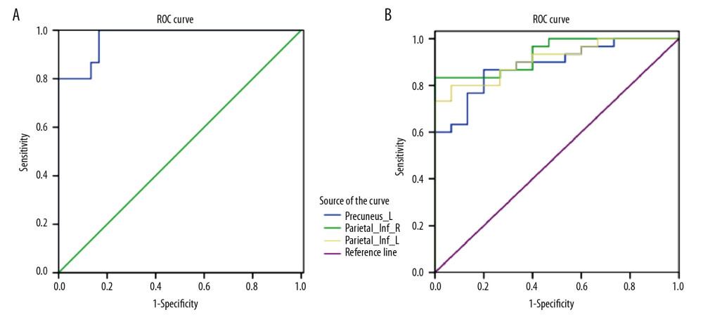 Figure 2. ROC curve analysis of the mean fALFF values for changed areas. The AUCs of fALFF values were as follows: left anterior cingulate (0.969) (A); left precuneus (0.887), right inferior parietal lobe (0.936), and left inferior parietal lobe (0.911) (B). ROC – receiver operating characteristic; AUC – area under the curve; fALFF – fractional amplitude of low-frequency fluctuation; MB – monocular blindness; HCs – healthy controls; Cingulum_Ant_L – left anterior cingulate; Precuneus–L – left precuneus; Parietal_Inf_R – right inferior parietal lobe; Parietal_Inf_L – left inferior parietal lobe.
 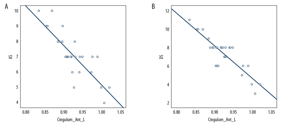 Figure 3. Correlations between the mean fALFF signal values of the Cingulum_Ant_L and score of anxiety scale and score of depression scale in MB patients. The mean fALFF signal value of the Cingulum_Ant_L showed negative correlations with the score of anxiety scale (r=−0.825, P<0.001) (A); The mean fALFF signal value of the Cingulum Ant L showed negative correlations with the score of depression scale (r=−0.871, P<0.001) (B). fALFF – fractional amplitude of low-frequency fluctuation; Cingulum_Ant_L – left anterior cingulate; MB – monocular blindness; AS – anxiety scale; DS – depression scale.
 Tables
Table 1. Demographics and clinical measurements of MB and HC groups.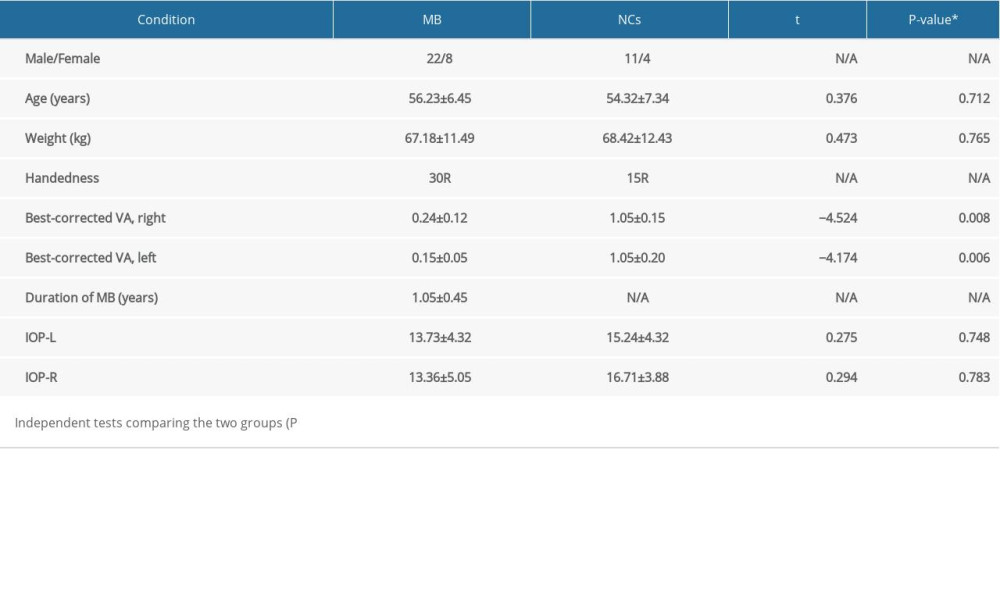 Table 2. Brain areas with significantly different fALFF values between MB and HCs groups.
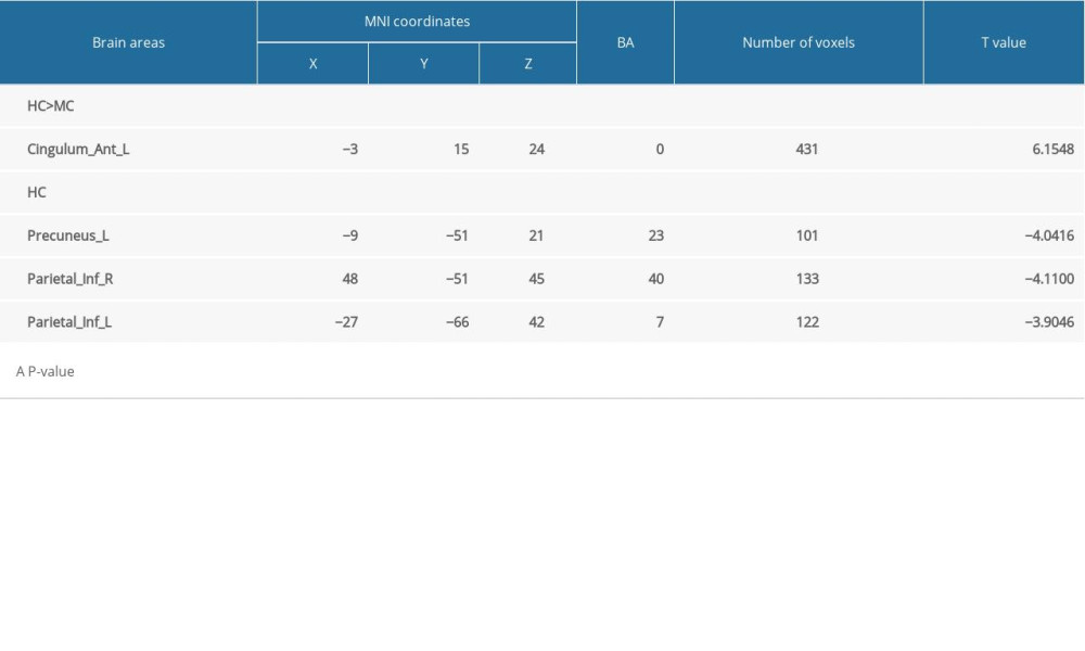 Table 3. Two sample t tests between mean fALFF values between MB patients and HCs-related brain regions.
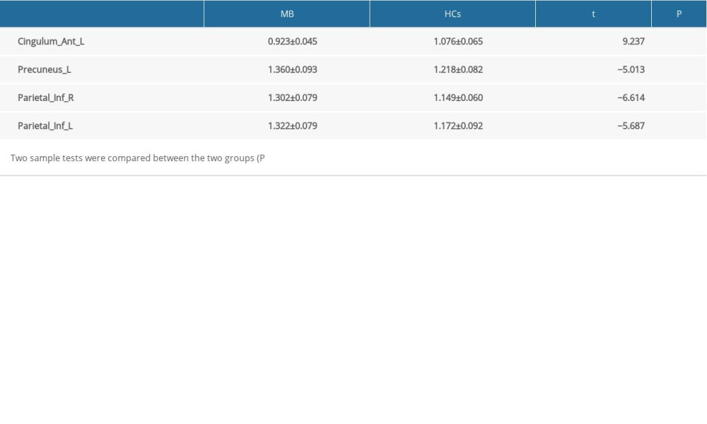 Table 4. The fALFF applied in neurodegenerative diseases.
 Table 5. Brain regions alternation and its potential impact.
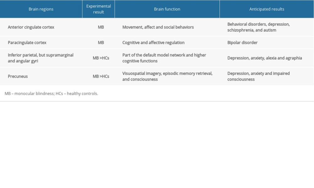
References
1. Tham YC, Lim SH, Shi Y, Trends of visual impairment and blindness in the singapore chinese population over a decade: Sci Rep, 2018; 8(1); 12224
2. WoldeKidan E, Daka D, Legesse D, Prevalence of active trachoma and associated factors among children aged 1 to 9 years in rural communities of Lemo district, southern Ethiopia: Community based cross sectional study: BMC Infect Dis, 2019; 19(1); 886
3. Chono I, Miyazaki D, Miyake H, High interleukin-8 level in aqueous humor is associated with poor prognosis in eyes with open angle glaucoma and neovascular glaucoma: Sci Rep, 2018; 8(1); 14533
4. Wang D, Wang E, Liu K: Sci Rep, 2017; 7(1); 7274
5. Huang H, He J, Johnson D, Deletion of placental growth factor prevents diabetic retinopathy and is associated with Akt activation and HIF1alpha-VEGF pathway inhibition: Diabetes, 2015; 64(1); 200-12
6. Rohwer K, Neupane S, Bittkau KS, Effects of crude fucus distichus subspecies evanescens fucoidan extract on retinal pigment epithelium cells-implications for use in age-related macular degeneration: Mar Drugs, 2019; 17(9); 538
7. Richard AI, Monocular blindness in Bayelsa state of Nigeria: Pan Afr Med J, 2010; 4; 6
8. Han L, Jia J, Fan Y, The vitrectomy timing individualization system for ocular trauma (VTISOT): Sci Rep, 2019; 9(1); 12612
9. Bales KL, Ianov L, Kennedy AJ, Autosomal dominant retinitis pigmentosa rhodopsin mutant Q344X drives specific alterations in chromatin complex gene transcription: Mol Vis, 2018; 24; 153-64
10. Chen JE, Glover GH, Functional magnetic resonance imaging methods: Neuropsychol Rev, 2015; 25(3); 289-313
11. Dormal G, Pelland M, Rezk M, Functional preference for object sounds and voices in the brain of early blind and sighted individuals: J Cogn Neurosci, 2018; 30(1); 86-106
12. Chan KC, Murphy MC, Bang JW, Functional MRI of sensory substitution in the blind: Conf Proc IEEE Eng Med Biol Soc, 2018; 2018; 5519-22
13. Voss P, Zatorre RJ, Early visual deprivation changes cortical anatomical covariance in dorsal-stream structures: Neuroimage, 2015; 108; 194-202
14. Burton H, Snyder AZ, Raichle ME, Resting state functional connectivity in early blind humans: Front Syst Neurosci, 2014; 8; 51
15. Maknojia S, Churchill NW, Schweizer TA, Graham SJ, Resting State fMRI: Going through the motions: Front Neurosci, 2019; 13; 825
16. Lee MH, Miller-Thomas MM, Benzinger TL, Clinical resting-state fMRI in the preoperative setting: Are we ready for prime time?: Top Magn Reson Imaging, 2016; 25(1); 11-18
17. Zou QH, Zhu CZ, Yang Y, An improved approach to detection of amplitude of low-frequency fluctuation (ALFF) for resting-state fMRI: Fractional ALFF: J Neurosci Methods, 2008; 172(1); 137-41
18. Huang X, Zhong YL, Zeng XJ, Disturbed spontaneous brain activity pattern in patients with primary angle-closure glaucoma using amplitude of low-frequency fluctuation: A fMRI study: Neuropsychiatr Dis Treat, 2015; 11; 1877-83
19. Krell D, Said Battistino F, Benafif S, Audit of CA125 follow-up after first-line therapy for ovarian cancer: Int J Gynecol Cancer, 2017; 27(6); 1118-22
20. Xiang J, Jia X, Li H, Altered spontaneous brain activity in cortical and subcortical regions in Parkinson’s disease: Parkinsons Dis, 2016; 2016 5246021
21. Chen Z, An Y, Zhao B, The value of resting-state functional magnetic resonance imaging for detecting epileptogenic zones in patients with focal epilepsy: PLoS One, 2017; 12(2); e0172094
22. Xu K, Liu H, Li H, Amplitude of low-frequency fluctuations in bipolar disorder: a resting state fMRI study: J Affect Disord, 2014; 152–54; 237-42
23. Turner JA, Damaraju E, van Erp TG, A multi-site resting state fMRI study on the amplitude of low frequency fluctuations in schizophrenia: Front Neurosci, 2013; 7; 137
24. Li Q, Huang X, Ye L, Altered spontaneous brain activity pattern in patients with late monocular blindness in middle-age using amplitude of low-frequency fluctuation: A resting-state functional MRI study: Clin Interv Aging, 2016; 11; 1773-80
25. Huang X, Zhou FQ, Hu YX, Altered spontaneous brain activity pattern in patients with high myopia using amplitude of low-frequency fluctuation: A resting-state fMRI study: Neuropsychiatr Dis Treat, 2016; 12; 2949-56
26. Liu Z, Tian J, Amplitude of low frequency fluctuation in primary open angle glaucoma: A resting state fMRI study: Conf Proc IEEE Eng Med Biol Soc, 2014; 2014; 6706-9
27. Huang X, Cai FQ, Hu PH, Disturbed spontaneous brain-activity pattern in patients with optic neuritis using amplitude of low-frequency fluctuation: A functional magnetic resonance imaging study: Neuropsychiatr Dis Treat, 2015; 11; 3075-83
28. Wang ZL, Zou L, Lu ZW, Abnormal spontaneous brain activity in type 2 diabetic retinopathy revealed by amplitude of low-frequency fluctuations: A resting-state fMRI study: Clin Radiol, 2017; 72(4); 340.e1-7
29. Bruner E, Preuss TM, Chen X, Rilling JK, Evidence for expansion of the precuneus in human evolution: Brain Struct Funct, 2017; 222(2); 1053-60
30. Ye Q, Zou F, Lau H, Causal evidence for mnemonic metacognition in human precuneus: J Neurosci, 2018; 38(28); 6379-87
31. Cavanna AE, The precuneus and consciousness: CNS Spectr, 2007; 12(7); 545-52
32. Wan CY, Wood AG, Chen J, The influence of preterm birth on structural alterations of the vision-deprived brain: Cortex, 2013; 49(4); 1100-9
33. Catani M, Robertsson N, Beyh A, Short parietal lobe connections of the human and monkey brain: Cortex, 2017; 97; 339-57
34. Clower DM, West RA, Lynch JC, Strick PL, The inferior parietal lobule is the target of output from the superior colliculus, hippocampus, and cerebellum: J Neurosci, 2001; 21(16); 6283-91
35. Sliwinska MW, James A, Devlin JT, Inferior parietal lobule contributions to visual word recognition: J Cogn Neurosci, 2015; 27(3); 593-604
36. Boldt R, Seppa M, Malinen S, Spatial variability of functional brain networks in early-blind and sighted subjects: Neuroimage, 2014; 95; 208-16
37. Devinsky O, Morrell MJ, Vogt BA, Contributions of anterior cingulate cortex to behaviour: Brain, 1995; 118(Pt 1); 279-306
38. Sohn MH, Albert MV, Jung K, Anticipation of conflict monitoring in the anterior cingulate cortex and the prefrontal cortex: Proc Natl Acad Sci USA, 2007; 104(25); 10330-34
39. Botvinick MM, Cohen JD, Carter CS, Conflict monitoring and anterior cingulate cortex: An update: Trends Cogn Sci, 2004; 8(12); 539-46
40. Huang X, Ye CL, Zhong YL, Altered regional homogeneity in patients with late monocular blindness: A resting-state functional MRI study: Neuroreport, 2017; 28(16); 1085-91
41. Fornito A, Whittle S, Wood SJ, The influence of sulcal variability on morphometry of the human anterior cingulate and paracingulate cortex: Neuroimage, 2006; 33(3); 843-54
42. Court H, McLean G, Guthrie B, Visual impairment is associated with physical and mental comorbidities in older adults: A cross-sectional study: BMC Med, 2014; 12; 181
Figures
Figure 1. Spontaneous cerebral activity in MB patients and HCs. Significant brain activity differences were observed in the left anterior cingulate and paracingulate gyri, left precuneus, right inferior parietal, but supramarginal and angular gyri and left inferior parietal, but supramarginal and angular gyri. The red or yellow denotes increased fAlFF values, and the blue areas indicate decreased fAlFF values, respectively (P,0.01 for multiple comparisons using Gaussian random field theory [z.2.3, P,0.01, cluster. 40 voxels, Alphasim corrected]). MB – monocular blindness; HCs – healthy controls; fALFF – fractional amplitude of low-frequency fluctuation; L – left; R – right.Figure 2. ROC curve analysis of the mean fALFF values for changed areas. The AUCs of fALFF values were as follows: left anterior cingulate (0.969) (A); left precuneus (0.887), right inferior parietal lobe (0.936), and left inferior parietal lobe (0.911) (B). ROC – receiver operating characteristic; AUC – area under the curve; fALFF – fractional amplitude of low-frequency fluctuation; MB – monocular blindness; HCs – healthy controls; Cingulum_Ant_L – left anterior cingulate; Precuneus–L – left precuneus; Parietal_Inf_R – right inferior parietal lobe; Parietal_Inf_L – left inferior parietal lobe.Figure 3. Correlations between the mean fALFF signal values of the Cingulum_Ant_L and score of anxiety scale and score of depression scale in MB patients. The mean fALFF signal value of the Cingulum_Ant_L showed negative correlations with the score of anxiety scale (r=−0.825, P<0.001) (A); The mean fALFF signal value of the Cingulum Ant L showed negative correlations with the score of depression scale (r=−0.871, P<0.001) (B). fALFF – fractional amplitude of low-frequency fluctuation; Cingulum_Ant_L – left anterior cingulate; MB – monocular blindness; AS – anxiety scale; DS – depression scale. Tables
Table 1. Demographics and clinical measurements of MB and HC groups.Table 2. Brain areas with significantly different fALFF values between MB and HCs groups.Table 3. Two sample t tests between mean fALFF values between MB patients and HCs-related brain regions.Table 4. The fALFF applied in neurodegenerative diseases.Table 5. Brain regions alternation and its potential impact.Table 1. Demographics and clinical measurements of MB and HC groups.Table 2. Brain areas with significantly different fALFF values between MB and HCs groups.Table 3. Two sample t tests between mean fALFF values between MB patients and HCs-related brain regions.Table 4. The fALFF applied in neurodegenerative diseases.Table 5. Brain regions alternation and its potential impact. In Press
Clinical Research
Effects of Single-Bout Endurance Exercise Intensity on Peripheral Neurotrophic Factors in Patients With Isc...Med Sci Monit In Press; DOI: 10.12659/MSM.952089
Review article
Anisodus tanguticus in Cancer Research: A Review of Traditional Use, Phytochemistry, Extraction Methods, an...Med Sci Monit In Press; DOI: 10.12659/MSM.952999
Clinical Research
Nasal Mucociliary Clearance and Its Relationship With Disease Severity in Patients With Multiple SclerosisMed Sci Monit In Press; DOI: 10.12659/MSM.952850
Clinical Research
Modified Thoracoabdominal Nerves Block Through the Perichondrial Approach vs Subcostal Transversus Abdomini...Med Sci Monit In Press; DOI: 10.12659/MSM.953976
Most Viewed Current Articles
17 Jan 2024 : Review article 14,176,570
Vaccination Guidelines for Pregnant Women: Addressing COVID-19 and the Omicron VariantDOI :10.12659/MSM.942799
Med Sci Monit 2024; 30:e942799
13 Nov 2021 : Clinical Research 3,762,188
Acceptance of COVID-19 Vaccination and Its Associated Factors Among Cancer Patients Attending the Oncology ...DOI :10.12659/MSM.932788
Med Sci Monit 2021; 27:e932788
14 Dec 2022 : Clinical Research 2,466,310
Prevalence and Variability of Allergen-Specific Immunoglobulin E in Patients with Elevated Tryptase LevelsDOI :10.12659/MSM.937990
Med Sci Monit 2022; 28:e937990
16 May 2023 : Clinical Research 708,927
Electrophysiological Testing for an Auditory Processing Disorder and Reading Performance in 54 School Stude...DOI :10.12659/MSM.940387
Med Sci Monit 2023; 29:e940387