01 January 2020: Clinical Research
A Novel Instant 3-Dimensional Printing System for Postoperative Fracture Patients: A Comparative Cohort Study
Lang Chen1ACE, Yuan Xiong2ACE, Bobin Mi2B, Chenchen Yan2C, Xudong Xie2F, Zexi He2F, Wu Zhou2BG*, Guohui Liu2AGDOI: 10.12659/MSM.928240
Med Sci Monit 2021; 27:e928240


Abstract
BACKGROUND: Traditional plaster (TP) is a widely used auxiliary fixation (AF) approach for postoperative fracture patients. However, patient discomfort and inconvenience to clinicians has limited its application. We introduce a novel instant 3-dimensional printing appliance system (3D-AS) to address such issues.
MATERIAL AND METHODS: Twenty-seven postoperative fracture patients were divided randomly between a TP group and a 3D-AS group, and analyzed retrospectively. Radiographic images during follow-up were evaluated for fracture healing and fracture reduction quality. The range of motion (ROM) was recorded to assess motor performance. Patient pain was assessed using the Visual Analogue Scale (VAS). Complications were also compared between the 2 groups.
RESULTS: The patients comprised 17 men and 10 women with ages ranging from 21 to 69 years (mean age: 47.35). All patients completed a follow-up visit (range: 14–19 months, mean: 13.59 months). Although no significant difference was found between general characteristics (P>0.05) and the time of fracture union (P>0.05), significant differences between groups were seen in complications (P<0.05), VAS (P<0.01), patient satisfaction (P<0.05), and ROM for the upper joints (P<0.05).
CONCLUSIONS: Our study suggests that 3D-AS provides better upper-limb ROM and more comfortable healing for postoperative fracture patients, indicating that it can be recommended for use in such patients.
Keywords: Auxiliary Fixation, Fracture Healing, Imaging, Three-Dimensional, Cohort Studies, Fractures, Bone, Health Care Costs, Patient Satisfaction, Printing, Three-Dimensional, Surveys and Questionnaires
Background
Recently, traumatic bone fracture incidence has increased with developments in transportation [1]. Patients with fractures often suffer serious pain due to the displacement of the fracture end, and serious secondary injury, including nerve injury, blood vessel injury, and osteofascial compartment syndrome, which may occur should the fracture end not be fixed appropriately [2]. In addition to temporary preoperative fixation, postoperative auxiliary fixation (AF) is also important for fracture patients. To the best of our knowledge, the use of traditional plaster (TP) is amongst the most widely used approaches to AF [3]. However, the disadvantages of TP should not be dismissed. First, TP is bulky, increasing the burden and discomfort of patients [4]. Secondly, TP is formed from a material that is not breathable, and consequently often gives rise to bedsore [5]. Thirdly, artifacts may be created with TP; therefore, TP must be removed before any X-ray examination. This increases both the workload of medical staff and patient burden [6]. Fourthly, plaster solidification is protracted, requiring technicians to handle fixation [7]. Finally, plaster removal is inconvenient, and TP cannot be reused [8].
Given such disadvantages, attention has turned to other approaches better able to address these issues [9]. Recently, 3-dimensional (3D) printing has been introduced, and found to be useful in many areas of medicine [10]. For example, Zhou et al. reported the application of a 3D-printed template to assist sacroiliac screw placement, improving the operation’s safety profile [11]. Moreover, a 3D-printed brace was recently used for adolescent idiopathic scoliosis patients [12]. However, an important bottleneck to the wider application of 3D printing in medicine is the availability of a human-suitable, safe, and environmentally friendly 3D printing material. Additionally, as the extrusion speed of a current 3D printer is approximately 60 grams per hour, there can be a very long wait for each 3D-printed product [13]. For example, a wrist joint weighs over 300 grams, so patients can wait 5 or more hours before fixation. Three-dimensional printing materials are typically also very expensive, and production costs are high [14]. To address such issues, we introduced a novel instant 3D printing splint system (3D-AS) for postoperative fracture patients. In the current study, 3D-AS was undertaken using an instant 3D printer (BiYing-3D instant printer, Wuhan Biying Biotechnology Co., Ltd). A biodegradable corn starch-based material was designed and used as the printing material; the cost of this material is similar to that of TP. Using the 3D-AS approach, a 3D-printed splint can be constructed in 5–10 minutes.
Material and Methods
PATIENT ELIGIBILITY:
Between August 2018 and May 2019, 27 postoperative fracture patients with AF were included in the study. Ethics approval and informed consent to participate were obtained from each patient. Inclusion criteria were: (1) closed fracture patients; (2) postoperative fracture patients with TP or 3D-AS for AF; (3) aged over 18 years; (4) full mental competence; (5) American Society of Anesthesiology (ASA) score <level 3. Exclusion criteria were: (1) open fracture patients with external fixation; (2) fracture patients receiving non-surgical treatment; (3) postoperative fracture patients without AF; (4) ASA score >level 3; (5) under 18 years.
3D-AS PROTOCOL:
3D-AS was performed using an instant 3D printer (BiYing-3D instant printer, Wuhan Biying Biotechnology Co., Ltd, Wuhan, China). A biodegradable corn starch-based material was designed and used as the 3D-AS printing material. The 3D-printed splint was able to be constructed rapidly; in 5–10 minutes. The whole 3D-AS procedure comprised 4 steps: 3D scanning, slicing administration, 3D printing, and wearing. First, for 3D scanning, stereolithography (STL) file format was used by both the designing software and the printing device. The STL file uses triangular faces to approximate an object’s surface. The smaller the triangular face, the greater the surface resolution. The Python Lex-Yacc (PLY) scanner reads 3-dimensional data from a file and produces a VRML- or WRL-format file, often used as input for full-color printing (Figure 1A). Secondly, for slicing, the printer reads cross-sections from the file, printing it layer-by-layer from a liquid, powder, or flake, and then stitches these sections together to create a single final entity. The technology is characterized by its ability to make objects of almost any shape (Figure 1B). Thirdly, for 3D printing, the 3D printer’s resolution is adequate for most tasks, although curved surfaces can be rough. Higher resolution structures can be obtained by printing a slightly larger object, and then grinding surfaces smooth (Figure 1C–1E). Finally, clinicians provide the instant 3D-printed AF to the patient, assisting him or her to wear the AF with optimal tightness (Figure 1F). As 3D-printed splints are constructed based on the individual characteristics of each patient, the fit of 3D-AS is better than that of TP. Furthermore, the splint is printed immediately before handling the affected limb. Hence, patients just need help to put on their appliances, rather than needing to go through the shaping and maintaining procedures required with TP. An example of a 3D-AS is shown in Figure 2.
EVALUATION INDEX:
The general characteristics of the included patients and their postoperative outcomes were compared. Radiographic images, taken during follow-up, were used to evaluate fracture healing and fracture reduction quality. The range of motion (ROM) in both upper-limb joints (elbow and wrist) and both lower-limb joints (knee and ankle) was used to assess patient motor performance at their final follow-up visit. Patient satisfaction was evaluated using an investigator-designed questionnaire (see Table 1). Patient pain in both groups was assessed after surgery using the Visual Analogue Scale (VAS). Complications including incision infection, non-union, and fragment displacement were compared between groups.
STATISTICAL ANALYSIS:
Fisher’s exact test was used to compare categorical variables between the TP group and the 3D-AS group, as data were limited. A Kolmogorov-Smirnov test was performed to test whether continuous variables complied with normal distribution. An independent group
Results
The 27 patients in the study were treated using TP or 3D-AS. The 2 cohorts had similar characteristics (see Table 2): average age (46.46±11.33 years
Similarly, no statistically significant difference was seen in the time to fracture union (18.08±3.00 weeks
Discussion
In postoperative fracture patients, AF helps prevent postoperative complications [15]. However, critical concerns about AF should not be ignored [16]. First, widely used AF approaches, including TP, plaster bandage, and splint, all have standardized production, so it is impossible to achieve a perfectly fitted AF for an affected limb, leading to potential chronic discomfort in patients. In our retrospective study, a statistically significant difference was found in VAS scores after surgery between the groups. When compared with 3D-AS, TP increased discomfort for fracture patients. Similarly, patient satisfaction in the 3D-AS group was greater than in the TP group, indicating a more comfortable experience in the 3D-AS group. Secondly, current AF approaches cover wounds and surgical incisions, severely hindering monitoring during healing. A statistically significant difference in the incidence of incision infection was seen between the groups, suggesting incision management was superior in the 3D-AS group. Thus, 3D-AS is the method of choice for surgical patients, based on our results. Existing AF approaches require complex handling by experienced healthcare professionals, particularly for TP and plaster bandage, which are time- and labor-intensive undertakings. As 3D-printed splints are constructed individually for each patient, their quality of fit is greater than that of TP, with the splint being printed essentially instantly prior to the handling of an affected limb. Hence, using 3D-AS, the only help patients need is help with putting on the splint, rather than requiring technicians to shape and maintain fixation, as with TP.
Another issue is fragment displacement that affects joint ROM. Four patients in the TP group had fragment displacement after surgery, all with upper-limb fractures (3 distal radius fractures and 1 olecranon fracture). Although displacement of the fracture fragment depends on the rigidity and choice of implants, other factors may also contribute to fragment displacement. First, the wrist and elbow are not weight-bearing joints, compared with the ankle and knee joints, and as such they are much more flexible. Thus, earlier joint exercise is needed in patients with these 2 affected joints, compared with lower-limb fracture patients, who are at greater risk of postoperative fragment displacement.
Patients wearing TP experience more discomfort than 3D-AS patients according to our study. This may reduce compliance in TP patients, due to discomfort, leading perhaps to earlier TP removal or inappropriate TP fixation. A statistically significant difference was also seen in ROM of the upper joints. The more comfortable, less painful experience of 3D-AS patients was crucial to a better clinical outcome; the 3D-AS patients were more active during joint function exercise in the affected limb, and exhibited better clinical compliance.
The manufacture of the 3D-AS was performed using an instant 3D printer (BiYing-3D instant printer, Wuhan Biying Biotechnology Co., Ltd, Wuhan, China). In previous cases, patients may have suffered secondary pain due to the use of inappropriate printing materials. Therefore, if material appropriate for the human body, which is safe and environmentally friendly, can be designed, it should become a key factor in determining how 3D printing is applied in AF [17]. A corn starch-based biodegradable material was designed and used as the printing material for 3D-AS in our study. The cost of this material is close to that of TP while meeting the requirements above. The resulting printed product is non-stimulating, colorless, odorless, and waterproof, allowing it to be worn while bathing. The weight of the printed product is approximately 1/6 that of TP, and is 20 times as durable as TP. Moreover, the 3D printer was equipped with a more efficient materials inlet, which avoids silk blocking during the printing process. The printing speed is also a critical index, and 3D-AS is rapidly formed. After near-instant printing is completed, affected limbs can be fixed rapidly: the printing time of an ordinary wrist joint was completed within 10 minutes. Accurate printing can be achieved with 3D-AS, greatly reducing costs. Due to the porous design, more comfort was reported by patients. Owing to effective avoidance of wounds and incisions, 3D-AS improves the healing rate without altering management of dressing and wound treatment. Since the 3D-AS printing material was composed of corn starch-based material, there was no difference in the cost of intervention between the TP group and the 3D-AS group (Table 3,
Like all reported research, our study has some limitations. Firstly, outcomes from surgical treatment depend mainly on the fixation technique, the rigidity of fixation, and soft-tissue handling; our study focused only on the comparative efficacy of TP versus 3D-AS in postoperative fracture patients. Secondly, this is a retrospective study with a relatively small cohort. Finally, the follow-up period was relatively short, precluding a definitive conclusion.
Conclusions
Our study suggests that, compared with TP, 3D-AS provides better upper-limb ROM and a more comfortable experience for postoperative fracture patients, indicating that 3D-AS can be robustly recommended for such patients.
Figures
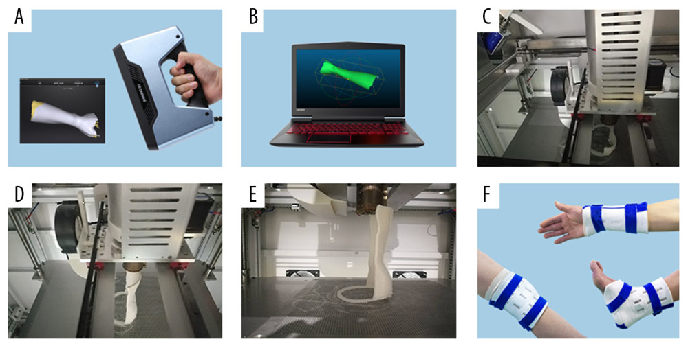 Figure 1. (A) An input file is produced by a scanner for further full-color printing. (B) The printer reads the cross-section in the file and then creates a single entity. (C–E) The printing process of the 3-dimensional printing appliance system. (F) Application of the 3-dimensional printing appliance system.
 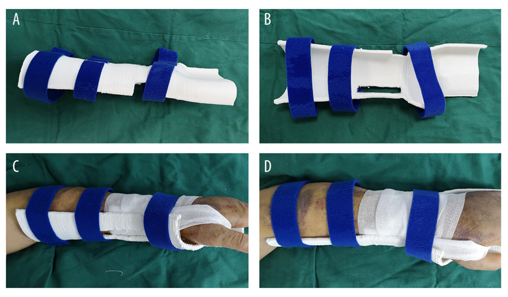 Figure 2. (A–D) Application of the 3-dimensional printing appliance system for distal radius fracture.
 References
1. Puzio TJ, Murphy PB, Gazzetta J, The electric scooter: A surging new mode of transportation that comes with risk to riders: Traffic Inj Prev, 2020; 21; 175-78
2. McVeigh LG, Perugini AJ, Fehrenbacher JC, Assessment, quantification, and management of fracture pain: From animals to the clinic: Curr Osteoporos Rep, 2020; 18(5); 460-70
3. Chen Y, Lin H, Yu Q, Application of 3D-printed orthopedic cast for the treatment of forearm fractures: Finite element analysis and comparative clinical assessment: Biomed Res Int, 2020; 2020 9569530
4. Mulders MAM, Walenkamp MMJ, van Dieren S, Volar plate fixation in adults with a displaced extra-articular distal radial fracture is cost-effective: J Bone Joint Surg Am, 2020; 102; 609-16
5. Qiao F, Jiang F, Closed reduction of severely displaced radial neck fractures in children: BMC Musculoskelet Disord, 2019; 20; 567
6. Pedersen J, Mortensen SO, Rölfing JD, A protocol for a single-center, single-blinded randomized-controlled trial investigating volar plating versus conservative treatment of unstable distal radius fractures in patients older than 65 years: BMC Musculoskelet Disord, 2019; 20; 309
7. Saving J, Severin WS, Olsson K, Nonoperative treatment compared with volar locking plate fixation for dorsally displaced distal radial fractures in the elderly: A randomized controlled trial: J Bone Joint Surg Am, 2019; 101; 961-69
8. Hussain MS, Avilucea FR, Archdeacon MT, Effectiveness of the taut-line hitch knot in reducing and splinting lower extremity fractures: J Orthop Trauma, 2019; 33; e31-35
9. Wu G, Li B, Yang Y, Comparative study of surgical and conservative treatments for fifth metatarsal base avulsion fractures (type I) in young adults or athletes: J Orthop Surg (Hong Kong), 2018; 26 2309499017747128
10. Langerhuizen DWG, Doornberg JN, Janssen MMA, Do 3-D printed handheld models improve surgeon reliability for recognition of intraarticular distal radius fracture characteristics?: Clin Orthop Relat Res, 2020 [Online ahead of print]
11. Franch J, Barba A, Rappe K, Use of three-dimensionally printed b-tricalcium phosphate synthetic bone graft combined with recombinant human bone morphogenic protein-2 to treat a severe radial atrophic nonunion in a Yorkshire terrier: Vet Surg, 2020 [Online ahead of print]
12. Pasha S, 3D spinal and rib cage predictors of brace effectiveness in adolescent idiopathic scoliosis: BMC Musculoskelet Disord, 2019; 20; 384
13. Tian Y, Zhang J, Liu T, A comparative study of C2 pedicle or pars screw placement with assistance from a 3-dimensional (3D)-printed navigation template versus C-arm based navigation: Med Sci Monit, 2019; 25; 9981-90
14. Zhang M, Li J, Fang T, Evaluation of a three-dimensional printed guide and a polyoxymethylene thermoplastic regulator for percutaneous pedicle screw fixation in patients with thoracolumbar fracture: Med Sci Monit, 2020; 26; e920578
15. Guo Y, Tong L, Li S, External fixation combined with limited internal fixation versus open reduction internal fixation for treating ruedi-allgower type III pilon fractures: Med Sci Monit, 2015; 21; 1662-67
16. Hack J, Kranz Y, Knauf T, Stability of internal versus external fixation in osteoporotic pelvic fractures – a biomechanical analysis: Injury, 2020 [Online ahead of print]
17. Wu W, Xu W, Wan C, Preoperative plan with 3D printing in internal and external fixation for complex tibial plateau fractures: Orthop Surg, 2019; 11; 560-68
Figures
Figure 1. (A) An input file is produced by a scanner for further full-color printing. (B) The printer reads the cross-section in the file and then creates a single entity. (C–E) The printing process of the 3-dimensional printing appliance system. (F) Application of the 3-dimensional printing appliance system.Figure 2. (A–D) Application of the 3-dimensional printing appliance system for distal radius fracture. Tables
 Table 1. Patient satisfaction survey.
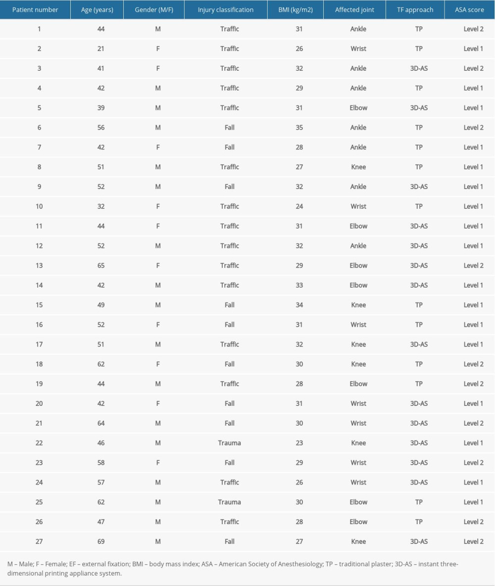 Table 2. General characteristics of the included patients.
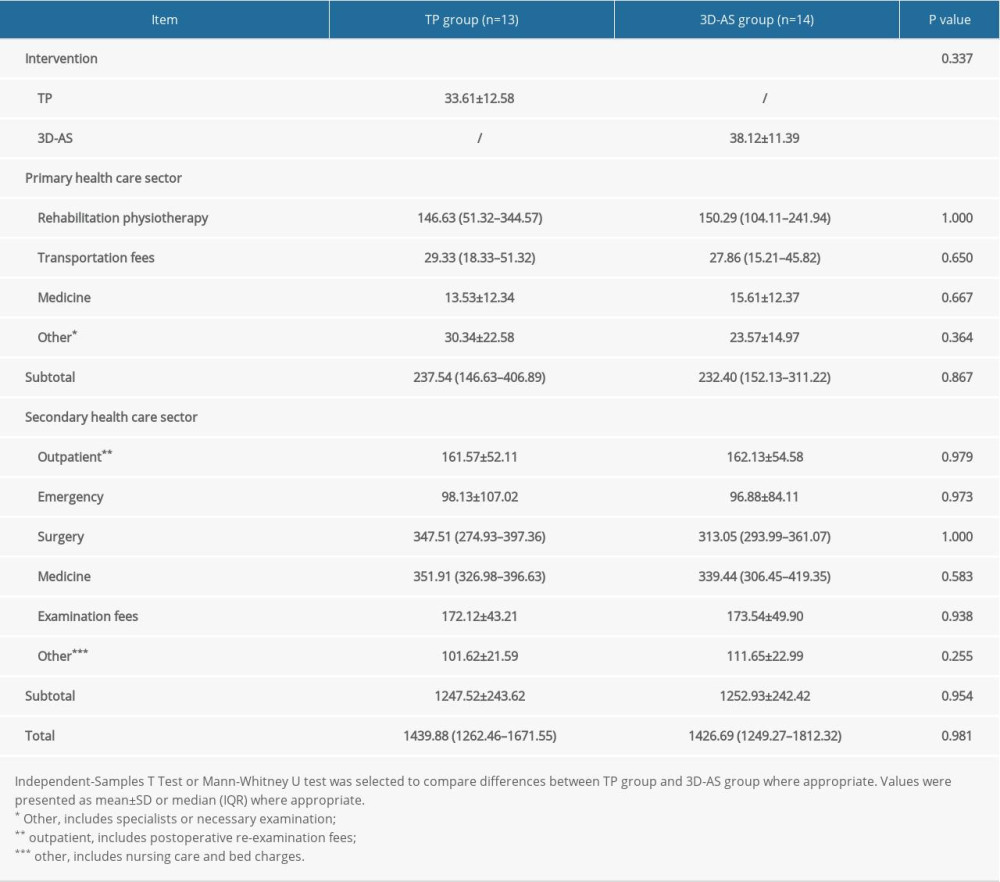 Table 3. Costs of the intervention; primary health care sector and secondary health care sector in the study (in United States dollars).
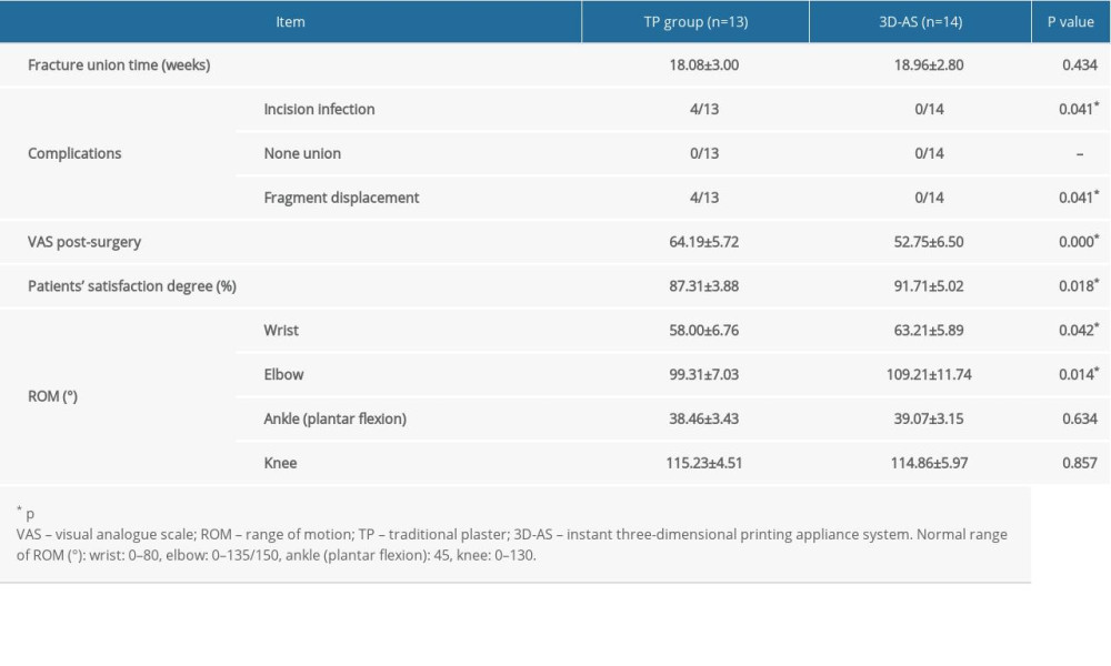 Table 4. Clinical outcomes of the included patients.Table 1. Patient satisfaction survey.Table 2. General characteristics of the included patients.Table 3. Costs of the intervention; primary health care sector and secondary health care sector in the study (in United States dollars).Table 4. Clinical outcomes of the included patients.
 In Press
Clinical Research
Effect of Dexmedetomidine Hydrochloride Nasal Spray on Anxiety and Sleep in Patients Undergoing Gynecologic...Med Sci Monit In Press; DOI: 10.12659/MSM.952465
Clinical Research
Prognostic Value of Mortality Scoring Systems in Patients With Severe Burns: Identifying Key Predictors of ...Med Sci Monit In Press; DOI: 10.12659/MSM.951713
Laboratory Research
Evaluation of the Trueness and Precision of Cast, Milled-Cast, Milled, and 3D-Printed Post-and-Core Techniq...Med Sci Monit In Press; DOI: 10.12659/MSM.953491
Clinical Research
Outcomes After Minimally Invasive Intramedullary Nail Fixation and Locking Plate Fixation Among Patients Wi...Med Sci Monit In Press; DOI: 10.12659/MSM.952670
Most Viewed Current Articles
17 Jan 2024 : Review article 14,176,214
Vaccination Guidelines for Pregnant Women: Addressing COVID-19 and the Omicron VariantDOI :10.12659/MSM.942799
Med Sci Monit 2024; 30:e942799
13 Nov 2021 : Clinical Research 3,757,839
Acceptance of COVID-19 Vaccination and Its Associated Factors Among Cancer Patients Attending the Oncology ...DOI :10.12659/MSM.932788
Med Sci Monit 2021; 27:e932788
14 Dec 2022 : Clinical Research 2,466,153
Prevalence and Variability of Allergen-Specific Immunoglobulin E in Patients with Elevated Tryptase LevelsDOI :10.12659/MSM.937990
Med Sci Monit 2022; 28:e937990
16 May 2023 : Clinical Research 708,809
Electrophysiological Testing for an Auditory Processing Disorder and Reading Performance in 54 School Stude...DOI :10.12659/MSM.940387
Med Sci Monit 2023; 29:e940387