04 March 2022: Clinical Research
Oxidative Properties of Blood-Derived Extracellular Vesicles in 15 Patients After Myocardial Infarction
Vytautas Žėkas1ABCDEG*, Reet Kurg2ADG, Kristiina Kurg2CDEF, Daiva Bironaitė3ABCDE, Mantas RadzevičiusDOI: 10.12659/MSM.935291
Med Sci Monit 2022; 28:e935291


Abstract
BACKGROUND: In this study, we investigated the yield and composition of extracellular vesicles (EVs) derived from 40- to 60-year-old healthy male controls and post-myocardial infarction (post-MI) patients’ blood samples and assessed their pro-inflammatory and oxidative-related properties. Our study aimed to determine the EV yield and composition differences between both groups and to find out if there were differences between EV-mediated oxidative stress reactions.
MATERIAL AND METHODS: Fifteen post-MI patients and 25 healthy individuals were included. EVs were isolated by ultracentrifugation and analyzed using nanotracking analysis (NTA), western blotting and fluorescent flow cytometry (FFC). Oxidative stress (OS) in blood samples was identified by measuring malondialdehyde concentration from serum, while EVs-induced OS was measured in the human vein endothelium cells (HUVEC) using H2DCFDA (2’,7’-dichlorodihydrofluorescein diacetate) fluorescence as a marker.
RESULTS: We found higher EVs concentration in healthy controls than in the post-MI group (7.07±3.1 E+10 ml vs 3.1±1.9 E+10 ml, P<0.001) and a higher level of CD9-positive exosomes (MFI 275±39.5 vs 252±13, P<0.001). Post-MI patients’ EVs carry pro-oxidative nicotinamide adenine dinucleotide phosphate (NADPH) oxidases isoforms NOX1 (NADPH oxidase 1), NOX5 (NADPH oxidase 5) and NOX2 (NADPH oxidase 2) and anti-oxidative thioredoxin, extracellular signal-regulated kinases 1/2 (ERK1/2), and protein kinase B (Akt B). In the post-MI EVs, there was a higher predominance of enzymes with anti-oxidative effects, leading to weaker OS-inducing properties in the HUVEC cells.
CONCLUSIONS: We conclude that post-MI patient blood sample EVs have stronger anti- than pro-oxidative properties and these could help fight against post-MI consequences.
Keywords: atherosclerosis, Blotting, Western, Extracellular Vesicles, Flow Cytometry, Oxidative Stress, Adult, exosomes, Female, Follow-Up Studies, Humans, Male
Background
According to the American Heart Association, cardiovascular diseases (CVDs) remain one of the main causes of death worldwide [1]. CVDs comprise multiple maladies such as coronary artery disease, hypertension, stroke, and peripheral vascular disease. Atherosclerosis (AS) is the main cause of morbidity and mortality in CVD patients [1]. AS is a lipid-driven inflammatory disease of arterial intima in which the balance of pro- and anti-inflammatory mechanisms dictate the final clinical outcome [2]. AS is considered one of the most important pathologies in the modern world but the detection of early changes in the vasculature and prognosis of this disease is still lacking [3]. Oxidative stress plays an important role in atherosclerosis by causing the production of reactive oxygen species (ROS), which are important for the pathogenesis of myocardial infarction (MI) and post-stroke survival [4]. In subclinical AS, oxidative stress can induce endothelial cell activation, permeability changes, recruitment of various inflammatory cells, and other important processes [5]. Combined, these processes eventually facilitate damage to the endothelium and induce the progression of AS [5]. EVs are closely associated with damaging ROS signaling to the endothelium [6]. In CVD patients’ blood, EVs can be released by blood cells, such as platelets, erythrocytes, and leukocytes; and by heart and blood vessel cells such as cardiomyocytes, cardiac fibroblasts, and endothelial cells [7]. In general, EVs are particles released from various types of cells, have a membrane composed of lipid bilayer transmembrane proteins, carry soluble proteins, and do not replicate [7]. EVs have been identified in a diverse range of human biofluids including serum and plasma as well as urine, saliva, breast milk, amniotic fluid, ascites fluid, cerebrospinal fluid, and bile [8–10]. EVs can be classified as exosomes or microvesicles. The term ‘exosome’ is used for vesicles 40–100 nm in size that are released as a consequence of multivesicular endosome fusion with the plasma membrane, while ‘microvesicle’ refers to EVs that are generally up to ~1000 nm in diameter and are shed from the plasma membrane [8,11,12]. Small vesicles of 100 nm may also bud from the cell surface [13]. Membrane vesicles originate from endosomal and plasma membranes and are released into the extracellular environment by various cells [8]. They have an important role in intercellular communication, carrying cytosolic proteins, lipids, and RNA [8]. A systematic review compared studies on the cardioprotection effect of EVs [14], showing that EVs from the investigated sources mediated the protection using different pathways by carrying proteins or nucleic acids responsible for cell survival. For instance, in an oxidative stress environment, EVs from cardiac progenitor cells were found in greater quantity and at a higher count and carrying different cargo than control [14]. When researching the effects of EVs on cardiomyocytes from MI patients and donors, only donor EVs had positive protective effects in preventing cell death. The authors of the aforementioned systemic review showed that EVs cardioprotective effects observed in different studies from different sources are inconsistent. It would seem that EVs, in general, are protective, but it is nearly impossible to properly compare the studies because of differences in purification methods and the EVs experiments themselves [14], which is a problem that calls for the standardization of research techniques. The ultimate goal would be to use the EVs as a therapeutic or diagnostic tool in routine clinical work. Serum and plasma are attractive sources for EV isolation since blood sampling is a minimally invasive procedure [15]. However, the isolation and purification of EVs from human blood are complicated due to high serum viscosity, high abundance of various proteins, and non-EV lipid particles [10]. All of these serum features significantly impact the yield of EVs from human serum [10]. Currently, available purification methods do not allow full discrimination between exosomes and microvesicles, and, as such, purified EVs might include both fractions [16]. However, there are some cell surface markers, such as CD9, CD63, and CD81, which are thought to ensure the recognition of exosomes [17].
Ultracentrifugation (UC) is a classical method for EV purification yielding a large amount of EVs [10]. The most common methods for distinguishing EVs based on their size are scanning and transmission electron microscopy; atomic force microscopy, which is based on EV surfaces imaging; dynamic light scattering, which is based on fluctuations of scattered light intensity; and nanoparticle tracking analysis (NTA), which directly visualizes individual particles by scattering of illuminated laser beam [16]. EV detection methods based on their surface markers, such as immunoaffinity-capture by beads, enzyme-linked immunosorbent assay (ELISA), and conventional flow cytometry, can be also used to analyze EV subpopulations expressing specific antigens [18].
In this study, we isolated EVs, mainly exosomes, from blood samples from 40- to 60-year-old men who were either healthy (healthy controls) or had previously had a myocardial infarction (post-MI group). We investigated the EV quantity, composition, and oxidative stress-related properties in human endothelial cell lines (HUVEC) for both isolated EV groups.
Our study aimed to determine the differences in the quantity and composition of EV between healthy and post-MI patients and to investigate possible differences in EV-mediated oxidative stress reactions between the 2 groups.
Material and Methods
PATIENTS:
The study protocol was approved by the local Vilnius University Bioethics Committee (No.: 158200-18-990-495 and 158200-15-807-319). All procedures followed were in accordance with the ethical standards of the responsible committee on human experimentation (institutional and national) and with the Helsinki Declaration of 1964 and its later amendments. Informed consent was obtained from all patients for inclusion in the study.
HEALTHY SUBJECTS:
Healthy 40- to 60-year-old men were examined between December 2015 and July 2017. Healthy subjects had no prior history of acute cardiovascular disease and were not under any treatment for cardiovascular diseases.
POST-MI GROUP:
The test group included 15 men who had been treated for MI either at the Cardiac Intensive Care or at the Intensive Care hospital units, or the first cardiology ward of Vilnius University Santaros Klinikos Hospital 3 months prior to inclusion. Patients were enrolled between February and May of 2018 and were 40 to 60 years old, had had MI for the first time in their life, and coronary angiography confirmed coronary occlusion of more than 50% in at least 2 arteries. Coronary catheterization and coronary angioplasty were performed on all patients. At the time of blood collection, patients were under antihypertensive treatment and received aspirin and statins. Twelve patients had primary arterial hypertension. Altogether, only 1 patient had systemic inflammation, diabetes mellitus, and primary arterial hypertension at the same time.
BLOOD COLLECTION:
We drew 4 ml of blood into plasma sodium citrate tubes. To produce platelet-free plasma, the plasma tubes were then subjected to differential centrifugation. Plasma tubes were centrifuged at 3000 g for 10 min, and three-quarters of recovered plasma was transferred into a sterile tube and centrifuged again at 10 000 g for 30 min. After centrifuging, the plasma was transferred to separate tubes and kept at −80°C until ultracentrifugation. The post-MI patients’ blood was collected 3 months after the MI, which is a regular follow-up period.
ULTRACENTRIFUGATION:
The frozen plasma was thawed and up to a 100 μl of each blood plasma sample was diluted to a total volume of 1 ml with PBS (phosphate-buffered saline, pH=7.4) and samples were centrifuged at 1200 g for 30 min at 4°C (Pico 21, Thermo Scientific) to remove cells and debris. Then, 3 ml of PBS was added to the supernatant and it was further centrifuged at 120 000 g for 1.5 h at 4°C using the Optima™ L-90K Ultracentrifuge with installed rotor SW55Ti. The EVs were re-suspended in 300 μl of Dulbecco’s PBS (DPBS) (Sigma-Aldrich). During the purification of exosome samples, some material could be lost, and there was no procedure in place to assure compensation of the loss of material during purification. The Bradford protein assay (Bio-Rad Laboratories; USA) was used to measure the concentration of EVs in each sample. BSA (bovine serum albumin) was used as a standard.
CLINICAL CHEMISTRY ANALYSIS:
The concentrations of C reactive protein (CRP), glucose, total cholesterol, triglycerides (TG), high-density lipoprotein cholesterol (HDL-C), and low-density lipoprotein cholesterol (LDL-C) were measured from patients’ and healthy subjects’ blood using routine techniques of a clinical chemistry laboratory (Architect ci8200, Abbott, Chicago, Illinois).
MALONDIALDEHYDE:
Malondialdehyde (MDA) is a marker of lipid peroxidation and is often used to represent the level of oxidative stress in biological samples [19]. We mixed 50 μl of thawed serum with 750 μl of 0.44 M phosphoric acid solution, 250 μl of 42 mM thiobarbituric acid solution, and 450 μl of deionized water. The prepared samples were incubated at 100°C for 60 min. After incubation, the samples were rapidly cooled in an ice bath, and 500 μl of the sample was diluted in a methanol solution (1: 1). The blood serum sample was mixed by shaking and then centrifuged for 3 min at 10 000 g. After centrifugation, 500 μl of the centrifuged serum sample was added to the chromatographic vial, and serum sample was analyzed via the high-performance liquid chromatography (HPLC) method. Malondialdehyde concentration was measured by a Shimadzu Nexera X2 UHPLC system (Shimadzu). Data were collected and processed using LabSolutions software (Shimadzu).
NANOPARTICLE TRACKING ANALYSIS:
Nanoparticle tracking analysis (NTA) was performed with a ZetaView nanoparticle analyzer (Particle Metrix GmbH; Germany). The 102-nm polystyrene beads were used to calibrate the apparatus before each session. We recorded 11 measurements for each case twice and the results were averaged in at least 1 dilution in Dulbecco’s PBS (Corning, Inc., NY, USA). ZetaView Software 8.04.02 (Particle Metrix GmbH) was used to analyze images, settings were set to default image evaluation, and we used the following camera acquisition parameters: sensitivity set to 85, shutter set to 70, and frame rate set to 30. Each sample was measured 3 times. Concentrations of the samples of extracellular vesicles were expressed as the number of EVs per 1 ml of diluents; in this case, PBS.
FLUORESCENT FLOW CYTOMETRY:
For the identification of EVs with special surface antibodies by fluorescent flow cytometry (FFC) analysis, we used the method described by Mellisho et al [20]. EV sample volume was adjusted to the total protein concentration. A vial containing only PBS was used as a negative control. To all EV samples and the negative control, 5 μl of latex beads was added and left overnight at 4°C. In the morning, 1 μl of 2M glycine was added to each EV sample and the negative control vial and kept for 45 min at room temperature. Then, 2 washing steps at 4000 rpm for 5 min at 4°C were performed using a 0.5% PBS/BSA blocking solution, and EV samples were incubated for 20 min at room temperature. EV samples were incubated with antibodies for 1 h at room temperature and centrifuged once at 4000 rpm for 5 min at 4°C. The supernatant was removed until 100 μl was left in the tubes; then, 400 μl of PBS was added and the pellet was carefully re-suspended. This solution was tested on an LSRII flow cytometer (BD, San Jose, California).
WESTERN BLOTTING:
For immunoblot analysis, all samples were first lysed in sodium dodecyl sulfate (SDS)-sample buffer, heated at 100°C for 5 min, and loaded onto 10% sodium dodecyl sulfate-polyacrylamide gel (SDS-PAGE). After electrophoresis, the proteins were transferred onto a polyvinylidene difluoride (PVDF) membrane (Millipore Corp.) and blocked for 30 min overnight with 2% non-fat dry milk or 3% bovine serum albumin (BSA) in TBST (Tris-Buffered Saline Tween 20) buffer (20 mM Tris pH 7.5, 0.15 M NaCl, 0.01% Tween20). The primary antibodies used were anti-TSG101 (tumor susceptibility gene 101) (Abcam, Germany) and anti-thioredoxin (Abcam, Germany). The concentration of primary antibody was always 1: 1000. Secondary goat anti-rabbit antibody (LabAS, Tartu, Estonia) was used at the dilution of 1: 10 000. The signal was visualized by adding the enhanced chemiluminescence (ECL) substrate (Amersham ECL Western Blotting Detection Reagents) for 1 min, and the film was exposed for 10 min, after which it was scanned in using Epson Expressions 1680 software. Two healthy control samples and 2 post-MI samples were chosen for further analysis to detect NADPH (nicotinamide adenine dinucleotide phosphate) oxidases (NOX), protein kinase B (Akt B), and extracellular signal-regulated kinases 1/2 (ERK 1/2) with the primary antibody being anti-NOX2 (Abcam, Germany), anti-NOX5 (ThermoFisher, USA), anti-NOX1 (ThermoFisher, USA), anti-Akt B (ThermoFisher, USA), and anti-ERK 1/2 (ThermoFisher, USA). All other western blot steps were done in the same manner as before.
OXIDATIVE STRESS IN CELL CULTURE:
To determine the oxidative stress in the cell culture, we used the method described by Wei et al [21]. EVs were added to the pooled HUVEC cells, which were at up to the 5th passage (Corning, USA, catalog number: 354151) and incubated for 20 h. EV concentration was equalized in each sample of HUVEC cells by using NTA data. After the incubation, every sample was stained with 5 μM H2DCFDA (2′,7′-dichlorodihydrofluorescein diacetate) in the dark for 30 min. After staining, cells were lifted with trypsin and washed once with complete medium EGM-2 BulletKit (Lonza, Germany) and centrifuged at 180 g for 7 min. After washing, samples were measured with flow cytometer FACS Canto II (BD, USA) using FITC (fluorescein isothiocyanate) channel at excitation 490 nm and emission 520 nm. Collected data was analyzed using FACS Diva software. Data showed that post-MI EVs on average gave less H2DCFDA fluorescence intensity in the sample when compared to EVs of the healthy group.
STATISTICAL ANALYSIS:
Variables were tested by the paired non-parametric Wilcoxon test and Spearman test for the correlation coefficient. The confidence level for the test was set at 0.05; all
Results
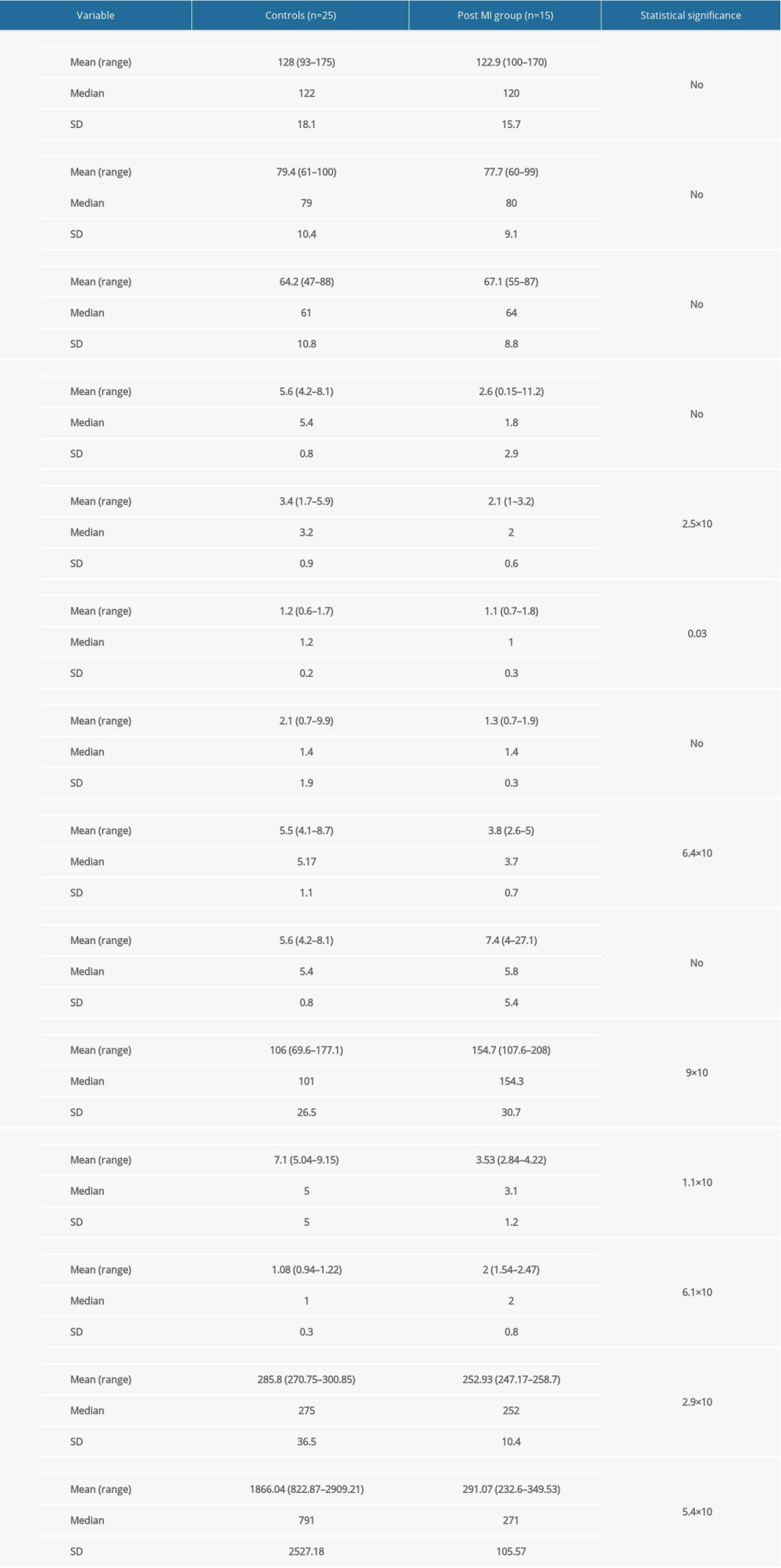
Post-MI patients had a significantly elevated concentration of blood serum MDA (106±26.5 vs 154.7±30.7, P<0.001) and had lower levels of low-density lipoprotein cholesterol (3.4±0.9 vs 2.1±0.6, P<0.001), high-density lipoprotein cholesterol (1.2±0.2 vs 1.1±0.3, P=0.03), and total cholesterol concentrations (5.5±1.1 vs 3.8±0.7, P<0.001) compared to healthy controls (Table 1).
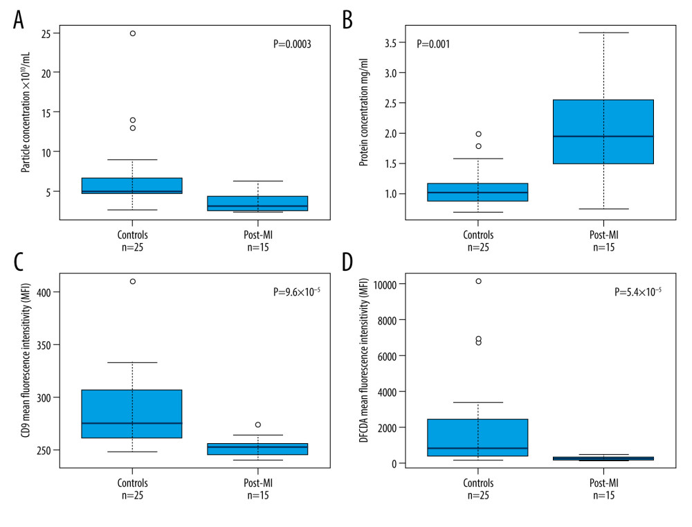
According to nanoparticle tracking analysis (Zeta View, Particle Metrix, Germany), there was a statistically significant difference in blood particle concentration between the healthy control group and the post-MI group (7.07±3.1×10¹⁰/ml vs 3.1±1.9×10¹⁰/ml,
The CD9 level on exosome membranes was tested by FFC (Figure 1C). Comparing CD9 median fluorescence intensity (MFI) and the percentage of positive events revealed that healthy human blood samples had a higher number of CD9-positive exosomes than post-MI group individuals (MFI 275±39.5 vs 252±13,
To determine anti-oxidative EV properties, thioredoxin level was identified by western blotting. Thioredoxin was detected in almost all samples, but at a noticeably higher level in post-MI EV samples than in healthy individuals. Similar results were obtained with the cell survival-regulating enzymes: extracellular signal kinases 1/2 (ERK 1/2) and protein kinase B (Akt B) were detected in all EV samples, but were found at higher levels in post-MI EVs samples.
The investigation of pro-oxidative NADPH oxidases (NOX1, NOX2, and NOX5 isoforms) in EV samples showed a strong upregulation of all 3 NOX isoforms in post-MI EVs, but only NOX1 and NOX5 isoforms were obtained in the healthy EVs. Oxidative stress testing of isolated EVs was done by adding them to the endothelial cell culture (HUVEC) and measuring the production of ROS through H2DCFDA fluorescence intensity. The negative (using cell growth medium) and positive controls (using 1M hydrogen peroxide) were used to define and normalize the results. Data showed that post-MI patient exosomes gave a lower oxidative stress response than the EVs of the healthy group, since post-MI exosomes on average gave less H2DCFDA fluorescence intensity in the sample when compared to the healthy group (
Discussion
LIMITATIONS OF THE STUDY:
Since this is a pilot study, only a small sample of patients was collected. We plan to conduct further research based on the present study, but with a larger sample size and including both men and women, which could generate more accurate results. Another limitation is the implementation of the data collection method. There was no procedure to prevent material loss during the purification of exosomes. The ultracentrifugation method used here is known for losing sample material during purification. But we chose this method since the idea was to use the least expensive and most easily accessible method that could be employed in the clinical laboratory.
Conclusions
We compared biochemical markers (the yield and inflammatory and oxidative properties of EVs) isolated from both healthy controls and post-MI patients’ blood samples. The isolated EV yield from post-MI patients was lower when compared to healthy controls. Despite the inflammatory origin of post-MI EVs, which was determined by showing a higher level of MDA in post-MI patients’ blood, and as they carried pro-oxidant enzymes NOX1, NOX5, and NOX2, post-MI EVs caused less oxidative stress in endothelial cell culture experiments. Higher levels and better release of thioredoxin, ERK1/2, and Akt B might have strongly contributed to this effect. We conclude that there are quantitative and qualitative differences between the EVs of healthy vs post-MI individuals. Healthy controls EVs, as well as post-MI patient EVs, carry both pro-oxidant and anti-oxidant enzymes, but post-MI EVs have a stronger anti-oxidative effect on the endothelium, which could help improve the post-MI condition.
References
1. Benjamin EJ, Muntner P, Alonso AAmerican Heart Association Council on Epidemiology and Prevention Statistics Committee and Stroke Statistics Subcommittee, Heart Disease and Stroke Statistics-2019 Update: A Report From the American Heart Association: Circulation, 2019; 139(10); e56-e528
2. Bäck M, Yurdagul A, Tabas I, Inflammation and its resolution in atherosclerosis: Mediators and therapeutic opportunities: Nat Rev Cardiol, 2019; 16(7); 389-406
3. Wang J, Tan GJ, Han LN, Novel biomarkers for cardiovascular risk prediction: J Geriatr Cardiol, 2017; 14(2); 135-50
4. Moris D, Spartalis M, Spartalis E, The role of reactive oxygen species in the pathophysiology of cardiovascular diseases and the clinical significance of myocardial redox: Ann Transl Med, 2017; 5(16); 326
5. Burtenshaw D, Kitching M, Redmond EM, Reactive oxygen species (ROS), intimal thickening, and subclinical atherosclerotic disease: Front Cardiovasc Med, 2019; 6; 89
6. Saeed-Zidane M, Linden L, Salilew-Wondim D, Cellular and exosome mediated molecular defense mechanism in bovine granulosa cells exposed to oxidative stress: PLoS One, 2017; 12(11); e0187569
7. de Freitas RCC, Hirata RDC, Hirata MH, Aikawa E, Circulating extracellular vesicles as biomarkers and drug delivery vehicles in cardiovascular diseases: Biomolecules, 2021; 11(3); 388
8. Raposo G, Stoorvogel W, Extracellular vesicles: Exosomes, microvesicles, and friends: J Cell Biol, 2013; 200(4); 373-83
9. Théry C, Amigorena S, Raposo G, Clayton A, Isolation and characterization of exosomes from cell culture supernatants and biological fluids: Curr Protoc Cell Biol, 2006; Chapter 3(Unit 3); 22
10. Brennan K, Martin K, FitzGerald SP, A comparison of methods for the isolation and separation of extracellular vesicles from protein and lipid particles in human serum: Sci Rep, 2020; 10(1); 1039
11. Harding C, Heuser J, Stahl P, Endocytosis and intracellular processing of transferrin and colloidal gold-transferrin in rat reticulocytes: Demonstration of a pathway for receptor shedding: Eur J Cell Biol, 1984; 35(2); 256-63
12. Pan BT, Teng K, Wu C, Electron microscopic evidence for externalization of the transferrin receptor in vesicular form in sheep reticulocytes: J Cell Biol, 1985; 101(3); 942-48
13. Booth AM, Fang Y, Fallon JK, Exosomes and HIV Gag bud from endosome-like domains of the T cell plasma membrane: J Cell Biol, 2006; 172(6); 923-35
14. Wendt S, Goetzenich A, Goettsch C, Evaluation of the cardioprotective potential of extracellular vesicles – a systematic review and meta-analysis: Sci Rep, 2018; 8(1); 15702
15. Perakis S, Speicher MR, Emerging concepts in liquid biopsies: BMC Med, 2017; 15(1); 75
16. Rupert DLM, Claudio V, Lässer C, Bally M, Methods for the physical characterization and quantification of extracellular vesicles in biological samples: Biochim Biophys Acta Gen Subj, 2017; 1861(1 Pt A); 3164-79
17. Corrado C, Raimondo S, Chiesi A, Exosomes as intercellular signaling organelles involved in health and disease: Basic science and clinical applications: Int J Mol Sci, 2013; 14(3); 5338-66
18. Kowal J, Arras G, Colombo M, Proteomic comparison defines novel markers to characterize heterogeneous populations of extracellular vesicle subtypes: Proc Natl Acad Sci USA, 2016; 113(8); E968-77
19. Khoschsorur G, Winklhofer-Roob BM, Rabl H, Evaluation of a sensitive HPLC method for the determination of Malondialdehyde, and application of the method to different biological materials: Chromatographia, 2000; 52; 181-84
20. Mellisho EA, Velásquez AE, Nuñez MJ, Identification and characteristics of extracellular vesicles from bovine blastocysts produced in vitro: PLoS One, 2017; 12(5); e0178306
21. Wei F, Wang A, Wang Q, Plasma endothelial cells-derived extracellular vesicles promote wound healing in diabetes through YAP and the PI3K/Akt/mTOR pathway: Aging (Albany NY), 2020; 12(12); 12002-18
22. Ross R, Atherosclerosis – an inflammatory disease: N Engl J Med, 1999; 340(2); 115-26
23. Stocker R, Keaney JF, Role of oxidative modifications in atherosclerosis: Physiol Rev, 2004; 84(4); 1381-478
24. Papac-Milicevic N, Busch CJ, Binder CJ, Malondialdehyde epitopes as targets of immunity and the implications for atherosclerosis: Adv Immunol, 2016; 131; 1-59
25. Sproston NR, Ashworth JJ, Role of C-reactive protein at sites of inflammation and infection: Front Immunol, 2018; 9; 754
26. Berk BC, Weintraub WS, Alexander RW, Elevation of C-reactive protein in “active” coronary artery disease: Am J Cardiol, 1990; 65(3); 168-72
27. Rautou PE, Vion AC, Amabile N, Microparticles, vascular function, and atherothrombosis: Circ Res, 2011; 109(5); 593-606
28. Puhm F, Boilard E, Machlus KR, Platelet extracellular vesicles: Beyond the blood: Arterioscler Thromb Vasc Biol, 2021; 41(1); 87-96
29. Davidson SM, Andreadou I, Barile L, Circulating blood cells and extracellular vesicles in acute cardioprotection: Cardiovasc Res, 2019; 115(7); 1156-66
30. Deddens JC, Vrijsen KR, Colijn JM, Circulating extracellular vesicles contain miRNAs and are released as early biomarkers for cardiac injury: J Cardiovasc Transl Res, 2016; 9(4); 291-301
31. Ge X, Meng Q, Zhuang R, Circular RNA expression alterations in extracellular vesicles isolated from murine heart post ischemia/reperfusion injury: Int J Cardiol, 2019; 296; 136-40
32. Ramkumar S, Raghunath A, Raghunath S, Statin therapy: Review of safety and potential side effects: Acta Cardiol Sin, 2016; 32(6); 631-39
33. Suades R, Padró T, Alonso R, Lipid-lowering therapy with statins reduces microparticle shedding from endothelium, platelets and inflammatory cells: Thromb Haemost, 2013; 110(2); 366-77
34. Kulshreshtha A, Singh S, Ahmad M, Simvastatin mediates inhibition of exosome synthesis, localization and secretion via multicomponent interventions: Sci Rep, 2019; 9(1); 16373
35. Mobarrez F, Egberg N, Antovic J, Release of endothelial microparticles in vivo during atorvastatin treatment; A randomized double-blind placebo-controlled study: Thromb Res, 2012; 129(1); 95-97
36. Zu L, Ren C, Pan B, Endothelial microparticles after antihypertensive and lipid-lowering therapy inhibit the adhesion of monocytes to endothelial cells: Int J Cardiol, 2016; 202; 756-59
37. Arslan F, Lai RC, Smeets MB, Mesenchymal stem cell-derived exosomes increase ATP levels, decrease oxidative stress and activate PI3K/Akt pathway to enhance myocardial viability and prevent adverse remodeling after myocardial ischemia/reperfusion injury: Stem Cell Res, 2013; 10(3); 301-12
38. Tan Y, Cheng H, Su C, PI3K/Akt signaling pathway ameliorates oxidative stress-induced apoptosis upon manganese exposure in PC12 cells: Biol Trace Elem Res, 2021; 200(2); 749-60
39. Madrigal-Matute J, Fernandez-Garcia CE, Blanco-Colio LM, Thioredoxin-1/peroxiredoxin-1 as sensors of oxidative stress mediated by NADPH oxidase activity in atherosclerosis: Free Radic Biol Med, 2015; 86; 352-61
40. Poznyak AV, Grechko AV, Orekhova VA, NADPH oxidases and their role in atherosclerosis: Biomedicines, 2020; 8(7); 206
41. Fan LM, Douglas G, Bendall JK, Endothelial cell-specific reactive oxygen species production increases susceptibility to aortic dissection: Circulation, 2014; 129(25); 2661-72
42. Rosc-Schlüter BI, Häuselmann SP, Lorenz V, NOX2-derived reactive oxygen species are crucial for CD29-induced pro-survival signalling in cardiomyocytes: Cardiovasc Res, 2012; 93(3); 454-62
Tables
In Press
Clinical Research
Comparative Effectiveness of a Nurse-Led Care Model vs Usual Care in Rheumatoid Arthritis: A Longitudinal C...Med Sci Monit In Press; DOI: 10.12659/MSM.953211
Clinical Research
Impact of Treatment Modality on Pain, Sexual Function, and Psychological Well-Being in Patients With Bartho...Med Sci Monit In Press; DOI: 10.12659/MSM.952422
Clinical Research
Association Between Radiographic Knee Osteoarthritis, Pre-Fracture Mobility, and Hip Fracture Patterns in O...Med Sci Monit In Press; DOI: 10.12659/MSM.952678
Clinical Research
Association Between Total Cholesterol–to–High-Density Lipoprotein Ratio and Gestational Hypertension: A Cas...Med Sci Monit In Press; DOI: 10.12659/MSM.952395
Most Viewed Current Articles
17 Jan 2024 : Review article 14,176,084
Vaccination Guidelines for Pregnant Women: Addressing COVID-19 and the Omicron VariantDOI :10.12659/MSM.942799
Med Sci Monit 2024; 30:e942799
13 Nov 2021 : Clinical Research 3,757,530
Acceptance of COVID-19 Vaccination and Its Associated Factors Among Cancer Patients Attending the Oncology ...DOI :10.12659/MSM.932788
Med Sci Monit 2021; 27:e932788
14 Dec 2022 : Clinical Research 2,466,116
Prevalence and Variability of Allergen-Specific Immunoglobulin E in Patients with Elevated Tryptase LevelsDOI :10.12659/MSM.937990
Med Sci Monit 2022; 28:e937990
16 May 2023 : Clinical Research 708,768
Electrophysiological Testing for an Auditory Processing Disorder and Reading Performance in 54 School Stude...DOI :10.12659/MSM.940387
Med Sci Monit 2023; 29:e940387