02 March 2022: Clinical Research
A Radiographic Study of the Root-to-Crown Ratio of Natural Permanent Teeth in 81 Saudi Adults
Abdulelah Sameer Sindi1ADEF, Fuad Al Sanabani23ADFG, Bandar M.A. Al-Makramani23ABCDE, Khurshid Mattoo2ABDE*, Hafiz Adawi2BDEG, Hosain Al-Mansour4CDEF, Fahad M. Albakri5BDE, Mohammed M. Al Moaleem2BDEG, Mohamed Sobhy2ADEF, Hatem Abdu Humadi6BCDE, Mossab Ahmad Hamzi6BCDE, Essa Mosa Agili6BCDE, Shailesh JainDOI: 10.12659/MSM.936085
Med Sci Monit 2022; 28:e936085
Abstract
BACKGROUND: The ratio between a tooth root and its crown is an essential diagnostic parameter that determines treatment options. This radiographic study used panoramic dental radiographs or orthopantograms to measure the mean root (R)-to-crown (C) ratios (R/C) of the permanent teeth in 81 Saudi adults.
MATERIAL AND METHODS: A total of 81 panoramic radiographs of Saudi adult patients (40 males and 41 females) previously treated in the College of Dentistry, Saudi Arabia, aged 16-35 years, were selected. The crown height and root length for each tooth were measured on the digital panoramic radiographs. The correlation coefficient (intra-class) was calculated to assess the intra-examiner reproducibility and a good agreement was achieved (ICC=0.79-0.89).
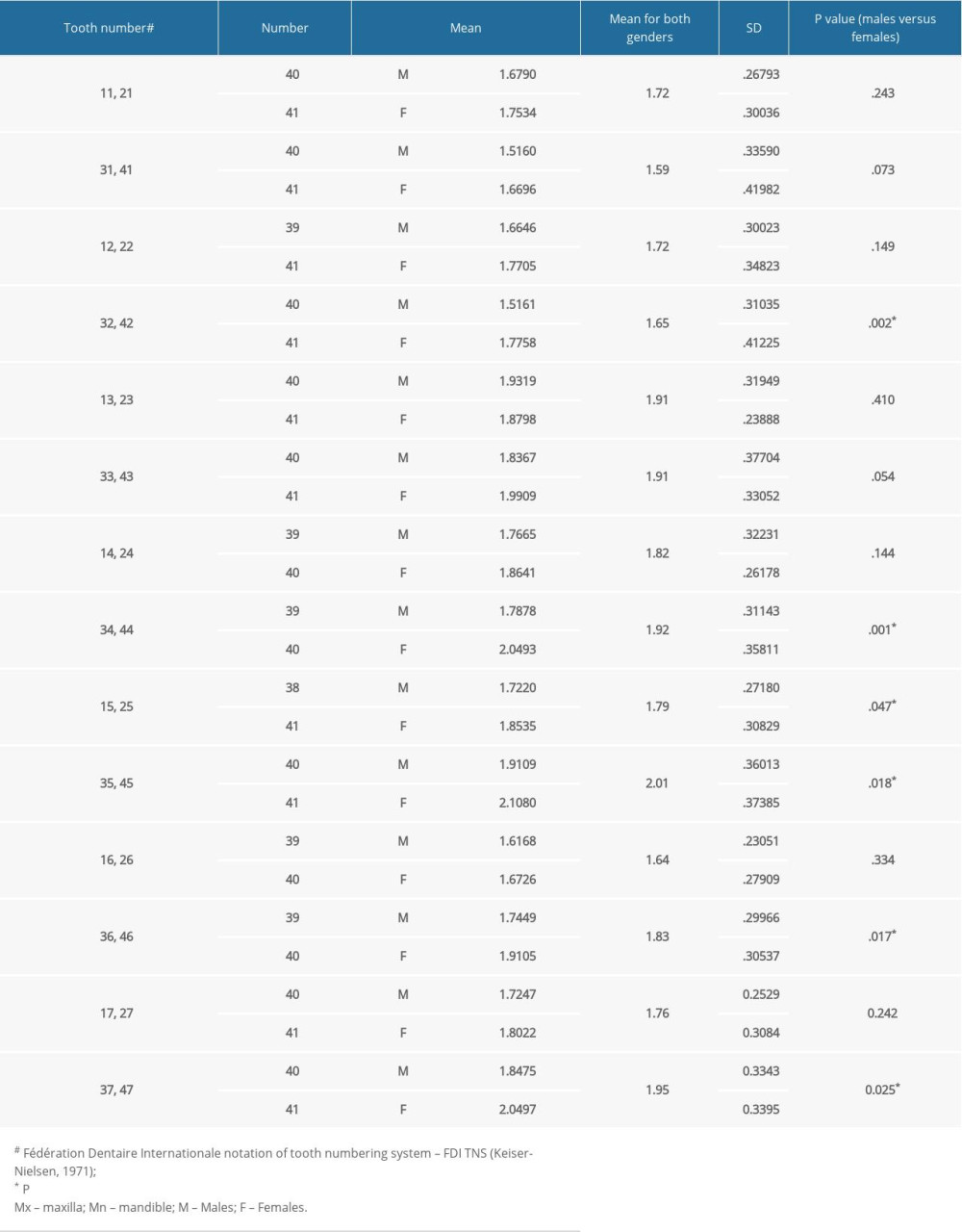
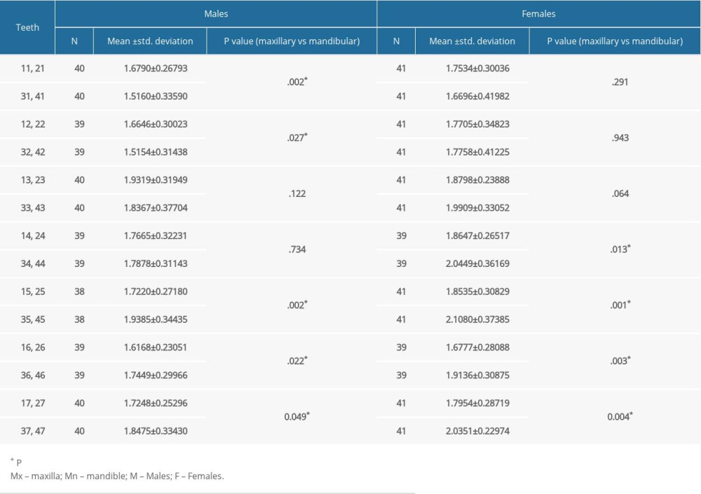
RESULTS: For both males and females, the highest mean R/C ratio was for maxillary canine (1.91) and for mandibular second premolar (2.0) while the lowest R/C ratio was for maxillary first molar (1.64) and for mandibular central incisor (1.59). Except for the maxillary second premolar, no significant differences in R/C ratios were observed for maxillary arch. In the mandibular arch, the R/C ratio for lateral incisor, both premolars, and molars differed significantly (P<0.05). Among males, statistically significant differences between teeth existed in R/C ratios for central and lateral incisors, second premolar, and both molars (P>0.05). For females, significant differences between teeth in R/C ratios were observed for both premolars and both molars (P>0.05).
CONCLUSIONS: This study supports the findings from previous studies that orthopantograms can be used to calculate root/crown ratios, which varies between males and females and the dental arch among Saudi adults.
Keywords: Radiography, Bitewing, Radiographic Image Interpretation, Computer-Assisted, Dental Abutments, Periodontium, Adolescent, Adult, Dentition, Permanent, Female, Follow-Up Studies, Humans, Male, Radiography, Panoramic, Reproducibility of Results, Saudi Arabia, Tooth Crown, young adult
Background
The formulation of a competent fixed prosthodontic treatment plan for the restoration and/or replacement of lost natural teeth is the foundation for long-lasting service of dental prostheses. A plethora of factors affect the selection of a fixed dental prosthesis (FDPs), including the condition of the prospective abutment, biomechanics, esthetic requirements, the patient’s desire, economics, clinician’s skills, laboratory support, and the patient’s motivation and anticipated cooperation [1]. The supporting abutment teeth and the overlying FDP should have acceptable mesial and distal wall heights to counteract the dislodging horizontal/vertical/rotational forces [2]. Conversely, single-unit crowns are more likely to dislodge in a facial-to-lingual/palatal direction. Teeth with short clinical crowns may not be an ideal abutment. The use of intentional surgery (crown lengthening), increasing vertical dimension, or multiple abutments in such cases must be considered [3].
The extent of the roots within the supporting tissues (alveolar bone) must be evaluated for many factors, especially the root/crown (R/C) ratio. The precise valuation of the R/C ratio is an important parameter during diagnosis and selection of the potential abutments for different types of prosthetic restorations, either FDPs or removable partial dentures (RPDs) [4]. On the contrary, the term crown height space is used to describe alterations in the implant/abutment ratio, which are to be considered while designing an implant restoration. The R/C ratio determines the root length embedded in the alveolar bone, relative to crown length, which exists outside the alveolar bone as determined radiographically [5]. The R/C ratio represents the biomechanical equivalent of the principles of the lever that categorizes and differentiates a particular tooth into a class I lever system. The portion of the tooth inside the bone (alveolar) is the resistant arm, the portion of tooth outside alveolar bone is the effort arm and the fulcrum or center of rotation of class I lever is that portion (point) of the root where the root is embedded in the alveolar bone [6,7]. If the fulcrum of the class I lever moves apically as a result of alveolar bone loss, the crown length (effort arm) will be increased and the root length (resistance arm) will be decreased, and the tooth will thus be more vulnerable to the harmful effect of lateral forces [8].
Knowing the R/C ratios of normal dentition that has been assessed objectively (radiographs) acts as a reference for various dental estimations, which are essential for designing dental prosthesis, carrying tooth movement for orthodontic corrections, and correcting skeletal discrepancies during orthognathic procedures. The estimation of the accurate R/C ratio in partially edentulous situations can be the difference between saving a tooth or extracting the same in case of tooth-supported overdentures [9]. In summary, knowledge about the normal R/C ratio will help in increasing the longevity and selecting the most appropriate prosthetic abutment. It can be used by clinicians as a prognostic index for predicting the outcome of their treatment. It has been found that the magnification factor of digital panoramic radiograph on the R/C ratio is insignificant because crowns and roots of teeth are frequently in the similar vertical plane [10]. Furthermore, the R/C ratio can be assessed in digital panoramic radiograph with acceptable reproducibility to regulate the advancement of root resorption during orthodontic corrections [11]. Alterations in R/C ratio take place during orthodontic tooth movements like up-righting. Keeping track of changes in the R/C ratio through radiographs has been considered to be a reliable method [11]. The advantage of image quality and clarity in the digital panoramic system over conventional panoramic radiographs is well established [12]. Using software and digital panoramic radiographs, measurements between various landmarks can be easily done at chairside without consuming much time or resources. Several studies have evaluated R/C ratios, but have shown conflicting results for different populations. A study in the Egyptian population found no significant difference between R/C ratios of right and left teeth for males and females [13]. In the Malay population, R/C ratios for males and females were similar but were significantly different to that of the antagonist teeth [14]. The range of R/C ratios in the Korean population also was found to vary between males and females, [15] while in the Iranian population, the R/C ratios were similar [16]. These studies show that while there may be less variations between males and females, the variations in R/C ratio may vary within the arch and between a single type or group of teeth. There is no previously published study from the Kingdom of Saudi Arabia that assessed the absolute or relative values of the R/C ratio of natural permanent teeth. Hence, the aim of this study was to assess R/C ratios in males and female and assess variations in the R/C ratio by sex, tooth type, and dental arch. The main objectives of the study were: to determine the average values of root to crown ratios using digital panoramic radiographs in Saudi adults (southern red coast); to determine the variation of root to crown ratios by sex; and to determine the variation of root/crown ratios of individual tooth type and against its antagonist tooth type. Hence, this radiographic study used digital panoramic dental radiographs to measure the root/crown ratio of the permanent teeth in 81 Saudi adults.
Material and Methods
ETHICS:
The present clinical study was duly approved by the Ethics Committee of the College of Dentistry and its affiliated university (vide Reference No. CODJU-1718I) as part of student research under the deanship of academic research. Any research conducted on humans and animals is performed with strict adherence to the standards laid down in the Helsinki declaration. All subjects who undergo their treatment in the college hospital are required to give a written informed consent before any diagnostic or treatment procedure is started.
STUDY DESIGN:
This study was performed between year 2019 and 2020 at one of the accredited universities of the Kingdom. The study used a retrospective, exploratory, non-interventional approach on a cross-sectional population sample that had reported to the Outpatient Department of the college.
SAMPLE PREPARATION, SELECTION, AND GROUPING:
A total of 81 digital panoramic radiographs of Saudi adult patients (40 males and 41 females), aged 16–35 years were selected based on the following inclusion/exclusion criteria. Most permanent natural dentitions are intact until the age of 35 years, after which tooth loss results in occlusal changes (supraeruption, mesial migration), thereby altering the R/C ratio. Sample-size requirements (reliability, flexibility, and efficiency) for research were determined to be 81 (±5% accuracy and an alpha of 0.05 [95% confidence interval]). The inclusion criteria were: cases with complete case history details; normal, healthy, permanent natural dentition without any evidence of previous/existing malocclusion, with an ideal class 1 molar and canine relationship; no evidence of proximal and/or extensive caries or restorations; no evidence of attrition; no history of developmental disturbances in deciduous/permanent natural dentition; good-to-excellent-quality radiographs; and no evidence of periodontal disease. These criteria were used to minimize the effect of alterations in the R/C ratio, which can even be caused by large untreated proximal caries. An essential criterion that was the basis of selection of cases was the contact between adjacent teeth, which is important to maintaining the natural R/C ratio. Exclusion criteria were: history of previous orthodontic treatment (with or without orthognathic surgery), previous surgery, maxillofacial trauma, crowding or spacing, soft and hard tissue pathology, intrabony lesions attached to root, and hypercementosis or dilacerations.
MEASUREMENTS AND DATA EVALUATION, COLLECTION, AND ANALYSIS:
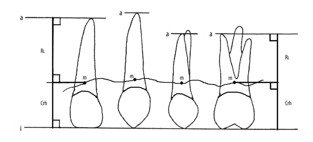
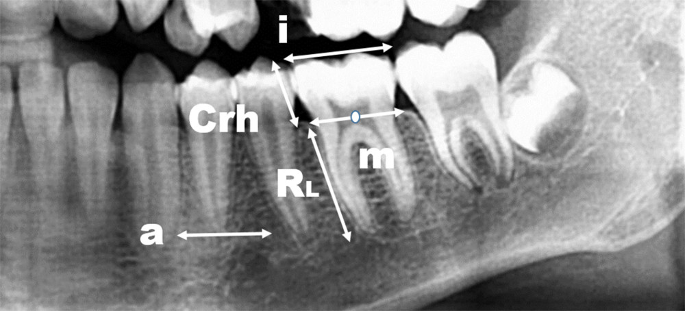
All digital pantographs used for this study were taken through a digital orthopantomogram (Gendex GXDP-700 Series OPG System, KaVo, Germany), viewed on a computer using dental imaging software (6.14.7.3, Carestream Health, Inc, 2014), which is a modern version of the classical orthopantomograph, with the chief advantage being reduced radiation exposure and immediate computer screen display, as well as the ability to be stored in all available formats. Measurement of the crown height and root length was done using a previously described method [15]. The crown heights and root lengths were expressed in millimeters (mm) and calibrated. The crown height (Crh) was considered to be a perpendicular line from a definite point (m) to a reference line on the incisal and occlusal surface (i), while the point ‘m’ was the center (midpoint) of a tangent line that connected the distal and proximal bone (Figures 1, 2). Any tooth with a single tip at the incisal/occlusal surface (like canines and premolars) that would have a line that forms a tangent to the tip (incisal/occlusal) was positioned at right angles to the particular tooth (long axis). For teeth possessing an incisal edge or multiple cusps, a line that followed the edge or buccal cusps was considered. For measuring root length (RL), the perpendicular distance between (m) and the apical reference line (a) was considered (Figure 2). For permanent teeth having multiple roots, the measured length extended to the apex of the longest root toward the buccal side. In cases where the only single root was noticeable, the visible root was measured.
Intra-examiner reproducibility was assessed by re-measurement of the radiographs by the same assessor after 2 weeks. For reproducibility by an examiner (inter examiner reliability), another examiner trained to perform the same exercise measured the dimensions of the same teeth. The intra-class correlation coefficient (ICC) was calculated and an agreement was achieved.
STATISTICAL ANALYSIS:
Descriptive statistics were used to calculate the mean and standard deviation. The independent
Results
The correlation coefficient for intra-examiner calibration ranged from 0.79 to 0.89, which indicates a good reliability of the measurement method. The means of R/C ratios, related descriptive statistics and their respective statistical significances (
Discussion
The present study was designed to investigate the root-to-crown ratio of permanent natural teeth, since there are no existing studies done on Saudi adults. The main findings of the study are that for both sexes, maxillary canines and mandibular second premolars had a highest R/C ratio, while the maxillary first molar and mandibular central incisor had the lowest R/C ratio. Also, in the maxillary arch, only the second premolar showed significant differences in R/C ratios, while in the mandibular arch, lateral incisor, both premolars, and molars showed significant differences. The findings show that among males, there were statistically significant differences between antagonist teeth existing in R/C ratios for central and lateral incisors, second premolar, and both molars. For females, significant differences were observed for both premolars and both molars (
The measurement of R/C ratios was made from digital panoramic radiograph because it is a routine radiograph that can be taken for each patient as a diagnostic aid during an examination, diagnosis, and treatment planning in either daily practice or in research. Furthermore, digital panoramic radiographs are less technique-sensitive, more economical, and involve low radiation exposure. The drawback of panoramic radiographs is that the horizontal dimensions (variables) are unreliable, but the vertical and angular variables are considered to be acceptable if and when a patient’s head is placed in a head-stabilizing holder [17]. In this study, as the crown and root of permanent teeth lie almost on the vertical plane, except for the palatal roots, the palatal roots were, therefore, excluded to minimize the magnification effect of digital panoramic radiograph on R/C ratios [18].
In the current study, the means R/C ratios varied from 1.59 to 2, which was higher than the result reported among the Korean population, which varied from 1.29 to 1.89 [15]. However, the result of the current study was less than the result reported among White, Malay, and Iranian populations [11,14,16]. The variation in the result when compared to other studies could be attributed to ethnic differences and different measurement methods used. The results of the present study revealed that the highest R/C ratio was for mandibular premolars, which is similar to the finding among White populations and the Korean populations [11,15]. The lowest R/C ratio in the maxillary arch was for the permanent maxillary first molar, which is also in agreement with results obtained on Korean and White populations. The lowest R/C ratio for maxillary first molars could be explained by the long crown of maxillary molars and the exclusion of the palatal root, which is the longest root of maxillary molars. The short crowns of mandibular premolars could explain the higher ratios.
In the present study, the R/C ratios of mandibular second premolars and molar teeth were significantly higher than that of maxillary teeth in both sexes and the first premolar in females. This was in a limited agreement with the study done in a Finnish population [11], which found that the mean R/C ratio of all mandibular teeth, except for the lateral incisors in males and the second molars in both sexes, was also substantially higher than in maxillary teeth [11].
The results of this study also show that among Saudi adults, there were less sex differences in the mean R/C ratio of maxillary teeth (except for the maxillary second premolar), while lateral incisors and all posterior teeth in the mandibular arch had significant sex differences. Such findings show the need to consider sex when determining the R/C ratio clinically for mandibular teeth. These results differ mainly from the study done in the Korean population [15], where more differences existed in the maxillary teeth (maxillary canine, maxillary first and second premolar, maxillary first and second molars) than in the mandibular teeth. In the same study, the mean R/C ratios were also found to differ significantly for maxillary canines and maxillary first premolars in males and central and lateral incisors and canines in females.
We also found that the R/C ratios of mandibular lateral incisors, premolars, and first molars were significantly higher in females than in males. This result is inconsistent with the findings of a study done in a Finnish population [11].
While the ideal R/C ratio for a tooth to serve as an abutment for a fixed prosthesis must have a value close to 2, our study reports that such a ratio is rare in natural teeth of Saudi adults. If one keeps a minimum ratio of 1.5, then most of the teeth in both sexes fulfill the minimum criteria for the R/C ratio.
The results of this study are limited by its sample size, which is very small. The study is also limited by the difficulty of comparing data with previous results due to different assessment methods used in other studies. The radiographic technique used for evaluating the R/C ratio does not take into consideration ethnicity-associated differences. Still, the study will be useful for evaluation of tooth anomaly (developed or acquired), comparison with other populations, and as a diagnostic index. In academic and clinical use, the specific results of this study should raise awareness of differences between the sexes and among arches and individuals.
Conclusions
Within the scope and limitations of this study, one can conclude that the mean R/C ratios among Saudi adults range between 1.64 to 1.91 for maxillary teeth and from 1.59 to 2 for mandibular teeth for both sexes. The R/C ratios for males range from 1.62 to 1.93 (maxillary arch) and 1.52 to 1.94 (mandibular arch), while for females it ranges from 1.68 to 1.88 (maxillary arch) and 1.67 to 2.11 (mandibular arch). Except for the maxillary second premolar, no significant sex differences in mean R/C ratios existed for maxillary arch, while all mandibular posterior teeth exhibited sex differences in addition to mandibular lateral incisors. Posterior teeth generally exhibit differences in root-to-crown ratio irrespective of sex in the studied population. This study supports the findings from previous studies that digital panoramic dental radiography using orthopantomogram can be used to calculate root/crown ratios, which varies between sexes and according to the measurement of the dental arch.
Figures
 Figure 1. Radiographic method for measuring crown height and root length in the assessment of the root/crown (R/C) ratio. a – apical level, i – incisal/occlusal reference line, RL – root length, Crh – crown height, m – the midpoint of the line connecting the mesial and distal proximal bone. Root length in mm=measured perpendicular from point m to point a. Crown height in mm=measured perpendicular from point m to i. (Figure created using MS Paint, version 20H2 (OS build 19042,1466), windows 10 Pro, Microsoft corporation).
  Figure 2. Radiographic landmarks used to measure root (length) and crown (height) in the assessment of the root-crown (R/C) ratio. a – apical level, i – incisal/occlusal reference line, RL – root length, Crh – crown height, m – the midpoint of the line connecting the mesial and distal proximal bone. Root length in mm=measured perpendicular from point m to point a. Crown height in mm=measured perpendicular from point m to i. (Figure created using MS Paint, version 20H2 (OS build 19042,1466), MS PowerPoint, windows 10 Pro, Microsoft corporation).
 Tables
Table 1. Mean root to crown ratios with respective standard deviations (SD) for permanent natural teeth from male and female subjects. Table 2. Mean root to crown ratios for individual type of teeth and their relative differences against their opposing antagonist tooth between males and female subjects.
References
1. Subhashini MR, Abirami G, Jain AR, Abutment selection in fixed partial denture – a review: Drug Invention Today, 2018; 10(1); 111-15
2. Chansoria S, Chansoria H, Abutment selection in fixed partial denture: IOSR Journal of Dental and Medical Sciences (IOSR-JDMS), 2018; 17(3); 4-12
3. Rosenstiel SF, Land MF, Fujimoto J: Contemporary of fixed prosthodontics-e-book, 2016; 342-44, Elsevier Health Sciences
4. Carr AB, Brown DT, McGivney GP: McCracken’s removable partial prosthodontics, 2005; 189-229, St Louis, MO, Mosby/Elsevier
5. Newman MG, Takei HH, Carranza FA: Carranza’s Clinical Periodontology, 2002; 481-83, Philadelphia, WB Saunders Co
6. Wilson TG, Kornman KS: Fundamentals of periodontics, 2003; 531-39, Chicago, Quintessence
7. Bosshardt DD, The periodontal pocket: Pathogenesis, histopathology and consequences: Periodontology 2000, 2018; 76(1); 43-50
8. Tonetti MS, Greenwell H, Kornman KS, Staging and grading of periodontitis: Framework and proposal of a new classification and case definition: J Periodontol, 2018; 89; S159-72
9. Mattoo KA, Deep A, Determining the need of a coping and/or its number/type in a tooth supported overdenture: Journal of Advanced Medical and Dental Sciences Research, 2020; 8(10); 46-49
10. Stramotas S, Geenty JP, Darendeliler MA, The reliability of crown-root ratio, linear and angular measurements on panoramic radiographs: Clin Orthod Res, 2000; 3(4); 182-91
11. Hölttä P, Nyström M, Evälahti M, Alaluusua S, Root-crown ratios of permanent teeth in a healthy Finnish population assessed from panoramic radiographs: Eur J Orthod, 2004; 26(5); 491-97
12. Angelopoulos C, Bedard A, Katz JO, Digital panoramic radiography: An overview: Semin Orthod, 2004; 10(3); 194-203
13. Tawfik WA, Soliman NL, El Batran MM, Root-crown ratios of permanent teeth in a sample of Egyptian population-panoramic radiographs assessment: Egypt Dent J, 2005; 51; 1535
14. Othman N, Taib H, Mokhtar N, Root-crown ratios of permanent teeth in Malay patients attending HUSM Dental Clinic: Archives of Orofacial Sciences, 2011; 6(1); 21-26
15. Yun HJ, Jeong JS, Pang NS, Radiographic assessment of clinical root-crown ratios of permanent teeth in a healthy Korean population: J Adv Prosthodont, 2014; 6(3); 171-76
16. Haghanifar S, Moudi E, Abbasi S, Root-crown ratio in permanent dentition using panoramic radiography in a selected Iranian population: J Dent (Shiraz), 2014; 15(4); 173-79
17. Tepedino M, Cornelis MA, Chimenti C, Cattaneo PM, Correlation between tooth size-arch length discrepancy and interradicular distances measured on CBCT and panoramic radiograph: An evaluation for miniscrew insertion: Dental Press J Orthod, 2018; 23; 39e1-13
18. Özalp Ö, Tezerişener HA, Kocabalkan B, Comparing the precision of panoramic radiography and cone-beam computed tomography in avoiding anatomical structures critical to dental implant surgery: A retrospective study: Imaging Sci Dent, 2018; 48(4); 269-75
Figures
 Figure 1. Radiographic method for measuring crown height and root length in the assessment of the root/crown (R/C) ratio. a – apical level, i – incisal/occlusal reference line, RL – root length, Crh – crown height, m – the midpoint of the line connecting the mesial and distal proximal bone. Root length in mm=measured perpendicular from point m to point a. Crown height in mm=measured perpendicular from point m to i. (Figure created using MS Paint, version 20H2 (OS build 19042,1466), windows 10 Pro, Microsoft corporation).
 Figure 2. Radiographic landmarks used to measure root (length) and crown (height) in the assessment of the root-crown (R/C) ratio. a – apical level, i – incisal/occlusal reference line, RL – root length, Crh – crown height, m – the midpoint of the line connecting the mesial and distal proximal bone. Root length in mm=measured perpendicular from point m to point a. Crown height in mm=measured perpendicular from point m to i. (Figure created using MS Paint, version 20H2 (OS build 19042,1466), MS PowerPoint, windows 10 Pro, Microsoft corporation).
 Tables
 Table 1. Mean root to crown ratios with respective standard deviations (SD) for permanent natural teeth from male and female subjects.
 Table 2. Mean root to crown ratios for individual type of teeth and their relative differences against their opposing antagonist tooth between males and female subjects.
 Table 1. Mean root to crown ratios with respective standard deviations (SD) for permanent natural teeth from male and female subjects.
 Table 2. Mean root to crown ratios for individual type of teeth and their relative differences against their opposing antagonist tooth between males and female subjects.
 In Press
05 Mar 2024 : Clinical Research
Role of Critical Shoulder Angle in Degenerative Type Rotator Cuff Tears: A Turkish Cohort StudyMed Sci Monit In Press; DOI: 10.12659/MSM.943703
06 Mar 2024 : Clinical Research
Comparison of Outcomes between Single-Level and Double-Level Corpectomy in Thoracolumbar Reconstruction: A ...Med Sci Monit In Press; DOI: 10.12659/MSM.943797
21 Mar 2024 : Meta-Analysis
Economic Evaluation of COVID-19 Screening Tests and Surveillance Strategies in Low-Income, Middle-Income, a...Med Sci Monit In Press; DOI: 10.12659/MSM.943863
10 Apr 2024 : Clinical Research
Predicting Acute Cardiovascular Complications in COVID-19: Insights from a Specialized Cardiac Referral Dep...Med Sci Monit In Press; DOI: 10.12659/MSM.942612
Most Viewed Current Articles
17 Jan 2024 : Review article
Vaccination Guidelines for Pregnant Women: Addressing COVID-19 and the Omicron VariantDOI :10.12659/MSM.942799
Med Sci Monit 2024; 30:e942799
14 Dec 2022 : Clinical Research
Prevalence and Variability of Allergen-Specific Immunoglobulin E in Patients with Elevated Tryptase LevelsDOI :10.12659/MSM.937990
Med Sci Monit 2022; 28:e937990
16 May 2023 : Clinical Research
Electrophysiological Testing for an Auditory Processing Disorder and Reading Performance in 54 School Stude...DOI :10.12659/MSM.940387
Med Sci Monit 2023; 29:e940387
01 Jan 2022 : Editorial
Editorial: Current Status of Oral Antiviral Drug Treatments for SARS-CoV-2 Infection in Non-Hospitalized Pa...DOI :10.12659/MSM.935952
Med Sci Monit 2022; 28:e935952


