17 October 2020: Clinical Research
Brain-Derived Neurotrophic Factor Inhibits the Wound-Healing and Cell Proliferative Ability of Human Airway Epithelial Cells in Asthmatic Children
Shuguang Jing1AB, Xinghua Li1CE, Wei Liu2DE, Xia Li1F*DOI: 10.12659/MSM.923680
Med Sci Monit 2020; 26:e923680


Abstract
BACKGROUND: Asthma is a chronic disease with high morbidity rates. Brain-derived neurotrophic factor (BDNF) has been proven to induce airway hyper-responsiveness, but the function of BDNF in the wound-healing process of asthmatic human airway epithelial cells (HAECs) remains unclear. This study investigated the effects of BDNF in asthmatic children with injured HAECs.
MATERIAL AND METHODS: HAECs were obtained from healthy children and asthmatic children through bronchoscopy, and then cultured in air-liquid (ALI) interface with or without BDNF. A mechanical injury model was established for the wound-healing assay. Quantitative real-time polymerase chain reaction (qRT-PCR) assay was performed to measure BDNF mRNA expressions, while western blot assay was used for the measurement of BDNF and CCND1 protein expressions. Cell proliferation of impaired HAECs was assayed in a ³H-thymidine incorporation experiment.
RESULTS: The mRNA and protein levels of BDNF were overexpressed, and the wound-healing ability of HAECs decreased in asthma samples. Also, the cell proliferation of HAECs was suppressed in the asthmatic injury model and the injury-induced increase of CCND1 protein expressions was inhibited in asthma. Although mRNA and protein expressions of BDNF remained unchanging in healthy HAECs, there was an increase in impaired asthmatic HAECs. Upregulating BDNF led to a decrease in wound-healing ability of HAECs in both healthy children and children with asthma. Simultaneously, overexpressed BDNF reduced the CCND1 protein expressions in healthy HAECs, but had little impact on asthmatic HAECs.
CONCLUSIONS: Brain-derived neurotrophic factor (BDNF) inhibited wound-healing and cell proliferative ability of human airway epithelial cells (HAECs) in asthmatic children.
Keywords: Asthma, Brain-Derived Neurotrophic Factor, Adolescent, Child, Gene Expression Regulation, Respiratory Mucosa, Wound Healing
Background
Asthma is a chronic inflammation of the airway that involves multiple inflammatory cells like eosinophils, mastocytes, and thymus-dependent lymphocytes [1]. In addition, aberrant mucus that plugs the airway and hyper-responsiveness also play pivotal roles in the pathophysiology of asthma [2–4]. Human airway epithelial cells (HAECs) are the first barriers in the airway. Studies have shown that airway epithelium dysfunction promotes mucosal permeability of foreign substances, over-releases epithelial cytokines, and stimulates dendritic cells, which is potentially relevant to the development of mild, moderate, and severe asthma [5,6]. The symptoms of asthma often present as recurrent wheezing, chest distress, dyspnea, or cough, and most children can have asthma symptoms relieved without treatment, or treatment with drug therapy, targeted biologic therapy, or by environment control [7]. However, the morbidity rates of asthma still continue to rise, which creates health burdens for more and more children, not only physically, but also financially [8]. Novel and more effective treatments for asthma are still needed.
Brain-derived neurotrophic factor (BDNF) is a member of the neurotrophins family. Mature BDNF mediated by TrkB receptor plays a regulatory role in neuronal differentiation, structure, and function [9]. Changes in BDNF expression levels and activities have been reported in the development of numerous neurodegenerative disorders [10]. Evidence shows that the suppression of BDNF-TrkB signaling and reinforcement of the neuropeptide Y (NPY) system can affect epilepsy treatment [11]. BDNF can also downregulated neuroprotective actions during hyperglycemia, contributing to the vulnerability of retinal neurons and to diabetic retinopathy [12]. In addition, reports have shown that BDNF has an important effect on asthma. Research has shown that BDNF can partially regulate neuronal hyper-reactivity in an allergic airway inflammation model of mice [13]. In addition, smooth muscle-derived BDNF have been shown to mediate airway hyper-responsiveness modulated by tropomyosin-related kinase B signaling pathway during allergic airway inflammation [14]. Moreover, the upregulated BDNF gene expressions and its mature isoform have been verified to be involved in airway hyper-responsiveness, asthma severity, and inflammatory signature [15]. Although many studies have shown the close correlation between BDNF and airways in asthma, whether BDNF has regulatory effects on repair of injured HAECs in asthma remains unclear.
To the best of our knowledge, this is the first study to investigate the function of BDNF in repairing the damaged epithelial barrier in asthma to discover new methods for asthma prevention, control, and treatment.
Material and Methods
BIOINFORMATICS ANALYSIS:
GSE43696 microarray data of HAECs in patients with bronchial asthma were obtained from the Gene Expression Omnibus (GEO) database (
SPECIMEN COLLECTION:
A total of 8 samples (5 males and 3 females, age 7–14 years) in a healthy control (HC) group and 24 samples (15 males and 9 females, age 6–14 years) in an asthma group were collected from October 2017 to October 2018 in Liaocheng People’s Hospital. Informed consent was obtained for all patients and permission was given for use of their tissues in clinical research. The clinical trial program was reviewed and approved by the Ethics Committee of Liaocheng People’s Hospital (LPH201612001). All individuals underwent bronchoscopy, and then epithelial brushings were obtained from bronchi according to previously published guidelines [16,17].
CELL ISOLATION AND CELL CULTURE IN AIR-LIQUID (ALI) INTERFACE:
Collected bronchial brushes were placed in serum-free Bronchial Epithelial Cell Growth Medium (BEGM, Lonza, NJ, USA) containing 100 units/mL of penicillin (TargetMol, Boston, NY, USA) and 100 μg/mL of streptomycin (TargetMol, Boston, NY, USA), centrifuged, washed, and then re-suspended in fresh BEGM for further experiments. HAECs (5×10⁵ per well) were transferred onto 0.4-μm supporting membranes of Transwell pre-carpeted with collagen from human placenta (also known as Collagen Type 4, Sigma, CA, USA). BEGM was added under the membranes. Then, cells were incubated under conditions of 5% CO2 at 37°C until reaching 80% confluence, with medium changed every other day. Next, medium on the membranes was removed, and air-liquid interface (ALI) medium, consisting of BEGM and Dulbecco’s modified Eagle’s medium (DMEM, Gibco, California, USA) with or without 50 ng of BDNF (Cat. 2837, Tocris, MN, USA) in a 1: 1 ratio, was placed under the membranes and changed every other day. Incubation was conducted with 5% CO2 at 37°C.
WOUND-HEALING ASSAY:
HAECs were incubated for 15 days until reaching 100% fusion rate. A 10-μL pipette tip was used to draw horizontal lines at the bottom of each culture plates crossing the culture hole. Medium was removed and phosphate-buffered saline (PBS; Gibco, USA) was used to wash away the cells that crossed. Next, serum-free BEGM containing 1% B27 (Gibco, CA, USA), 100 units/mL of penicillin, 100 μg/mL of streptomycin, and 2 mmol/L glutamine (Thermo Scientific, CA, USA) was added, followed by incubation with 5% CO2 at 37°C for 24 h. The wound closure was then measured and analyzed.
QUANTITATIVE REAL-TIME POLYMERASE CHAIN REACTION (QRT-PCR):
Total mRNAs were extracted from HAECs in the HC group and asthma group using an RNAqueous™-4PCR Total RNA Isolation Kit (Invitrogen, CA, USA). Then, the first-strand cDNA was synthesized with SuperScript™ IV Reverse Transcriptase (Invitrogen, CA, USA). Next, 25 μL ABsolute QPCR Mix, SYBR Green, and ROX (Thermo Scientific, CA, USA) was used for qPCR in the QuantStudio™ 5 Real-Time PCR System (Applied Biosystems, CA, USA). The conditions were: 1 cycle at 95°C for 15 min, followed by 40 cycles at 95°C for 15 s, 50°C for 30 s, and 72°C for 30 s. The relative expression of each mRNA was calculated by comparative cycle threshold (CT) method (2−ΔΔCT) [18]. The primers (BDNF) were:
WESTERN BLOT ANALYSIS:
The whole proteins were extracted using the Genomic DNA Isolation Kit (Biovision, San Francisco, CA, USA) from HAECs in the HC group and asthma group, followed by detection of protein concentrations using a Bicinchoninic Protein Assay kit (BCA, Pierce, Rockford, IL, USA). Then, 50 μg of the protein was transferred onto 10% sodium dodecyl sulfate-polyacrylamide gel electrophoresis (SDS-PAGE, Solarbio, Beijing, China) and then transferred onto polyvinylidene fluoride (PVDF) membranes. The membranes were blocked with 5% non-fat dried milk for 2 h, followed by incubation with the primary antibodies at 4°C overnight. The primary antibodies were: recombinant anti-BDNF antibody (EPR1292; 1: 1000, ab108319, Abcam, USA), recombinant anti-cyclin D1 antibody (SP4; 1: 25, ab16663, Abcam, USA), and anti-GAPDH antibody (6C5 as loading control; 1: 500, ab8245, Abcam, USA), with GAPDH serving as the internal reference. Next, the corresponding secondary antibodies goat anti-mouse IgG H&L (HRP; 1: 2000, ab205719, Abcam, USA) and goat anti-rabbit IgG H&L (HRP; 1: 2000, ab205718, Abcam, USA) were added at room temperature for 1 h Finally, the blots were developed by using Pierce™ ECL Western Blotting Substrate (Thermo Scientific, CA, USA).
:
3H-thymidine incorporation assay was performed after 7 days of ALI culture to detect cell proliferative ability of healthy and asthmatic HAECs in the wound model. Cells were cultured in Roswell Park Memorial Institute-1640 (RPMI-1640) medium (0.2 mL/well, Hyclone, UT, USA), then phytohemagglutinin (Sigma, CA, USA) was added to each well. After 56-h incubation, 20 μL 3H-TdR (Isotope Research Institute of China Atom Science Research Institute, Beijing, China) was added for another 72-h incubation. All the incubations were conducted with 5% CO2 at 37°C. Scintillation solution consisting of 2,5-diphenyl oxazole (PPO, Sigma, USA), 1,4-bis-2(5-phenyloxazoyl)benzene (POPOP, Sigma, USA), and dimethylbenzene (Thermo Scientific, CA, USA) was then added. The liquid-scintillation counting system Beckman LS-9800 (Beckman, CA, USA) was used for measurement of radioactivity of HAECs at 3 h and 24 h after wounding.
STATISTICAL ANALYSIS:
All the experimental data from this study are expressed as mean±standard deviation (mean±SD) and were analyzed by Statistical Product and Service Solutions (SPSS, NDtimes, Beijing, China). The
Results
BDNF WAS OVEREXPRESSED IN ASTHMATIC SAMPLES AND GROUPS:
According to bioinformatics analysis, we found that the gene expressions of BDNF was significantly higher in asthmatic samples. Then, qRT-PCR was performed to detect the relative messenger RNA (mRNA) expression levels of BDNF in HAECs. We found that BDNF mRNA expressions were much higher in the asthma group than in the healthy control (HC) group (P<0.001; Figure 1A). BDNF-related protein expressions in HAECs were measured through western blot analysis. BDNF protein expressions in the HC1 group were different from that in HC2 and HC3 groups. Protein expression levels of BDNF in the Asthma1, Asthma2, and Asthma3 groups were greatly overexpressed compared with HC groups (P<0.001; Figure 1B).
WOUND-HEALING AND CELL PROLIFERATIVE ABILITY OF HAECS WAS INHIBITED IN THE MECHANICAL INJURY MODEL OF ASTHMA:
Wound-healing assay showed that the wound closure of HAECs in the asthma group was remarkably led than in the HC group (P<0.01; Figure 2A). We performed a 3H-thymidine incorporation experiment to test cell proliferative ability. HAECs had similar radioactivity in the HC and the asthma groups at 3 h after wounding. Nevertheless, the counts per minute (CPM) of asthmatic HAECs cultured for 24 h in the mechanical injured model were significantly lower than in the HC group (P<0.001, Figure 2B). Western blot assay was then conducted to measure CCND1 protein expressions. Figure 2C shows that the relative protein expressions of CCND1 in the scratch group were much higher than that in the control group, and the CCND1 protein expressions in asthmatic HAECs were inhibited in comparison with the HC group (P<0.001).
UPREGULATION OF BDNF INHIBITED WOUND HEALING:
qRT-PCR and western blot analysis showed that the relative gene expression levels of BDNF in the mechanical injury model of asthmatic HAECs were significantly higher than in the control group and the HC group (P<0.001; Figure 3A, 3B). Moreover, the upregulation of BDNF greatly suppressed the wound-healing ability of HAECs, which was more notable in the HC group (P<0.001; Figure 3C). The relative protein expression levels of CCND1 were inhibited with the increase of BDNF in the HC group compared with the asthma group (P<0.001; Figure 3D).
Discussion
Asthma is a chronic inflammatory disease of the airway in which various cells (e.g., eosinophils, mast cells, lymphocytes, neutrophils, and airway epithelial cells) and neurotransmitters are involved. The etiology of asthma is complicated, and there are many factors that induce it and participate in it [2,19,20].
Neurotrophic factor is considered to be a bridge between the neurogenic inflammatory response of the airway induced by the interaction of nerves and immune mechanisms. The stimulation of allergen and infection causes the high expression and over-secretion of airway neurotrophic factor and its receptor, which leads to changes in neuroplasticity and abnormal differentiation and participates in airway hyper-responsiveness [14,21]. BDNF is an important neurotrophic factor. In recent years, many studies have proven that BDNF can be produced by a variety of immune inflammatory cells, which in turn affects immune inflammatory cells [22,23]. BDNF levels in inflammatory diseases, autoimmune diseases, and allergic diseases are significantly increased [24–26]. In the present study, we tested the gene expression of BDNF through qRT-PCR and western blot experiments. The results were consistent with previous studies. The relative mRNA expression levels and protein expression levels of BDNF were significantly overexpressed in the asthma group compared to the HC group, suggesting that BDNF is essential in the development of asthma.
The first defense of the respiratory system against external pathogens is the airway mucosa, and the protective barrier on the surface of the airway mucosa is the airway epithelium, which is composed of a variety of cells [27]. Changes in the morphology and function of airway epithelial cells occur in the early stages of asthma and are in a continuous and abnormal state of injury and repair [28]. Airway epithelial cells play an important role in immune regulation and maintain the stability of the airway mucosal microenvironment. Airway hyper-responsiveness is characterized by excessive or premature contraction of the airway in response to various stimuli when the epithelial structure in intact, but its structural integrity can be damaged if the airway epithelium participates in the airway inflammatory response [29]. Airway remodeling is the main pathological basis of irreversible airflow obstruction and progressive decline of lung function in asthma. The typical pathological features are injury and proliferation of airway epithelial cells [2,27,28]. All of the above evidence shows that intervention in the abnormal repair process after airway epithelial cell injury is one of the key points affecting airway remodeling of asthma. Therefore, in the present study, the mechanical injury model of asthmatic HAECs was established
Previous studies have shown that BDNF can partially regulate the inflammation, mucus secretion, and hyper-responsiveness of the airway [11–14], but it has been unclear whether BDNF affects airway epithelial barrier repair. In the healthy samples, we found little change of the BDNF mRNA expression levels in the mechanical injury model. Intriguingly, mRNA expression of BDNF in asthmatic injured HAECs was greatly promoted. Similarly, the protein expressions of BDNF in healthy and injured HAECs remained unchanged, but significantly increased in asthmatic and injured samples. Next, BDNF was upregulated in the HAECs. The degree of wound closure was remarkably decreased with the overexpression of BDNF, showing that the increase in BDNF suppressed the repair ability of injured epithelial cells. Furthermore, our experiments showed that the protein expression levels of CCND1 were inhibited by the overexpressed BDNF in healthy samples.
Conclusions
BDNF gene expression of human airway epithelial cells was upregulated in asthma, which hampered the wound-healing and cell proliferative ability of airway epithelium. Thus, downregulation of the expressions of BDNF has potential to be an effective way to repair the airway epithelium damage in asthma, which could benefit many asthmatic patients.
Figures
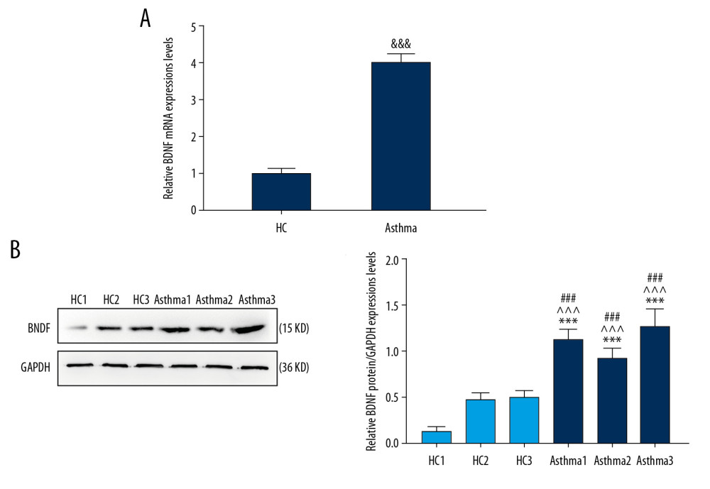 Figure 1. The expression of BDNF in HAECs of asthmatic children was higher than that in the healthy control group. (A) qRT-PCR was used to detect the mRNA expressions of BDNF in newly separated asthmatic HAECs and healthy control HAECs. (B) The protein expression of BDNF in asthmatic and healthy controls was measured through western blot. &&& P<0.001 versus HC, *** P<0.001 versus HC1, ^^^ P<0.001 versus HC2, ### P<0.001 versus HC3, n=3. BDNF – brain-derived neurotrophic factor; qRT-PCR – quantitative real-time reverse transcription polymerase chain reaction; mRNA – messenger RNA; HAECs – human airway epithelial cells; HC – healthy controls.
 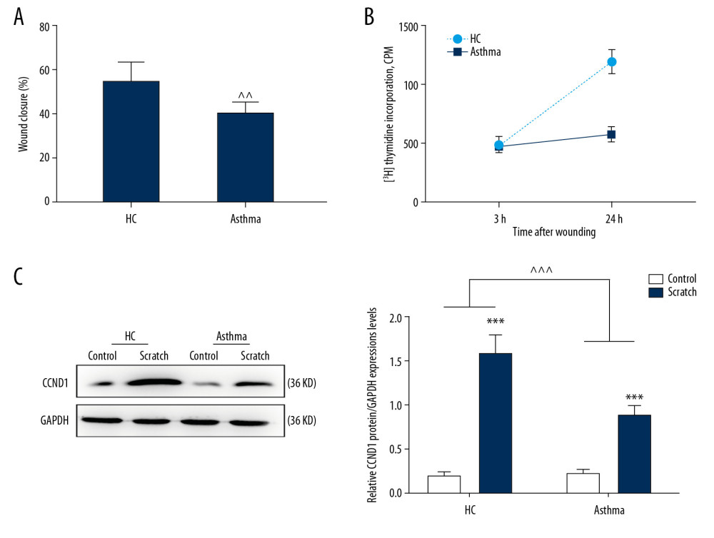 Figure 2. Wound closure and cell proliferation after wounding is impaired in asthmatic HAECs. (A) A mechanical injury model was established to assess wound closure. (B) Cell proliferation of asthmatic and normal HAECs was assayed through 3H-thymidine incorporation experiments. (C) The protein expression of CCND1 was measured via western blot. ^^ P<0.01, ^^^ P<0.001 versus HC, *** P<0.001 versus Control, n=3. HAECs – human airway epithelial cells; HC – healthy controls.
 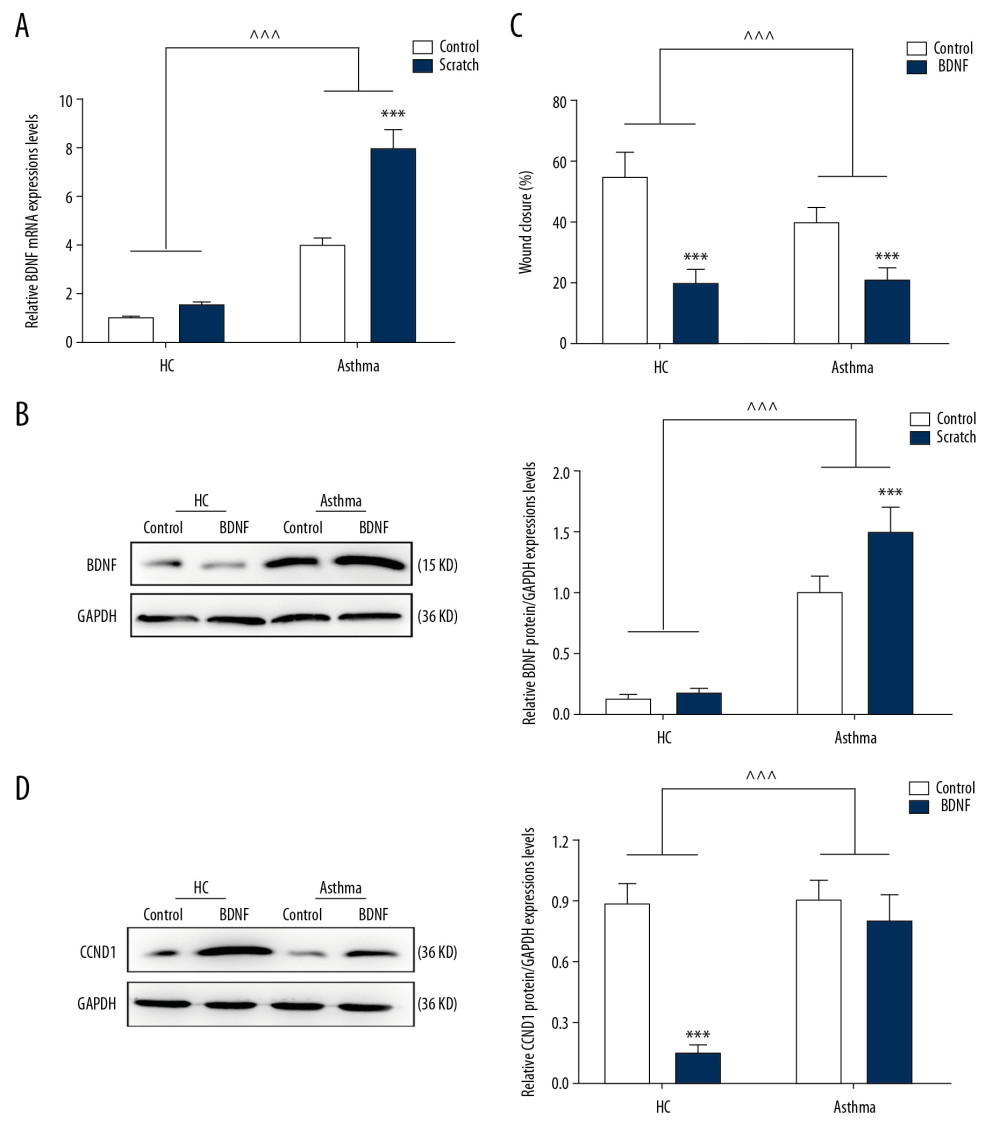 Figure 3. Upregulation of BDNF inhibited the wound from healing. (A) The mRNA expression level of BDNF in asthmatic and normal HAECs was tested through qRT-PCR. (B) Western blot analysis was performed to measure the relative BDNF protein expressions. (C) Wound closure in the asthmatic group and normal group was tested after exogenous BDNF was added. (D) CCND1 protein expression was assayed via western blot. *** P<0.001 versus Control, ^^^ P<0.001 versus HC, n=3. BDNF – brain-derived neurotrophic factor; mRNA – messenger RNA; qRT-PCR – quantitative real-time reverse transcription polymerase chain reaction; HC – healthy controls.
 References
1. Hashimoto SAirway inflammation in asthma: Arerugi, 2017; 66(3); 168-72 [in Japanese]
2. Mims JW, Asthma: Definitions and pathophysiology: Int Forum Allergy Rhinol, 2015; 5(Suppl 1); S2-6
3. Fahy JV, Dickey BF, Airway mucus function and dysfunction: N Engl J Med, 2010; 363(23); 2233-47
4. Chapman DG, Irvin CG, Mechanisms of airway hyper-responsiveness in asthma: the past, present and yet to come: Clin Exp Allergy, 2015; 45(4); 706-19
5. Loffredo LF, Abdala-Valencia H, Anekalla KR, Beyond epithelial-to-mesenchymal transition: Common suppression of differentiation programs underlies epithelial barrier dysfunction in mild, moderate, and severe asthma: Allergy, 2017; 72(12); 1988-2004
6. Gon Y, Hashimoto S, Role of airway epithelial barrier dysfunction in pathogenesis of asthma: Allergol Int, 2018; 67(1); 12-17
7. Castillo JR, Peters SP, Busse WW, Asthma exacerbations: Pathogenesis, prevention, and treatment: J Allergy Clin Immunol Pract, 2017; 5(4); 918-27
8. Asher I, Pearce N, Global burden of asthma among children: Int J Tuberc Lung Dis, 2014; 18(11); 1269-78
9. Hempstead BL, Brain-derived neurotrophic factor: three ligands, many actions: Trans Am Clin Climatol Assoc, 2015; 126; 9-19
10. Zuccato C, Cattaneo E, Brain-derived neurotrophic factor in neurodegenerative diseases: Nat Rev Neurol, 2009; 5(6); 311-22
11. Iughetti L, Lucaccioni L, Fugetto F, Brain-derived neurotrophic factor and epilepsy: A systematic review: Neuropeptides, 2018; 72; 23-29
12. Behl T, Kotwani A, Downregulated brain-derived neurotrophic factor-induced oxidative stress in the pathophysiology of diabetic retinopathy: Can J Diabetes, 2017; 41(2); 241-46
13. Braun A, Lommatzsch M, Neuhaus-Steinmetz U, Brain-derived neurotrophic factor (BDNF) contributes to neuronal dysfunction in a model of allergic airway inflammation: Br J Pharmacol, 2004; 141(3); 431-40
14. Britt RD, Thompson MA, Wicher SA, Smooth muscle brain-derived neurotrophic factor contributes to airway hyperreactivity in a mouse model of allergic asthma: FASEB J, 2019; 33(2); 3024-34
15. Watanabe T, Fajt ML, Trudeau JB, Brain-derived neurotrophic factor expression in asthma. association with severity and type 2 inflammatory processes: Am J Respir Cell Mol Biol, 2015; 53(6); 844-52
16. Hsu AC, Parsons K, Moheimani F, Impaired antiviral stress granule and ifn-beta enhanceosome formation enhances susceptibility to influenza infection in chronic obstructive pulmonary disease epithelium: Am J Respir Cell Mol Biol, 2016; 55(1); 117-27
17. Hsu AC, Parsons K, Barr I, Critical role of constitutive type I interferon response in bronchial epithelial cell to influenza infection: PLoS One, 2012; 7(3); e32947
18. Guenin S, Mauriat M, Pelloux J, Normalization of qRT-PCR data: the necessity of adopting a systematic, experimental conditions-specific, validation of references: J Exp Bot, 2009; 60(2); 487-93
19. Finkas LK, Martin R, Role of small airways in Asthma: Immunol Allergy Clin North Am, 2016; 36(3); 473-82
20. Bellanti JA, Settipane RA, Asthma, allergy, and psychiatric disease: Allergy Asthma Proc, 2015; 36(6); 415-17
21. Barrios J, Ai X, Neurotrophins in asthma: Curr Allergy Asthma Rep, 2018; 18(2); 10
22. van den Ameele S, Coppens V, Schuermans J, Neurotrophic and inflammatory markers in bipolar disorder: a prospective study: Psychoneuroendocrinology, 2017; 84; 143-50
23. Prakash YS, Martin RJ, Brain-derived neurotrophic factor in the airways: Pharmacol Ther, 2014; 143(1); 74-86
24. Stoll P, Wuertemberger U, Bratke K, Stage-dependent association of BDNF and TGF-beta1 with lung function in stable COPD: Respir Res, 2012; 13; 116
25. Lu B, Nagappan G, Lu Y, BDNF and synaptic plasticity, cognitive function, and dysfunction: Handb Exp Pharmacol, 2014; 220; 223-50
26. Bathina S, Srinivas N, Das UN, Streptozotocin produces oxidative stress, inflammation and decreases BDNF concentrations to induce apoptosis of RIN5F cells and type 2 diabetes mellitus in Wistar rats: Biochem Biophys Res Commun, 2017; 486(2); 406-13
27. Crystal RG, Randell SH, Engelhardt JF, Airway epithelial cells: Current concepts and challenges: Proc Am Thorac Soc, 2008; 5(7); 772-77
28. Nakagome K, Nagata M, Pathogenesis of airway inflammation in bronchial asthma: Auris Nasus Larynx, 2011; 38(5); 555-63
29. Smyth RL, The airway epithelium in health and disease: “Calm on the surface, paddling furiously underneath”: Thorax, 2009; 64(4); 277-78
30. Bendris N, Lemmers B, Blanchard JM, Cell cycle, cytoskeleton dynamics and beyond: the many functions of cyclins and CDK inhibitors: Cell Cycle, 2015; 14(12); 1786-98
Figures
Figure 1. The expression of BDNF in HAECs of asthmatic children was higher than that in the healthy control group. (A) qRT-PCR was used to detect the mRNA expressions of BDNF in newly separated asthmatic HAECs and healthy control HAECs. (B) The protein expression of BDNF in asthmatic and healthy controls was measured through western blot. &&& P<0.001 versus HC, *** P<0.001 versus HC1, ^^^ P<0.001 versus HC2, ### P<0.001 versus HC3, n=3. BDNF – brain-derived neurotrophic factor; qRT-PCR – quantitative real-time reverse transcription polymerase chain reaction; mRNA – messenger RNA; HAECs – human airway epithelial cells; HC – healthy controls.Figure 2. Wound closure and cell proliferation after wounding is impaired in asthmatic HAECs. (A) A mechanical injury model was established to assess wound closure. (B) Cell proliferation of asthmatic and normal HAECs was assayed through 3H-thymidine incorporation experiments. (C) The protein expression of CCND1 was measured via western blot. ^^ P<0.01, ^^^ P<0.001 versus HC, *** P<0.001 versus Control, n=3. HAECs – human airway epithelial cells; HC – healthy controls.Figure 3. Upregulation of BDNF inhibited the wound from healing. (A) The mRNA expression level of BDNF in asthmatic and normal HAECs was tested through qRT-PCR. (B) Western blot analysis was performed to measure the relative BDNF protein expressions. (C) Wound closure in the asthmatic group and normal group was tested after exogenous BDNF was added. (D) CCND1 protein expression was assayed via western blot. *** P<0.001 versus Control, ^^^ P<0.001 versus HC, n=3. BDNF – brain-derived neurotrophic factor; mRNA – messenger RNA; qRT-PCR – quantitative real-time reverse transcription polymerase chain reaction; HC – healthy controls. In Press
Clinical Research
Body Weight and Insulin Resistance Indicators Among ChildrenMed Sci Monit In Press; DOI: 10.12659/MSM.951434
Clinical Research
Comparison of Radiographic Cervical Sagittal Alignment Parameters in Patients With Nonspecific Neck Pain, D...Med Sci Monit In Press; DOI: 10.12659/MSM.952950
Clinical Research
Combined Fibrinogen and Urinary α1-Microglobulin as Predictors of Respiratory Tract Infection in Children w...Med Sci Monit In Press; DOI: 10.12659/MSM.951066
Database Analysis
Evaluation of Salivary Total Oxidant Status (TOS) and Total Antioxidant Status (TAS) in Orthodontic Patient...Med Sci Monit In Press; DOI: 10.12659/MSM.952052
Most Viewed Current Articles
17 Jan 2024 : Review article 14,175,576
Vaccination Guidelines for Pregnant Women: Addressing COVID-19 and the Omicron VariantDOI :10.12659/MSM.942799
Med Sci Monit 2024; 30:e942799
13 Nov 2021 : Clinical Research 3,756,620
Acceptance of COVID-19 Vaccination and Its Associated Factors Among Cancer Patients Attending the Oncology ...DOI :10.12659/MSM.932788
Med Sci Monit 2021; 27:e932788
14 Dec 2022 : Clinical Research 2,465,966
Prevalence and Variability of Allergen-Specific Immunoglobulin E in Patients with Elevated Tryptase LevelsDOI :10.12659/MSM.937990
Med Sci Monit 2022; 28:e937990
16 May 2023 : Clinical Research 708,651
Electrophysiological Testing for an Auditory Processing Disorder and Reading Performance in 54 School Stude...DOI :10.12659/MSM.940387
Med Sci Monit 2023; 29:e940387