08 November 2021: Clinical Research
Risk Factors of Neurological Complications in Severe Fever Patients with Thrombolytic Syndrome: A Single-Center Retrospective Study in China
Xiao Fei12ABCDEFG, Kai Fang3BCD, Xiuying Ni2BCD, Wan-hua Ren1ABCDEFG*DOI: 10.12659/MSM.932836
Med Sci Monit 2021; 27:e932836
Abstract
BACKGROUND: Severe fever with thrombocytopenia syndrome is a serious insect-borne infectious disease caused by the Huaiyangshanbanyang virus. We conducted a retrospective study to identify risk factors for neurological complications caused by the virus.
MATERIAL AND METHODS: We included 121 patients who had severe fever with thrombocytopenia syndrome and were admitted to our hospital from 2013 to 2020. Patients’ laboratory test results and clinical data were collected. Univariate and multivariate regression were used for statistical analysis.
RESULTS: Patients with neurological complications had higher mortality rates and longer hospital stays and disease duration than did patients without neurological complications. The neurological symptoms with the highest incidence rates were involuntary tremors (tongue and mandible), cognitive disorder, and limb tremors. Patients with neurological complications had a higher incidence of abnormal heart rhythms. Subcutaneous bleeding, pulmonary rales, percentage of neutrophils, increased lactate dehydrogenase and C-reactive protein levels, and decreased chloride ion concentration were closely related to the occurrence of neurological complications. The significant decrease in chloride ion concentration within 1 to 5 days of disease onset may be a risk factor for predicting the occurrence of neurological complications in patients with severe fever with thrombocytopenia syndrome.
CONCLUSIONS: Early monitoring of subcutaneous bleeding, pulmonary rales, electrocardiogram changes, and biochemical indicators in patients with severe fever with thrombocytopenia syndrome can predict the occurrence of neurological complications.
Keywords: Central Nervous System, SFTS Phlebovirus, Adolescent, Aged, 80 and over, Female, Humans, Length of Stay, Male, Nervous System Diseases, Risk Factors, Severe Fever with Thrombocytopenia Syndrome, young adult
Background
Severe fever with thrombocytopenia syndrome (SFTS) is an acute fatal infectious disease caused by the Huaiyangshanbanyang virus (BHAV), a
In humans, the clinical manifestations include fever, chills, anorexia, nausea, vomiting, abdominal pain, diarrhea, lymphadenopathy, and reduced leucocyte and platelet counts; the disease duration could be up to 49 days [14]. A previous meta-analysis reported a case mortality rate of 5% to 40% and an average mortality rate of 12.2% [15,16]. Relevant studies have been conducted in more than 20 provinces in China, with more than 5000 cases reported from 2009 to 2016 [17]. Patients with SFTS have also been reported in Japan, South Korea, Vietnam, and the United States [18–21]. Because of the heavy burden, lack of vaccines and effective therapies, and high mortality rates, the disease has become an important health issue.
The pathogenesis of neurological symptoms induced by BHAV is still unclear. Studies have shown that cytokines such as monocyte chemotactic protein-1 and interleukin-8 play an important role in the pathogenesis of viral invasion in the nervous system [22]. It has been confirmed that high mortality is closely related to neurological complications [23]. Early diagnosis and treatment of neurological complications may help to reduce mortality. There are many studies on the death-related risk factors of SFTS, but there are few studies on the risk factors of neurological complications. Therefore, we collected the clinical information of patients with SFTS to analyze the risk factors for neurological complications with the goal of detecting the neurological complications as soon as possible.
Material and Methods
SAMPLE SIZE:
A total of 121 patients with SFTS who were admitted to Weifang Yidu Central Hospital, Shandong, between 2013 and 2020 were enrolled in our study. Patients with suspected cases of SFTS were diagnosed with the disease if they fulfilled the following 2 criteria: (1) history of working, living, or traveling in hilly, forested, and mountainous areas during the epidemic season (from April to June) or with a history of being bitten by ticks 2 weeks before the disease onset, and (2) patients with the above mentioned epidemiological history who exhibited fever (armpit temperature above 37°C) and decreased peripheral blood platelet (reference range: 100–400×109/L) and white blood cell counts (reference range: 4–10×109/L). Blood samples were obtained from patients with suspected cases, and those who tested positive for the novel Bunyavirus nucleic acid were diagnosed with SFTS. The SFTS nucleic acid quantitative detection kit was used, and the polymerase chain reaction fluorescence probe method was used for detection, which was performed in the Weifang Center for Disease Control and Prevention. Using the presence or absence of neurological complications as a guide, patients with confirmed infection were divided into a group with neurological complications and a group without neurological complications. The experimental procedure is shown in Figure 1.
DATA COLLECTION:
The demographic factors, date of illness onset, admission date, death date, disease outcome, clinical presentations, physical examination, and laboratory parameters of these patients were retrospectively collected. Their case data were entered into the designed EpiData database, and were exported into an Excel spreadsheet.
STATISTICAL VALIDATION:
Statistical analysis included the use of the chi-squared test and
Results
DEMOGRAPHIC CHARACTERISTICS:
From 121 patients aged between 15 and 95 years, a total of 69 patients were diagnosed with neurological complications, of which 10 patients died from the disease, while the remaining 52 had less severe conditions. The mean age of the patients with neurological complications was significantly higher than that of patients without neurological complications; however, there was no significant difference in sex between the 2 groups. Patients in the neurological complications group had the disease between 4 and 34 days, but hospitalization ranged from 1 to 27 days. The median time to onset of neurological symptoms was 7.4 days. The later the neurological complications appeared, the longer the patient’s hospital stay and course of disease (P<0.01). The number of patients with a confirmed history of tick bites in the group without neurological complications was significantly higher than that in the neurological complication group (P<0.01). It appeared that there was no significant difference in the manifestation of neurological disorders in patients having comorbidities, such as hypertension, type 2 diabetes, heart disease, or cerebrovascular disease (Table 1).
CLINICAL SYMPTOMS, SIGNS, AND ELECTROCARDIOGRAM RESULTS:
Patients with neurological complications specifically refer to those who developed one or more of the following symptoms: (1) muscle and limb tremors (involuntary muscle tremors of the tongue, jaw, or limbs); (2) cognitive impairment, including decrease in directional force, memory, and computing power, unresponsiveness, inability to answer questions accurately, inability to cooperate with a simple command, inability to perform fine motor movement and aphasia, and verbal communication barriers (such as difficulty speaking, finding words, and naming objects); (3) consciousness-related problems, including drowsiness, lethargy, coma, and delirium; and (4) convulsions or seizures. Among all patients in this study with neurological disorders, 58 (84.1%) had muscle tremors, 49 (71%) had limb tremors, 28 (40.6%) had restlessness, 50 (72.5) had cognitive impairment, 26 (37.7%) had confusion, 5 (7.2%) had convulsions, 16 (23.2%) had drowsiness, 3 (4.3%) had lethargy, 18 (26.1%) had delirium, and 13 (18.8%) had coma. Ten (8.3%) patients with neurological complications died.
Among other clinical symptoms, gastrointestinal symptoms and subcutaneous bleeding commonly occurred in both groups. The gastrointestinal symptoms were gradually relieved 2 weeks after onset. Subcutaneous bleeding was manifested as petechiae or petechiae at the injection site, which gradually disappeared after the patients were discharged from the hospital.
Electrocardiogram (ECG) abnormalities, such as sinus bradycardia, premature beats, atrial fibrillation, and T-wave abnormalities, were observed in a greater number of patients with neurological complications than in those without neurological complications. These abnormalities were mainly manifested as cardiac rhythm abnormalities.
The probability of patients with neurological complications of having diarrhea, cough, sputum, subcutaneous bleeding, lymph node enlargement, pulmonary rale, gastrointestinal bleeding, sinus bradycardia, premature beats, atrial fibrillation, and T-wave abnormalities was significantly higher than that of patients without neurological complications (Table 2). Subcutaneous bleeding and pulmonary rale were closely related to the occurrence of neurological complications (Table 3).
LABORATORY INDICATORS:
Significant differences were observed between the 2 groups in routine laboratory results, including percentage of lymphocytes, erythrocyte count, platelet count, percentage of neutrophils and monocytes, biochemical measurements, such as aspartate aminotransferase (AST), lactate dehydrogenase (LDH), creatine kinase (CK), creatine kinase isoenzyme, sodium (Na), chloride (Cl), calcium (Ca), gamma-glutamyl transferase (GGT), alkaline phosphatase (ALP), direct bilirubin, and serum globulin. Significant differences were also observed in coagulation, including prothrombin time (PT), activated partial prothrombin time (APTT), and thrombin time (TT) and in other laboratory test results, such as C-reactive protein (CRP), procalcitonin, and brain natriuretic peptide (Table 4). Among them, the decrease in chloride (Cl) and increase in neutrophil percentage, LDH, CRP were significant (Table 3).
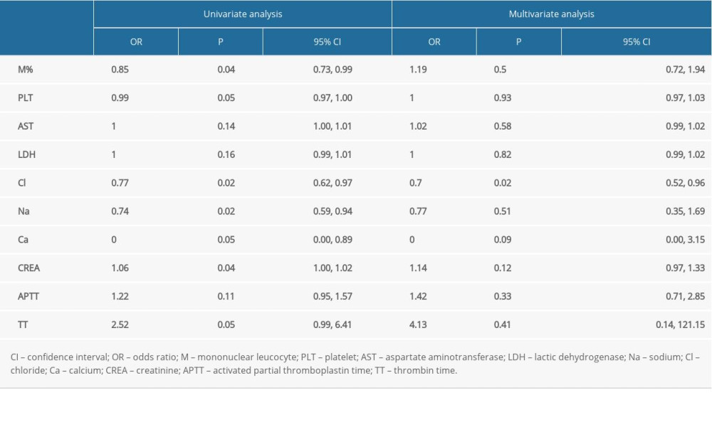
We selected several routine blood, inflammatory, myocardial enzyme, liver function, renal function, electrolyte, and routine blood coagulation indexes and drew a line chart (Figure 2). The red broken line represents the group with neurological complications, while the blue line represents the group without neurological complications. The 2 groups showed similar trends in the results of routine blood tests. In the routine blood test results, the neutrophil count increased 1 to 15 days after disease onset, while the lymphocyte count increased 15 days after disease onset. The platelet count decreased significantly 1 to 15 days after disease onset and gradually returned to normal. Among myocardial enzymes, the level of CK increased earlier than the level of LDH and AST in the group with neurological complications, and the levels of liver enzymes increased later than those of myocardial enzymes. The concentrations of Na, Cl, and Ca in both groups decreased significantly within 1 to 15 days after disease onset and then gradually returned to normal. The decrease in Cl concentration within 1 to 5 days after disease onset was closely related to the occurrence of neurological complications (Table 5). Among the coagulation indexes, the APTT and TT in patients with neurological complications were significantly higher than those in patients without neurological complications within 1 to 15 days of disease onset, suggesting that the coagulation function was affected. No significant difference was observed in blood urea nitrogen and creatinine levels between the 2 groups; however, the creatinine level in the group with neurological complications was significantly lower than that in the group without neurological complications 15 days after disease onset.
Discussion
SFTS was first reported in 2008 in Huaiyang Mountain, Henan Province, China [24]. The pathogen was isolated from the patient’s serum in 2010 and was then named as SFTS virus, which has since been renamed Huaiyangshanbanyang virus [2]. In addition to indirect effects induced by a cytokine storm, autopsy results in patients with SFTS with neurological symptoms suggested that SFTSV nucleocapsid protein-positive immunoblasts were detected in all organs examined, including in the central nervous system and vascular lumina of each organ. This indicates that the deterioration of central nervous system function can also be directly caused by BHAV infection [25].
The incidence of neurological complications associated with SFTS was 57.02% in the present study. The estimated percentage of neurological complications has been reported to range between 19% and 76% [22,26,27], which was consistent with the results of our study. We found that the neurological symptoms with the highest incidence rates were involuntary tremors (tongue and mandible), cognitive disorders, and limb tremors. In addition, there were consciousness disorders, epilepsy, coma, and other more serious symptoms. The symptoms of muscle limb tremors in patients with SFTS have been found by many researchers, and these symptoms often occur before the severe disturbance of consciousness [23,28]. In the present study, the incidence of these symptoms was more than 70%, so we added the above contents in the diagnosis of neurological complications, which was conducive to the early detection of neurological damage. Cui et al found that 19.1% of patients were clinically diagnosed with encephalitis during hospitalization, and the common mental abnormalities recorded included mental vagueness, irritability, convulsion, lethargy, and coma, excluding muscle and limb tremors; therefore, the incidence of neurological complications in their study was lower than that in our study [26]. However, we believe that encephalitis is not a neurological complication but is a kind of nervous system damage syndrome caused by systemic multiple organ damage.
Previous studies have showed that cerebrospinal fluid samples of patients, craniocerebral magnetic resonance imaging, and electroencephalogram have limited value in clinical application in the diagnosis of neurological complications [26,28]. Moreover, patients with disturbance of consciousness are often unable to complete these examination methods. We suggest that the onset of muscle tremors, limb tremors, cognitive impairment, and other symptoms should be considered as neurological complications, rather than waiting for a patient to develop serious symptoms or loss of consciousness. We also suggest that the basis of diagnosis cannot completely rely on examinations.
On the other hand, we found that patients with neurological complications were more likely to have lymph node enlargement, diarrhea, cough, expectoration, pulmonary rale, subcutaneous bleeding, and gastrointestinal bleeding. These symptoms mainly involved the respiratory system and the blood clotting system. Additionally, we found that spots of subcutaneous bleeding and pulmonary rales may indicate neurological complications, findings that have not been mentioned in previous studies. Thus, if a patient is suspected of having neurological complications, observing the extent of subcutaneous bleeding and pulmonary rales may be helpful in the diagnosis.
In addition, we also found that patients with neurological complications had a higher rate of sinus bradycardia, premature beats, atrial fibrillation, and T-wave abnormalities on ECG during hospitalization. Among them, sinus bradycardia, premature beats, and atrial fibrillation were all abnormal cardiac rhythms. All patients with abnormal ECG findings had no previous history of heart disease before they developed SFTS, and their ECG parameters gradually returned to normal after they recovered from SFTS, without any sequelae. This suggests that damage to the nervous system may affect changes in heart rhythm.
Similarly, liver enzyme, myocardial enzyme, and coagulation indexes also changed significantly in patients with neurological complications in the present study. Cl, LDH, and CRP were observed to be closely related to the occurrence of neurological complications, which was not completely consistent with the conclusions of previous studies [26]. According to previous research, the clinical course of SFTS is divided into 4 periods: a latent period (approximately 1 week), fever period (days 1–7 of onset), multiple organ dysfunction period (days 7–13 of onset), and decubation [29]. In the present study, the LDH of patients with neurological complications reached the peak at the multiple organ dysfunction period and return to normal at decubation, while the difference in LDH in the other group was mild (Figure 2). Similar differences can be seen in levels CK, AST, ALP, and GGT (Figure 3), and all of these indicators are risk factors associated with the severity of SFTS [30]. Contrarily, we found that CRP, which is commonly seen in acute infections or trauma, increased gradually and reached a peak at decubation in the neurologic complications group (Figure 2). This indicated that infection occurred in the recovery period of SFTS. The decrease in Na, Cl, and Ca occurred during the fever period and returned to the normal range during the recovery period (Figure 2). As for electrolyte disorders caused by SFTS, previous studies have confirmed that patients with SFTS usually have hyponatremia [31], which agrees our results. Of note, we also pointed out hypocalcemia and hypochloremia and when these changes took place. Furthermore, hypochloremia can be used as an indicator for early prediction of neurological complications, and this can provide a laboratory diagnostic basis for early diagnosis of the occurrence of nervous system complications.
Current treatments for SFTS include convalescent plasma [32], favipiravir [33,34], and ribavirin [35]. There is no specific drug for the treatment of nervous system symptoms. Gamma globulin is the first choice for the treatment of acute and chronic neuropathy [36]. Moreover, gamma globulin can affect the differentiation process of Schwann cells in the nervous system and improve their regenerative potential [37]. Studies have also shown that gamma globulin is effective in treating encephalitis caused by various viruses, such as the West Nile virus [38]. Therefore, gamma globulin may have a certain therapeutic effect on the neurological complications induced by BHAV, which needs to be confirmed in future studies.
Relatively mild symptoms, such as muscle tremors, limb tremors, and cognitive impairment, have been overlooked in many studies of neurological complications. However, these are actually the most common neurological symptoms of SFTS. We included these symptoms in the scope of neurological complications, and this is where we differed from previous studies. In previous studies, neurological complications were only diagnosed when patients suffered from disturbances of consciousness, coma, or convulsions. In addition, we found abnormalities in the cardiac rhythm in patients with neurological complications and that hypochloremia can be used as an early predictor of neurological complications.
Limitations of our study were that it was a retrospective study and lacked a rigorous experimental design. The diagnosis of neurological symptoms depended entirely on the judgment of the clinician, which may have resulted in delays in the detection of neurological symptoms in some patients.
Conclusions
In conclusion, the early diagnosis of SFTS with neurological complications cannot rely only on the observation of muscle tremors, limb tremors, and cognitive impairment. The occurrence of nervous system complications should also be indicated by the manifestations of subcutaneous bleeding points and lung rales. In addition, a change of ECG rhythm, increase of LDH, and significant decrease of Cl can be used as a basis for the early diagnosis of neurological complications.
Figures
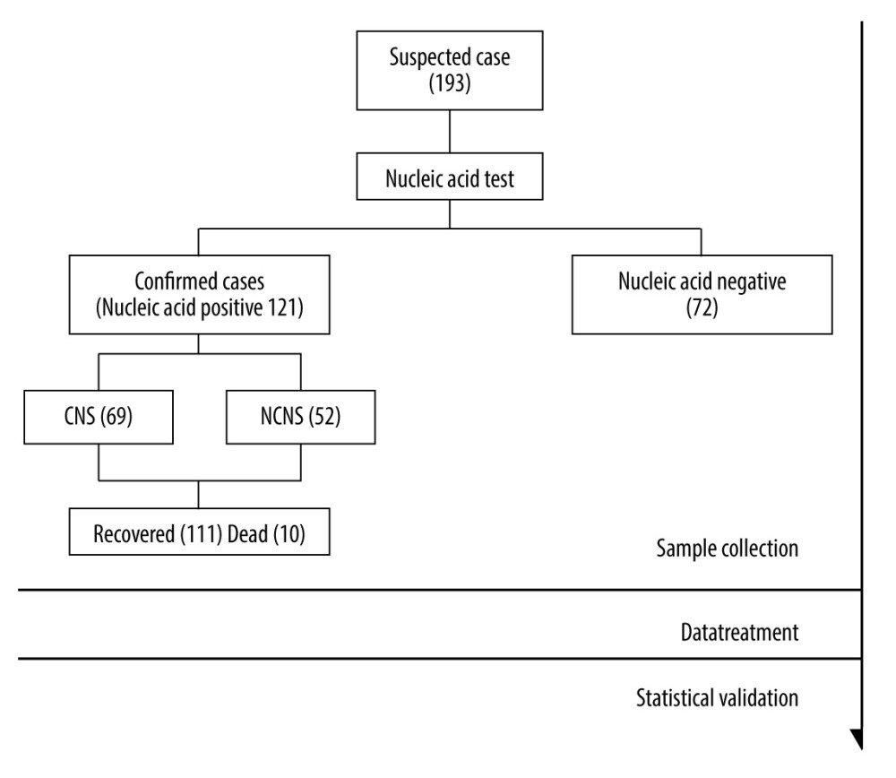 Figure 1. Research flowchart. Severe fever with thrombocytopenia syndrome nucleic acid tests were performed on the first or second day of admission. During hospitalization, ECG monitoring was performed daily and blood pressure and pulse were recorded. Clinical symptoms and signs of patients were recorded daily, with the focus on the observation of neurological symptoms. During the first and second weeks of hospitalization, routine blood, biochemical, urine, coagulation, and inflammatory indexes were checked daily. During the third and fourth weeks of hospitalization, laboratory tests were performed according to the patient’s condition. (Made by Microsoft office Word 2007, Microsoft USA).
 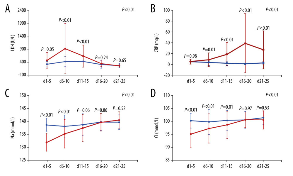 Figure 2. (A–D) The broken-line graph of lactic dehydrogenase, C-reactive protein, sodium, and chloride in confirmed cases of patients with severe fever with thrombocytopenia syndrome. The red broken line represents the group with neurological complications, and the blue line represents the group without neurological complications. (Made by Microsoft office Excel 2007, Microsoft USA).
 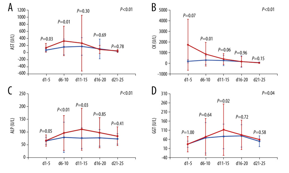 Figure 3. (A–D) The broken-line graph of aspartate aminotransferase, creatinine kinase, alkaline phosphatase, and gamma-glutamyl transferase in confirmed cases of patients with severe fever with thrombocytopenia syndrome. The red broken line represents the group with neurological complications, and the blue line represents the group without neurological complications. (Made by Microsoft office Excel 2007, Microsoft USA).
 Tables
Table 1. Demographic characteristics of patients with severe fever with thrombocytopenia syndrome.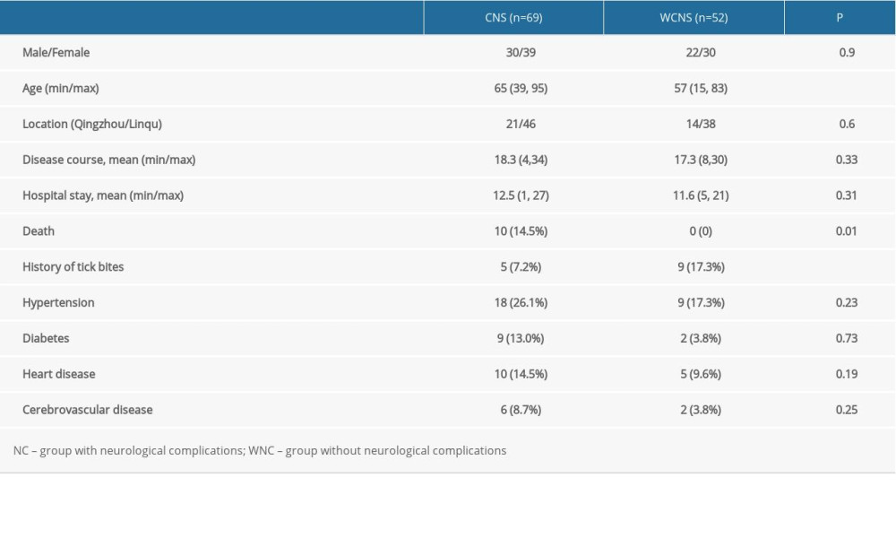 Table 2. Clinical symptoms and signs in patients with severe fever with thrombocytopenia syndrome.
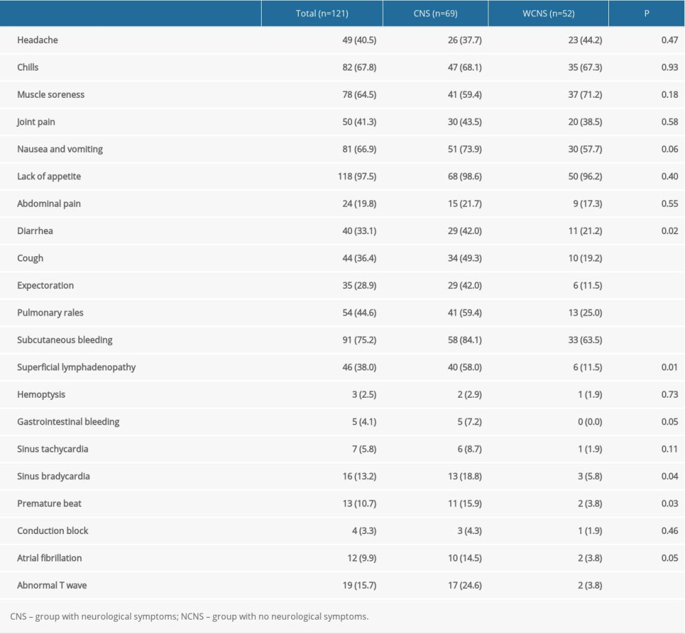 Table 3. Associations of clinical manifestation and laboratory parameters with neurological symptoms by multivariate logistic regression analysis.
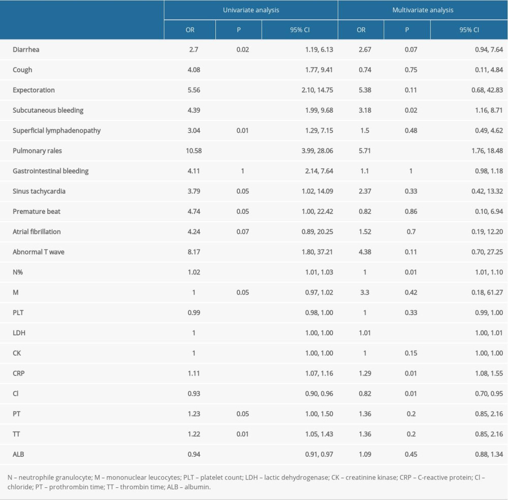 Table 4. Laboratory parameters of patients with severe fever with thrombocytopenia syndrome.
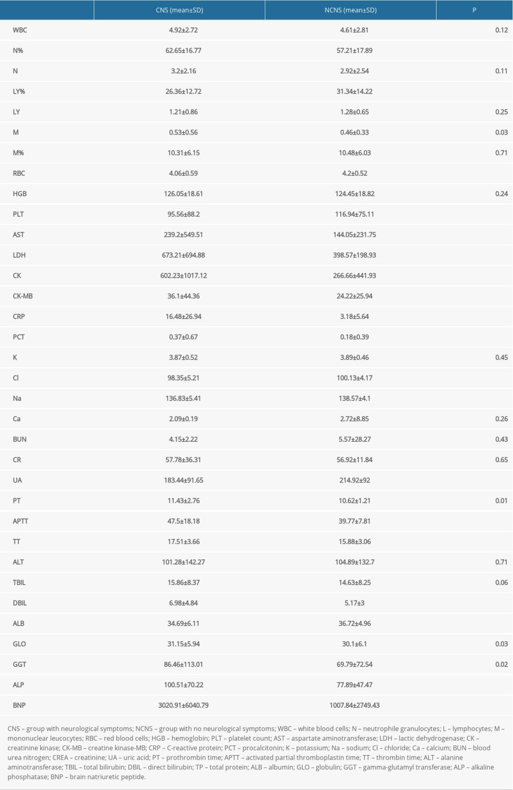 Table 5. Association between laboratory parameters in 1 to 5 days of disease onset with neurological symptoms by multivariate logistic regression analysis.
References
1. Maes P, Adkins S, Alkhovsky SV, Taxonomy of the order Bunyavirales: Second update 2018: Arch Virol, 2019; 164(3); 927-41
2. Yu XJ, Liang MF, Zhang SY, Fever with thrombocytopenia associated with a novel bunyavirus in China: N Engl J Med, 2011; 364(16); 1523-32
3. Yun SM, Song BG, Choi W, first isolation of severe fever with thrombocytopenia syndrome virus from Haemaphysalis longicornis ticks collected in severe fever with thrombocytopenia syndrome outbreak areas in the Republic of Korea: Vector Borne Zoonotic Dis, 2016; 16(1); 66-70
4. Yun SM, Park SJ, Park SW, Molecular genomic characterization of tick- and human-derived severe fever with thrombocytopenia syndrome virus isolates from South Korea: PLoS Negl Trop Dis, 2017; 11(9); e0005893
5. Yun Y, Heo ST, Kim G, Phylogenetic analysis of severe fever with thrombocytopenia syndrome virus in South Korea and migratory bird routes between China, South Korea, and Japan: Am J Trop Med Hyg, 2015; 93(3); 468-74
6. Gai Z, Liang M, Zhang Y, Person-to-person transmission of severe fever with thrombocytopenia syndrome bunyavirus through blood contact: Clin Infect Dis, 2012; 54(2); 249-52
7. Tang X, Wu W, Wang H, Human-to-human transmission of severe fever with thrombocytopenia syndrome bunyavirus through contact with infectious blood: J Infect Dis, 2013; 207(5); 736-39
8. Zhao L, Li J, Cui X, Distribution of Haemaphysalis longicornis and associated pathogens: Analysis of pooled data from a China field survey and global published data: Lancet Planet Health, 2020; 4(8); 320-29
9. Du Z, Wang Z, Liu Y, Ecological niche modeling for predicting the potential risk areas of severe fever with thrombocytopenia syndrome: Int J Infect Dis, 2014; 26; 1-8
10. Liu K, Cui N, Fang LQ, Epidemiologic features and environmental risk factors of severe fever with thrombocytopenia syndrome, Xinyang, China: PLoS Negl Trop Dis, 2014; 8(5); 2820
11. Ding S, Yin H, Xu X, A cross-sectional survey of severe fever with thrombocytopenia syndrome virus infection of domestic animals in Laizhou City, Shandong Province, China: Jpn J Infect Dis, 2014; 67(1); 1-4
12. Niu G, Li J, Liang M, Jiang X, Severe fever with thrombocytopenia syndrome virus among domesticated animals, China: Emerg Infect Dis, 2013; 19(5); 756-63
13. Kohl C, Brinkmann A, Radonić A, Zwiesel bat banyangvirus, a potentially zoonotic Huaiyangshan banyangvirus (Formerly known as SFTS)-like banyangvirus in Northern bats from Germany: Sci Rep, 2020; 10(1); 1370
14. Shin J, Kwon D, Youn SK, Characteristics and factors associated with death among patients hospitalized for severe fever with thrombocytopenia syndrome, South Korea, 2013: Emerg Infect Dis, 2015; 21(10); 1704-10
15. Saijo M, Pathophysiology of severe fever with thrombocytopenia syndrome and development of specific antiviral therapy: J Infect Chemother, 2018; 24(10); 773-81
16. Guo CT, Lu QB, Ding SJ, Epidemiological and clinical characteristics of severe fever with thrombocytopenia syndrome (SFTS) in China: An integrated data analysis: Epidemiol Infect, 2016; 144(6); 1345-54
17. He Z, Wang B, Li Y, Severe fever with thrombocytopenia syndrome: A systematic review and meta-analysis of epidemiology, clinical signs, routine laboratory diagnosis, risk factors, and outcomes: BMC Infect Dis, 2020; 20(1); 575
18. Kim KH, Yi J, Kim G, Severe fever with thrombocytopenia syndrome, South Korea, 2012: Emerg Infect Dis, 2013; 19(11); 1892-94
19. Takahashi T, Maeda K, Suzuki T, The first identification and retrospective study of severe fever with thrombocytopenia syndrome in Japan: J Infect Dis, 2014; 209(6); 816-27
20. Tran XC, Yun Y, Van An L, Endemic Severe fever with thrombocytopenia syndrome, Vietnam: Emerg Infect Dis, 2019; 25(5); 1029-31
21. McMullan LK, Folk SM, Kelly AJ, A new phlebovirus associated with severe febrile illness in Missouri: N Engl J Med, 2012; 367(9); 834-41
22. Park SY, Kwon JS, Kim JY, Severe fever with thrombocytopenia syndrome-associated encephalopathy/encephalitis: Clin Microbiol Infect, 2018; 24(4); 432.e1-.e4
23. Gai ZT, Zhang Y, Liang MF, Clinical progress and risk factors for death in severe fever with thrombocytopenia syndrome patients: J Infect Dis, 2012; 206(7); 1095-102
24. Xu B, Liu L, Huang X, Metagenomic analysis of fever, thrombocytopenia and leukopenia syndrome (FTLS) in Henan Province, China: Discovery of a new bunyavirus: PLoS Pathog, 2011; 7(11); 1002369
25. Kaneko M, Shikata H, Matsukage S, A patient with severe fever with thrombocytopenia syndrome and hemophagocytic lymphohistiocytosis-associated involvement of the central nervous system: J Infect Chemother, 2018; 24(4); 292-97
26. Cui N, Liu R, Lu QB, Severe fever with thrombocytopenia syndrome bunyavirus-related human encephalitis: J Infect, 2015; 70(1); 52-59
27. Kato H, Yamagishi T, Shimada T, epidemiological and clinical features of severe fever with thrombocytopenia syndrome in Japan, 2013–2014: PLoS One, 2016; 11(10); e0165207
28. Deng B, Zhou B, Zhang S, Clinical features and factors associated with severity and fatality among patients with severe fever with thrombocytopenia syndrome Bunyavirus infection in Northeast China: PLoS One, 2013; 8(11); e80802
29. Chen Y, Jia B, Liu Y, Risk factors associated with fatality of severe fever with thrombocytopenia syndrome: A meta-analysis: Oncotarget, 2017; 8(51); 89119-29
30. Liu J, Fu H, Sun D, Analysis of the laboratory indexes and risk factors in 189 cases of severe fever with thrombocytopenia syndrome: Medicine (Baltimore), 2020; 99(2); 18727
31. Xu X, Sun Z, Liu J, Analysis of clinical features and early warning indicators of death from severe fever with thrombocytopenia syndrome: Int J Infect Dis, 2018; 73; 43-48
32. Yoo JR, Kim SH, Kim YR, Application of therapeutic plasma exchange in patients having severe fever with thrombocytopenia syndrome: Korean J Intern Med, 2019; 34(4); 902-9
33. Song R, Chen Z, Li W, Severe fever with thrombocytopenia syndrome (SFTS) treated with a novel antiviral medication, favipiravir (T-705): Infection, 2020; 48(2); 295-98
34. Tani H, Fukuma A, Fukushi S, Efficacy of T-705 (Favipiravir) in the treatment of infections with lethal severe fever with thrombocytopenia syndrome virus: mSphere, 2016; 1(1); e00061-15
35. Liu W, Lu QB, Cui N, Case-fatality ratio and effectiveness of ribavirin therapy among hospitalized patients in china who had severe fever with thrombocytopenia syndrome: Clin Infect Dis, 2013; 57(9); 1292-99
36. Buttmann M, Kaveri S, Hartung HP, Polyclonal immunoglobulin G for autoimmune demyelinating nervous system disorders: Trends Pharmacol Sci, 2013; 34(8); 445-57
37. Tzekova N, Heinen A, Bunk S, Immunoglobulins stimulate cultured Schwann cell maturation and promote their potential to induce axonal outgrowth: J Neuroinflammation, 2015; 12; 107
38. Srivastava R, Ramakrishna C, Cantin E, Anti-inflammatory activity of intravenous immunoglobulins protects against West Nile virus encephalitis: J Gen Virol, 2015; 96(Pt 6); 1347-57
Figures
 Figure 1. Research flowchart. Severe fever with thrombocytopenia syndrome nucleic acid tests were performed on the first or second day of admission. During hospitalization, ECG monitoring was performed daily and blood pressure and pulse were recorded. Clinical symptoms and signs of patients were recorded daily, with the focus on the observation of neurological symptoms. During the first and second weeks of hospitalization, routine blood, biochemical, urine, coagulation, and inflammatory indexes were checked daily. During the third and fourth weeks of hospitalization, laboratory tests were performed according to the patient’s condition. (Made by Microsoft office Word 2007, Microsoft USA).
 Figure 2. (A–D) The broken-line graph of lactic dehydrogenase, C-reactive protein, sodium, and chloride in confirmed cases of patients with severe fever with thrombocytopenia syndrome. The red broken line represents the group with neurological complications, and the blue line represents the group without neurological complications. (Made by Microsoft office Excel 2007, Microsoft USA).
 Figure 3. (A–D) The broken-line graph of aspartate aminotransferase, creatinine kinase, alkaline phosphatase, and gamma-glutamyl transferase in confirmed cases of patients with severe fever with thrombocytopenia syndrome. The red broken line represents the group with neurological complications, and the blue line represents the group without neurological complications. (Made by Microsoft office Excel 2007, Microsoft USA).
 Tables
 Table 1. Demographic characteristics of patients with severe fever with thrombocytopenia syndrome.
 Table 2. Clinical symptoms and signs in patients with severe fever with thrombocytopenia syndrome.
 Table 3. Associations of clinical manifestation and laboratory parameters with neurological symptoms by multivariate logistic regression analysis.
 Table 4. Laboratory parameters of patients with severe fever with thrombocytopenia syndrome.
 Table 5. Association between laboratory parameters in 1 to 5 days of disease onset with neurological symptoms by multivariate logistic regression analysis.
 Table 1. Demographic characteristics of patients with severe fever with thrombocytopenia syndrome.
 Table 2. Clinical symptoms and signs in patients with severe fever with thrombocytopenia syndrome.
 Table 3. Associations of clinical manifestation and laboratory parameters with neurological symptoms by multivariate logistic regression analysis.
 Table 4. Laboratory parameters of patients with severe fever with thrombocytopenia syndrome.
 Table 5. Association between laboratory parameters in 1 to 5 days of disease onset with neurological symptoms by multivariate logistic regression analysis.
 In Press
15 Apr 2024 : Laboratory Research
The Role of Copper-Induced M2 Macrophage Polarization in Protecting Cartilage Matrix in OsteoarthritisMed Sci Monit In Press; DOI: 10.12659/MSM.943738
07 Mar 2024 : Clinical Research
Knowledge of and Attitudes Toward Clinical Trials: A Questionnaire-Based Study of 179 Male Third- and Fourt...Med Sci Monit In Press; DOI: 10.12659/MSM.943468
08 Mar 2024 : Animal Research
Modification of Experimental Model of Necrotizing Enterocolitis (NEC) in Rat Pups by Single Exposure to Hyp...Med Sci Monit In Press; DOI: 10.12659/MSM.943443
18 Apr 2024 : Clinical Research
Comparative Analysis of Open and Closed Sphincterotomy for the Treatment of Chronic Anal Fissure: Safety an...Med Sci Monit In Press; DOI: 10.12659/MSM.944127
Most Viewed Current Articles
17 Jan 2024 : Review article
Vaccination Guidelines for Pregnant Women: Addressing COVID-19 and the Omicron VariantDOI :10.12659/MSM.942799
Med Sci Monit 2024; 30:e942799
14 Dec 2022 : Clinical Research
Prevalence and Variability of Allergen-Specific Immunoglobulin E in Patients with Elevated Tryptase LevelsDOI :10.12659/MSM.937990
Med Sci Monit 2022; 28:e937990
16 May 2023 : Clinical Research
Electrophysiological Testing for an Auditory Processing Disorder and Reading Performance in 54 School Stude...DOI :10.12659/MSM.940387
Med Sci Monit 2023; 29:e940387
01 Jan 2022 : Editorial
Editorial: Current Status of Oral Antiviral Drug Treatments for SARS-CoV-2 Infection in Non-Hospitalized Pa...DOI :10.12659/MSM.935952
Med Sci Monit 2022; 28:e935952


