18 February 2025: Review Articles
Hydrogels in Oral Disease Management: A Review of Innovations in Drug Delivery and Tissue Regeneration
Li Dongwen AEF 1, Man Dapeng EF 1, Yu Jiazhi ABEF 1, Li Xiaoguang ABG 1*
DOI: 10.12659/MSM.946122
Med Sci Monit 2025; 31:e946122


Abstract
ABSTRACT: The oral cavity is an open and complicated structure with a variety of factors affecting topical oral medications. The complicated physical and chemical surroundings of the oral cavity can influence the action of free drugs. Thus, the drug delivery system can serve as a support structure or carrier. Hydrogels are prospective tissue engineering biomaterials that demonstrate immense potential for oral tissue regeneration and drug delivery. Hydrogels are crosslinked polymer chains with a 3-dimensional network structure that can take up larger volumes of liquid and have a soft, porous structure that closely resembles living tissue. Hydrogels protect the active drug from systemic and topical elimination, increase the bioavailability and absorption into cells, and release or modify the therapeutic drug release immediately after dosing. In this review, we introduce the classification of hydrogels, introduce the application of hydrogels in oral diseases (periodontal disease, endodontics, oral mucosal disease, alveolar surgery, oral cancer, maxillofacial bone defects, and oral implantation), summarize the synthesis methods and the applications of hydrogels, and discuss the possible directions of the future development of hydrogels, which will provide a new idea for the formulation and production of a more advantageous and efficient topical oral drug delivery system.
Keywords: Hydrogels, Oral Medicine, Regeneration, Humans, Drug Delivery Systems, Tissue Engineering, Mouth Diseases, Biocompatible Materials, Administration, Oral
Introduction
Oral health is an important component of overall health and has a critical effect on people’s wellbeing and quality of life. The prevalence of oral diseases is high and places a heavy burden on the healthcare system [1]. The oral cavity is an indivisible part of the alimentary system and consists of a variety of hard and soft organizations, including the teeth, oral mucosa, periodontal tissues, maxilla, and mandible [2]. A multitude of microbial organisms are located in the oral cavity and colonize diverse sites in the mouth, including the surface of the teeth and the periodontium, and form biological films that intrude into the oral tissues [3]. Oral diseases are pathologic alterations that happen in the hard and soft fabric of the mouth and maxillofacial area [4]. The most common oral diseases involve dental caries, periodontitis, pulp necrosis, oral mucositis, and jawbone abnormalities [5]. Dental caries is the result of dietary, sugar-driven buildup of biofilm and localized activation of acidity. Due to the regular intake of sugar, a mainly acidic condition is formed, which favors the growth of acidic bacteria in biofilms [6]. Periodontal disease consists of a series of infections which affect the structures that provide support for the teeth (gums, alveolar bone, and periodontal ligament), beginning with the localized inflammation of the gums, which is triggered by a biological film of microorganisms that forms on the teeth and gingiva [7]. Bacteria-host interactions result in an inflamed response. Given the complexity and diversity of oral health problems, there is a need for dental biomaterials that can effectively interface with a range of tissues, from soft gingival tissues to hard bone tissues.
The material must be capable of enduring the challenging conditions of the oral environment, which include sudden changes in temperature, pH swings caused by salivary and biological membranes, and the existence of a wide range of bacteria [8]. Bacterial infections usually cause oral diseases, and the treatment process is divided into 2 stages. The first is to fight infection and control the progression of inflammation; It then promotes tissue regeneration. Systemic medication had been the mainstay of therapy for the management of the infectious diseases of the oral cavity for a long time. Nevertheless, whole-body use of the drug can lead to problems like drug tolerance and hepatotoxicity. Hydrogels have a high water content, soft structure, and porosity that is very similar to living tissue. As hydrogel generation technology continues to improve, hydrogels can be better used in the buccal cavity [9]. With regard to topical oral medications, the effect of the oral environment on the medication should be considered [10]. The flow rate of saliva in the buccal cavity can interfere to some degree with the efficacy of local anesthesia. Salivary removal can thin and diminish the activity of the active components in the oral milieu. Hence, it is important to design carrier systems that can continuously release active ingredients at predicted concentrations that are effective. Hydrogels are composed of a 3-dimensional (3D) web, and due to their hydrophilic groups, hydrogels are capable of attracting large amounts of water and expanding in water [11]. The web usually consists of crosslinked chains of polymers and can also occasionally be formed from clusters of crosslinked colloids [12]. Due to their ability to absorb water, hydrogels are elastic and soft [13]. Due to a variation in the gel structure in the presence of internal or external stimuli, hydrogels allow for controlled drug release [14]. With a proper releasing regime, the activated drug is able to maintain a highly concentrated level in the area for a prolonged time.
Hydrogels typically have a porous structure, due to the many tiny pores and voids formed by their internal network. By adjusting the pore structure, it can be made to match the surrounding tissues and promote cell attachment and ingrowth [15]. The injectable nature of hydrogel stents is advantageous for the reconstruction of endodontic tissues. Due to the tiny volume and complex structure of endodontic canals, the injection of hydrogel is able to access all the root canals due to its own properties [16]. Therefore, the use of hydrogel materials in pulpal tissue has been extensively studied. Hydrogels have attracted much attention in the area of periodontal tissue engineering and jaw bone tissue engineering because of their malleability and ability to be effectively absorbed by the peripheral tissues. Additionally, hydrogels conform to the buccal mucosa and demonstrate an adequate modulus of elasticity comparable to that of soft tissues. In addition, hydrogels have antimicrobial properties. First, they can physically isolate bacteria and resist the incursion and transmission of hazardous microorganisms. Second, hydrogels can serve as active drug delivery vehicles, facilitating the release of a wide range of antibacterial compounds. These hydrogels will react to outside physical, chemical, and biological stimuli to initiate the liberation of the antimicrobial agent. Third, these hydrogels can display antibacterial properties by modulating physical and chemical properties, like the porosity structure and moisture content. Thus, hydrogels have emerged as a prospective material for the management of dental disorders [2].
Hydrogels imitate the biochemical properties of the extracellular matrix (ECM) [17] and can be used for the transport of drugs and cells, and are therefore considered as a potential biomaterial [17]. Natural hydrogels are highly biologically compatible and can be degraded to nontoxic coproducts that can react with biomolecules. They are, however, limited by their mechanical weakness and immunogenicity [18]. In comparison, synthetic hydrogels have improved stability but degrade into toxic byproducts. Hence, synthetic-natural compound hydrogels are proposed. These composite hydrogels are the most frequently used matrix for cell delivery injections [19]. Hydrogels can form 3D substrates that encapsulate either sensitive biologically active compounds or viable cells, which raises hopes for rejuvenation and restoration of the craniofacial and buccal tissues. This review first describes the staple categorization of hydrogels (Figure 1) and then presents the application of hydrogels in oral diseases (Figure 2).
In this review, we first introduce the classification of hydrogels, further introduce the application of hydrogels in oral diseases (periodontal disease, endodontics, oral mucosal disease, alveolar surgery, oral cancer, maxillofacial bone defects, and oral implantation), summarize the synthesis methods and the applications of hydrogels, and discuss the possible directions of the future development of hydrogels, which will provide new ideas for the formulation and production of a more advantageous and efficient topical oral drug delivery system.
Classification of Hydrogels
NATURAL HYDROGELS:
Hydrogels of natural origin including cellulose, chitosan, collagen, alginate, agarose, hyaluronic acid (HA), gelatin, and fibrin [20].
HA is one of the most frequently used natural polymers [21]. Research has shown the important role of hyaluronan in biological processes, including angiogenesis, ECM structure, inflammation, and healing of wounds [22]. Singh et al infused HA hydrogel as a root canal regeneration scaffold into the root canal to treat bad teeth and successfully induced sustained growth of the tooth roots [23]. Alginate is a nanosaccharide isolated from brown algae or bacteria [24]. Pan et al successfully regenerated alveolar bone and soft tissues with biomimetic polysaccharide hydogel/hydroxyapatite composite scaffolds, which provided a novel way to restore clinical bone defects [25]. Alginate-prepared scaffold materials exhibit excellent tooth differentiation, while the composite of alginate and hydroxyapatite could lead to induced periodontal stem cell polarization in vitro [26]. Chitosan is a long-chain cationic polysaccharide [27]. Chitosan/beta-glycerophosphate thermo-sensitive hydrogel has been applied as a pharmaceutical drug delivery system containing both bone morphogenetic protein-7 and ornidazole for sustained release for the therapy of periodontal diseases [28]. Chitosan/beta-glycerophosphate thermosensitive hydrogels were used to load aspirin to keep inflammation under control and to load erythropoietin to promote regeneration of the periodontium [29]. Naringin is a natively extracted polymethoxyflavonoid with anti-inflammatory properties that inhibit the inflammatory state of periodontitis, and the hydrogel contains carboxymethyl-hexanoyl chitosan [30]. Gelatin is a thermophilic copolymer with superior gel-sol transformation behavior and biocompatibility [31]. Studies have found that tailored hydrogels that imitate natural cells can boost the effectiveness of endodontic treatments, and methacrylic acid gelatin-based hydrogels with tunable physical and mechanical properties have been determined to be an efficacious strategy for promoting pulpal rejuvenation [32,33]. Collagen is the primary ingredient of ECM, with excellent coagulation, poor immunogenicity, and good biocompatibility, to support and protect organisms and their organs. Xie et al produced oxide amylose crosslinked hydrogel of collagen, having excellent thermal and mechanical properties, and studied its effect toward bioregulation. The findings indicated that these hydrogels possess tremendous promise for use as bioscaffolding materials in the medical field [34].
SYNTHETIC HYDROGELS:
In contrast to natural hydrogels, which have inferior mechanic properties, poor stabilization, and inefficient sterilization, the synthetic polymer hydrogels are of high magnetic weight and have consistent mechanical properties. Chow et al made a biodegradable hydrogel with bioactive and excellent mechanical performance for tissue engineering using polyethylene glycol (PEG) acrylate [35]. Polylactic acid is a bio-degradable synthesized polymer extracted from rich resources, and it is extensively applied in bio-medical areas because of its favorable biocompatibility, as well as lower virulence, bio-degradability, ease of processing, and environmental friendliness [36]. PEG is non-immunogenic, harmless, biodegradable, and hydrophilic and is a synthetic polymer with a wide range of biological applications [37]. Poly(d,l) lactic acid can be copolymerized with synthesized polymers such as PEG to form polylactic acid-PEG hydrogels, which can promote the differentiation of dental pulp stem cells (DPSCs) in vitro [38]. Gelatin methacrylate (GelMA) is a photocrosslinked hydrogel that incorporates the features of natural and synthetic bio-materials. GelMA’s photocrosslinking capability enables it to be a versatile option for gel manufacture, cell containment, and mechanical property conditioning [39]. The physiological nature of the GelMA hydrogel can be modified by varying the concentrate of GelMA or photocrosslinker, thereby making it appropriate for the encapsulation of cells at a temperature of 37°C. Ma et al developed an injectable crosslinkable hydrogel composed of GelMA/PEG dimethacrylate as a bio-scaffold to offer a proper surrounding for the growth of periodontal stem cells [40]. Sood et al created carboxymethyl cellulose-grafted polyhydrogels possessing antimicrobial characteristics for targeting drug delivery and antibiotic therapy [41]. The above synthetic methods modified the natural hydrogels in different degrees and increased the scope of application of hydrogels.
Synthetic hydrogels offer superior mechanical properties and stable mass stability, but the presence of unreactive monomers, initiators, or crosslinkers can cause inflammation or cytotoxicity [42]. Hydrogels can also be used in combination with other materials to improve the performance of composites [43,44].
Hydrogels in Oral Diseases
PERIODONTITIS:
Periodontitis is an inflamed disease marked by infection, a devastating immunological response, and pathologic dentine bone resorption [45]. In current periodontitis regimens, it is difficult to achieve antimicrobial and periodontal tissue regeneration effects, and topical medication has appeared as a prospective tactic for the management of periodontitis [46]. Nevertheless, the influence of oral environmental characteristics can lead to frequent topical medication, poor antimicrobial efficacy, and suboptimal periodontal regeneration [47]. Hence, it is scientifically significant to explore topical drug administration systems that can suppress pathological organisms and facilitate the regularization of periodontal tissues (Table 1).
Tan et al indicated a supramolecular hydrogel, stromal cell-derived factor 1 (SDF-1)/bone morphogenetic protein-2 (BMP-2)/amphiphilic peptide, Nap-Phe-Phe-Tyr (NapFFY), was prepared by co-assembling the hydrogel agent NapFFY together with SDF-1 and BMP-2. The experimental results showed that these 2 biological active factors could be optimally, simultaneously, and persistently liberated from the hydrogel, which could facilitate periodontal bone tissue’ proliferation and rebuild in an effective way. SDF-1/BMP-2/NapFFY hydrogels can be used as an alternative to clinical bone grafts for the repair of periodontal bone defects [48]. Jo et al discovered bioactive additives made up of proteins and propeptides were added to the hydrogel to promote bone regeneration through controlled release. A bi-active calcium-accumulating peptide was synthesized, which included collagen-binding motifs that can induce bone-forming differentiation. Using a tyrosine residue in calcium-accumulating peptide, the peptide was directly chemically conjugated to gelatin-based enzymatically crosslinked hydroxyphenylpropionic acid hydrogels under H2O2/amaranth peroxidase conditions. Human periodontal ligament stem cells (hPDLSCs) were loaded into calcium-accumulating peptide hydrogels to test the effect of accelerated bone formation. Calcium-accumulating peptide hydrogel indexes bone minerals surrounding the periodontal lodgement of stem cells and augments the expressivity of integumentary hallmarks in vitro. This hydrogel scaffold system enhances bone repair and can be used as a vehicle to transport stem cell bioactivation in tissue regeneration therapies [49].
The guided tissue regeneration technique is an efficacious method for repairing periodontal defects. The ideal barrier membrane should have appropriate antimicrobial and osteoinductive activities and good biodegradability. Wu et al showed zinc oxide nanoparticles were homogenized into ketone hydrogel (ChT-1% ZnO) using a single-step solubilization and rejuvenation process with alkali/urea solvent. ChT-1% ZnO has osteogenic and antibacterial effects in vitro and was further validated in vivo by a model of periodontal defects in rats [50]. Hypoxia-inducible factor 1α (HIF-1α) has a pivotal function in tissue regeneration. Preventing the degradation of HIF-1α by prolyl hydroxylase through pharmacological actions under normal hypoxic circumstances is becoming a prospective approach in renewable medicine. Nagai et al showed the ability of 1,4-dihydrophenyl lauric acid-4-keto-3-carboxylic acid (1,4-DPCA/hydrogel), an injectable prolyl hydroxylase inhibitor based on hydrogel, to facilitate the alveolar bone regeneration lost due to clinical periodontitis. Gingival HIF-1α protein expression and alveolar bone rejuvenation rate were considerably higher in hydrogel-injected mice than in the drug control group. 1,4-DPCA/hydrogel evoked an increase in bone remodeling, an elevation in the expansion of osteogenic genes, and a decrease in the expression of pro-inflammatory cytokine genes [51]. Glycogen synthase kinase-3 beta inhibitors (BIO) have been shown to be powerful inflammation modulators and bone-forming agents for the treatment of periodontitis. Almoshari et al synthesized pyrophosphorylated Pluronic F127 (F127-PPi) and blended it with plain F127 to formulate an injection-ready, temperature-controlled BIO hydrogel preparation PF127, which sticks to a rigid tissue and gradually liberates BIO for local curative actions. PF127 hydrogel binds more strongly to hydroxyapatite, the solubility of BIO in PF127 solution is significantly increased, and the degree of increase is proportional to the polymer concentration. Research has shown that thermostable PF127 hydrogel is an efficacious localized medication administration device for improved treatment of periodontitis and related diseases [52].
A cell-carrying aligned porous hydrogel scaffold was fabricated using chitosan and oxidized chondroitin sulfate as substrates, aiming to induce aligned regeneration of periodontal tissues. The well aligned porosity hydrogel group evoked a more organized periodic lens formation than did the plain hydrogel group. Wang et al suggested that the use of aligned mesoporous hydrogel scaffolds integrated with PDLSCs and gingival mesenchymal stem cells (GMSCs) – chitosan-oxidized chondroitin sulfate-PDLSC/GMSC composites – is promising to facilitate regeneration of endodontic tissues [53]. Chitosan/β-glycerophosphate hydrogels and chitosan/β-glycerophosphate/hydroxyapatite hydrogels were produced by the sol-gel approach. Chen et al showed that chitosan/β-glycerophosphate/hydroxyapatite hydrogel had good cytocompatibility as well as superiority in promoting the osteogenic differentiation of endodontic stem cells in vitro, which suggests that the binding of endodontic stem cells with this hydrogel has the application prospect in osseous tumor organization engineering [54]. Chien et al showed that injectable thermosensitive chitosan/gelatin/phosphoglycerol hydrogels provide a 3D milieu for grafted stem cells and enhance stem cell transport and implantation. The induced pluripotent stem cells (iPSCs)-BMP-6 hydrogel compound facilitated the formation of osteoblasts, neoconjunctive tissue polarization, and periodontal ligaments. The association of hydrogel-encapsulated iPSCs together with BMP-6 offers a new tactic to promote periapical rejuvenation. This combo would not only facilitate the transplantation of stem cells, but would also minimize the development of inflammation, resulting in a high probability of periodontal rejuvenation [55]. Amphoteric carboxymethyl-hexanoyl chitosan, β-glycerophosphate, and glycerin were made into a hydrogel, which gelated fast when exposed to a temperature of 37°C. The gelatinization of carboxymethyl-hexanoyl chitosan was then rapidly reduced to that of β-glycerophosphate. At the sites of naringin-loaded carboxymethyl-hexanoyl chitosan-β-glycerophosphate glycerol hydrogel, there was a significant reduction in periodic bond wastage and inflamed infiltrate. Chang et al suggested that naringin-loaded carboxymethyl-hexanoyl chitosan-β-glycerophosphate glycerol hydrogel can be used to suppress the development of the experimental periodontitis [56]. Thermosensitive chitosan hydrogel was prepared with an autoclavable chitosan flour and β-glycerophosphate (chitosan-PA/glycerophosphate). Zang et al showed that chitosan-PA/glycerophosphate hydrogels were characterized by short time of gelling, high viscosity, strong liquid absorption, suitable time of degradation, and mesoporous framework. Chitosan-PA/glycerophosphate thermosensitive hydrogel is a promising injectable tissue engineering scaffold with suitable physicochemical properties and biocompatibility [57]. Li et al showed chitosan nanoparticles containing bone morphogenetic protein-2 plasmid DNA (pDNA-BMP-2) were embedded into chitosan hydrogels containing α,β-glycerophosphate, called CS/CSn(pDNA-BMP-2)-GP. The results showed that the CS/CSn(pDNA-BMP-2)-GP composite system had superior biological characteristics and biocompatibility, suggesting its potency as a genetic delivering vector and regenerative scaffold for tissue regeneration for embedded periodontal restorations [58].
Anitua et al indicated the combination of gelatin and alginate (GA), enriched with either hydroxyapatite (GAHA), or hydroxyapatite and platelet-rich growth factors (PRGF; GAHAP), 3 different biological matrices, can provide a conducive micromilieu for human (h)DPSCs. The adhesion and chemotaxis of the cells were significantly enhanced by the addition of PRGF. The findings indicated that GAHA and GAHAP boosted the growth rate of cell protrophy and significantly stimulated osteogenic differentiation [59]. Ma et al showed nanoscale cell-loaded 3D hydrogel arrays with a gradient of ECM components were generated by manipulating the bulk proportions of the 2 types of hydrogels, like GelMA and PEG dimethacrylate. The behavior of PDLSCs in GelMA/PEG arrays (eg, cell viability, spreading) appeared to be dependent on the bulk ratio of GelMA/PEG, as the cell vibrance and splayed area declined with an increase in the ratio of PEG. This will help to investigate the interplay between cells and biomaterials in 3D space and boost the renewal of functional tissues [60]. hPDLCs and bone marrow stromal cells (BMSCs) were incubated with calix alginate hydrogel and a polylactic acid hydrogel, followed by examination of the proliferation of the cells. Expression of inflammation-related factor genes was observed in hPDLCs, and the expression of osteogenesis-related genes was visualized in BMSCs. Calcium alginate hydrogel caused less inflammation than did polylactic acid. The mineralized nodule count and the presentation of osteoblast related genes were remarkably increased in the hydrogel group, as opposed to the control group. The material showed good biocompatibility when implanted into the subcutaneous tissue. Calcium alginate hydrogel has better osteoinductive capacity than polylactic acid hydrogel. Chen et al suggested that a drug-containing calcium alginate hydrogel system can be used as a replacement material for bone defects of the oral cavity [61].
In the treatment of periodontitis, the continuous release of drugs has been a concern, and hydrogels can be used as a carrier to achieve this effect. Johnson et al suggested that antimicrobial-loaded hydrogels consisting of cellulose nanofibers and κ-carrageenan oligosaccharide nanoparticles can be used to treat periodontitis. This is a material with a high degree of thermostability, controllable emission, and water absorptivity. This ingredient has powerful antimicrobial activity toward periodontal pathogens. Malondialdehyde production was significantly increased, and biofilm formation and bacterial metabolic activity were reduced. It also decreases the reactive oxygen species and inflammatory factors produced by human gingival fibroblasts under inflammatory conditions. This hydrogel has antibacterial and anti-inflammatory characteristics and can be used to treat periodontitis [62]. Polysaccharide-based hydrogels are shaped by a dynamic crosslinked network of dynamic Schiff base bonds and dynamic ligand bonds, which provide a rapid gelation process, injectability, and superior self-healing properties. In particular, in the absence of external stimuli, the hydrogels shaped by the bi-dynamic network undergo a gel-sol transformation procedure. During the first phase of release of the drug, a continuous distribution characteristic of the drug was noticed, which was due to the process of spreading, whereas the complete release of the drug was attributed to the process of gel-sol transition. This suggests that injectable self-healing hydrogels having bi-dynamic bonds that modulate the gel-sol phase transition can be securely used for a controlled delivery therapy for periodontics. Guo et al suggested a polysaccharide-based hydrogel loading ginsenoside Rg1 and amniotic acid enhances periodontal regeneration in patients with periodontitis in vivo [63]. Morelli et al showed that micron-sized β-cyclodextrin hydrogels (bCD-Jef-MPs) suspended in a synthetic block polymer solution (Poloxamer 407) are highly hydrophilic and are enabled to undergo a fast, thermal-induced sol-gel stage transformation under body temperature. This system was demonstrated to realize sustainable releasing of antibacterial agents by in vitro experiments with a drug model of chlorhexidine gluconate [64].
ENDODONTICS:
Injectable hydrogels are of interest in pulpal regeneration because of their capability to infill inhomogeneous spaces, like pulpal hollows (Table 2). A new decellularized (d)ECM-loaded GelMA-5 wt% bioactive glass (BG) (Gel-BG) hydrogel can be used for dental pulp renewal. The effects of dECM levels on the physico-chemical properties and bioreactivity of Gel-BG hydrogels contacted with stem cells derived from stem cells from human exfoliated deciduous teeth (SHED) were investigated. Sadeghian et al indicated that the addition of 10 wt% of dECM dramatically increased the compressive strength of Gel-BG/dECM hydrogels, with proper bioactivation, rate of degradation, osteoinductivity, and mechanization, which is expected to be applied in clinical practice in the future [65]. Atila et al showed an injectable GelMA/thiolated pectin hydrogel, which carries electrospun core/shell fibers of melatonin-methylmethacrylate/tiglosporin-silk fibronectin, induces DPSC proliferation and pulpal differentiation by prolonging the release of tideglusib and melatonin, resulting in regeneration of vital dental pulp [66]. Athirasala et al used GelMA hydrogels with tunable physiological and mechanical properties to determine microenvironmental conditions to improve the movement, spreading, and multiplication of encapsulated osteoblast-like cells (OD21) as well as endothelial monolayers formed by endothelial colony-forming cells. The pre-vascularized pulp-like organization was constructed by injecting GelMA hydrogel prepolymer containing OD21 cells into the root canals of extraction teeth. The endothelial colony-forming cells that were seeded into the microchannels successively created monolayers and underwent vascular budding during 7 days of incubation, potentially yielding beneficial translational results [67]. Treated dentin matrix (TDM) has been used as a biologically active hydrogel for dentin rejuvenation in the dentin-pulp complex. Sedek et al investigated the synthesis, characterization, and transplantation optimization of syringable glycidyl methacrylate (GMA)/TDM hydrogel as a new photocrosslinked pulp sealant for dentin rejuvenation, photocrosslinking of gelatin (G)-GMA/TDM hydrogels with a novel bi-component photoinitiator system. The results showed that the G-GMA/TDM complex hydrogel formulated by the riboflavin/glycine photoinitiation system is a potentially biological active and bio-compatible system for cross-linking dentin regeneration as an active light pulp sealant [68]. Zhang et al reported a system of functional GelMA microspheres containing human platelet lysate, and Laponite nanoclay was prepared via the static microdroplet assay. The outcomes indicated that the GelMA/platelet lysate/Laponite microspheres remarkably improved the spreading, multiplication, and dentinogenic compartmentation of the wrapped hDPSCs, as compared with the pure GelMA microspheres. Furthermore, they facilitated microvessel generation and the registration of pulp-like tissues in vivo. This mixed microsphere system has tremendous future potential to accelerate pulp regeneration and microvessel formation in other tissues [69]. The formation of dentin restorations was induced by a straightforward UV approach using functionalized gelatin methacrylate containing gentian saponin R1 (Gel-MA/NGR1). The prepared hydrogels had appropriate interconnected porous microstructures and mechanical properties that facilitated cell adherence and multiplication. The hydrogels were characterized by hydrophilicity and sustainable distribution of the drug. Moreover, Gel-MA/NGR1 notably augmented dentin-forming compartmentation of mouse papilla cells by increasing the expansion levels of the dentin-forming signatures alkaline phosphatase and osteocalcin, as well as the level of extracellular matrix mineralization. Wang et al indicated that Gel-MA/NGR1 hydrogel facilitated restorative dentin production, suggesting that such hydrogel has significant promise as an endodontic cover material in inducing dentin production [70].
The microstructure, mechanical properties, rheological properties, and deformation of injection-ready dual-network chitosan hydrogels were investigated and compared with single-network chitosan hydrogels. The outcomes revealed that the dual-network hydrogels had desirable injectability, improved mechanical performance, and extended time for degradation. As a 3D system for cell culture, the properties of dual-network hydrogel could promote the dentinogenic differentiation and the mineralization of hDPSCs in vitro. Han et al suggest that dual-network glycol chitosan-based hydrogels are applicable to the rejuvenation of dental endodontic tissues [71]. Carvalho et al suggested chitosan hydrogel scaffolds (CS) were made functional to become a framework for loading human mastoid (SHED) stem cell secretions and assessing their biologic active functions and proangiogenic characteristics. Originally, the CS was enrolled in SHED secretions (CS-S), and the kinetics of the liberation of a few nutritional factors were evaluated. The impact of functioning scaffolds on apical papilla stem cells (SCAPs) was analyzed by proliferation and chemotaxis assays, and their vasculogenic potency was evaluated by a Matrigel canal creation assay with co-cultures of the human umbilical vein endothelial cells and SCAPs. SHEDs and SCAPs express a characteristic level of MSC signaling on surface markers. Cell multiplication increased markedly in the CS-S group, with faster chemotaxis and enhanced ability to develop vasculature-like constructs. CS-S offered a continuous and controllable liberation of nutrient factors, which paradoxically enhanced cell multiplication, tropism, and all angiogenic parameters in the coculture. Therefore, the functioning of the CS with a secretion carrier is a prospective promising platform for tissue engineering on the basis of cellular homing [72]. Zhang et al showed the effects of human endometrial-derived stromal cells (EnSCs) and titanium dioxide nanoparticles on pulp repair and rejuvenation in an ancient animal model were determined by investigating dentin production and organization of dentin regeneration. After obtaining and purifying collagenase, EnSCs were placed on 3D CSs with titanium dioxide nanoparticles. The binding of EnSCs with titanium dioxide nanoparticles and 3D chitosan support produced a synergistic effect, demonstrating an increase in the rate and quality of dentin formation [73]. Divband et al developed scaffolds laden with basic fibroblast growth factor (bFGF) and studied its effect on inducing angiogenesis in hDPSCs. Polycaprolactone (PCL)/CSs loaded with bFGF were demonstrated to possess the potency to facilitate the vasculogenesis of hDPSCs, which can supply the viability of the dentin-pulp composite as an initial requisite for pulpal regeneration program [74].
A systematic study of photocrosslinked hydrogels on the basis of HA and platelet lysate was performed. Platelet lysate, a growth factor engaged in orchestrating wound healing, is expected to provide specific biochemical cues for HA hydrogels to enhance recruitment, multiplication, and polarization of dental pulp cells. Almeida et al have shown that HA hydrogels containing polylactic acid promote cellular metabolism and spur the deposition of the mineralization matrix of hDPSCs, which clearly demonstrates the system’s potency in the restoration of destroyed pulp/dentin organization and regeneration of the dental pulp [75]. Silva et al investigated the viscoelastic and micostructural characterization of in situ created inventible HA hydrogels strengthened by cellulose nanocrystals as well as enrichment with platelet lysates. The cellulose nanocrystals increased the stabilization of the material, which prevented hydrolysis and enzymatic deterioration. The chemotaxis and persistence of platelet-derived growth factor and vascular endothelial growth factor released and derived from the polylactic acid-containing hydrogels were also improved. Hydrogels loaded with polylactic acid have preferential support properties for encapsulated human dental pulp cells. Finally, the polylactic acid-added hydrogel elicited chemotaxis and proangiogenic activity via facilitating recruitments of human dental pulp cells and cell budding in human dental pulp cells/human cord vein endothelial cells co-culture in vitro. An injection-ready HA/cellulose nanocrystal/platelet lysate hydrogel was identified with enhanced structural and biological features, which not only maintains platelet lysate delivery of chemotaxis and pro-angiogenic growth factors, but additionally enhances cell vitalization and angiogenic action. This hydrogel is very suitable as a controllable transport delivery system for growth factor, as well as a substrate to support cell culture and recruited and induced revascularization, with huge potency in regulating vascularized soft tissues, like dentin-endodontium composite [76]. Atila et al suggested tideglusib-containing injectable HA hydrogel and Rg1-containing chitosan microspheres were derived for vital pulp rejuvenation through the release of tideglusib and Rg1, with tideglusib triggering pulpal polarization by hDPSCs and Rg1 triggering pulpal revascularization by hDPSCs. Tideglusib and Rg1 were loaded in chitosan microspheres in HA and HA hydrogel, respectively. Hydrogel-cultured DPSCs showed odontogenic and vasculogenic differentiation at the level of gene presentation. This hydrogel has the potential to improve important pulp regeneration strategies [77].
Endodontic disease is a biofilm-mediated infection, and the main goal of treatment for endodontic disease is to eliminate bacterial biofilms in the root canal system. Therefore, in endodontic treatment, antibacterial properties play an important role. Bekhouche et al showed an novel fibrin hydrogel containing polylactic acid nanoparticles loaded with clindamycin (CLIN-PLA-NPs) conferred antimicrobial characteristics to the hydrogel. CLIN-PLA-NPs were fabricated using surface-active-agent-free nano-precipitation approach. The addition of CLIN-PLA-NP to fibrin hydrogels imparts antimicrobial and anti-biofilm properties to fibrin hydrogels while not influencing the viability and the function of the cells [78]. Another type of innovation is the cellularized fibrin hydrogel that has added chitosan, which gives this hydrogel antimicrobial properties. Chitosan in the fibrin network was found to have a potent antimicrobial effect, and DP-MSC showed similar survival rates, fibroblast-like patterns, growth rates, and type I/III collagen-producing ability. Ducret et al indicated that the inclusion of chitosan to fibrin hydrogel was beneficial in promoting the neogenesis of human dental pulp tissues because of its antimicrobial effect and because chitosan did not have significant adverse effects of dental pulp cells [79].
Direct pulp coverage is the procedure of using dental materials to treat exposure of the pulp in order to promote restorative dentin production and preservation of the pulp. To develop light-curing injection carriers for endodontic therapy, which includes direct pulpal coverage, PEG-maleate-citrate hydrogel was synthesized as a delivery carrier from PEG-maleate-citrate (45% w/v), acrylic acid (5% w/v), 2,2′-azobis(2-methylpropionamidine) dihydrochloride (0.1% w/v), and deionized water. The outcome indicated that the photo-curing time of hydrogel was equivalent to that of the compound resin. The cytotoxicity of hydrogels is similar to that of adhesive systems. In addition, calcium hydroxide hydrogels control the release of Ca(2+) [80]. Super molecular hydrogels produced by using low-molecular-weight gelling agents have attracted great interest in bio-medical applications. Yet, the limitations of in situ super molecular hydrogels are long gelation times and/or instability at high temperatures. Li et al suggested a stable Ag-isoG supramolecular hydrogel was constructed by an ultrafast in situ formation technique, and the hydrogelation process occurred within 1 s after the mixture of isoG and Ag+ at ambient environment conditions. This Ag-isoG hydrogel is stable even with exposure to high temperatures. Silver ions have significant antibacterial efficacy toward Staphylococcus aureus and Streptococcus oralis mutans. This hydrogel was in turn used in a root canal injury study, which showed a strong antibacterial action for Enterococcus faecalis, superior to that of ordinary calcium hydroxide pastes. This property promises Ag-isoG hydrogel as an acceptable alternative to intra-canal medications in root canal therapy [81]. The importance of the success of root canal treatment lies mainly in the radical removal of the biofilm of bacteria from the root canal. The effectiveness and biocompatibility of the materials used to sterilize the root canal are critical to the success of root canal treatment. In a study of a modification of nano-enzyme-loaded hydrogel (Fe3O4-CaO2 hydrogel), the results of in vitro experiments indicated that Fe3O4-CaO2 hydrogel releases reactive oxygen species, has excellent biocompatibility, and disrupts bacterial cell membranes. The outcome of in vivo experiments demonstrated that Fe3O4-CaO2 hydrogel could effectively remove root canal biomembrane inflammation and prevent further expansion of inflammation. In conclusion, Song et al showed Fe3O4-CaO2 hydrogel can be used as a novel and effective bio-compatible material for root canal disinfection. This study holds great promise for clinical root canal sterilization and can probably even be generalized to address other deep intractable incurable infections [82]. Bakhtiar et al showed fabrication of new injection-ready human amniotic membrane hydrogel stents crosslinked with matrix factors to promote the multiplication of DPSCs and optimize pulp renewal. Hydrogels crosslinked with low concentrations of genistein showed lower tooth discoloration. Hydrogels crossed-linked with 0.5 mM genistein degraded at a lower rate. Genistein crosslinking improves biodegradability and enhances biocompatibility of injectable human amniotic membrane hydrogels. Hydrogels wrapped with DPSCs are supportive of stem cell vitalization and growth. The highly revascularized pulp-like tissue formed by this bio-material shows the promising potential for pulp rejuvenation [83].
In recent years, the regeneration of dental pulp tissue has been realized by biological means and tissue engineering technology, so as to restore the physiological function of dental pulp while preserving the affected tooth. Transplantation of harvested stem cells shows great clinical potential in reconstruction of soft tissue niches in large animal models [84]. Siddiqui et al showed that placement of SLan, a self-assembled peptide hydrogel, in adult canine teeth following pulpotomy stimulates pulpal vascular regeneration, a non-cellular material-based method for facilitating pulp-like soft tissue rejuvenation in vivo [85]. RADA16-I is an ion-complexed self-assembling peptide that has a regularly collapsed minor configuration and assembles into well-ordered nanostructures. Dentonin is an extracellular matrix phophosphorylated glycoprotein with a fibrillated peptide motif that includes RGD and SGDG sequences. The combination of RAD and Dentonin produce a functioning self-assembled peptide RAD/Dentonin hydrogel support. Liu et al demonstrated that RAD/Dentonin could automatically assemble into a hydrogel with a β-sheet nanofibrous framework. RAD/Dentonin has good bio-compatibility and strengthens the adhesion growth, transport, dentinogenic derivation, and deposition of hDPSCs, and is expected to be used as a framework for pulpal tissue architecture [86].
ORAL MUCOSAL DISEASE:
Oral mucosal lesions are a cluster of disorders associated with high morbidity and uncontrollable symptoms, and over 25% of adolescent and young adults experience oral ulcers, which seriously impacts patients’ quality of life [87]. The oral mucosa has a moist and hyperdynamic surrounding [88], with constant flushing of the oral mucosa by endogenous saliva and exogenous food. Moreover, chewing, speaking, swallowing, and sometimes even changes in facial expression induce motions of the tongue and oral mucosa. These usually invalidate topical treatment strategies with protective materials and therapeutic agents because of their short residence time on the mucosal surface [89]. Commonly used topical medications are usually thinned or flushed out by saliva in less than an hour. From a patient-centered viewpoint, oral mucosal drug delivery is appealing because of its ability to be easily administered with no swallowing and increased patient security. Ideal oral mucosal restorative materials would be safe, usable, light, and pliable, and possess good moist-tissue adherence to withstand fluid washout and oral movement, to minimize discomfort to the patient. Several polymer-based bioadhesives were developed [90], but each product has significant limitations (Table 3).
A photostretchable cyclo-o-nitrophenyl modified HA was designed to produce 3 different reaction groups simultaneously under light irradiation at 395 nm: sulfhydryl, nitroso, and aldehyde groups. The thiol and nitroso groups generated in the procedure undergo rapid non-radiative crosslinking via S-nitrosylation [91], leading to the formation of thin gels within 5 s. At the same time, the photogenerated aldehydes are immobilized on the amino groups of the organization for in situ adhesion, thus creating a contained setting that prevents bacteria getting into the area of the wound. Furthermore, HA is a widely used biocompatible material with anti-inflammatory effects that promote M2 cell polarization [92]. One study improves on traditional treatments for oral mucosal diseases [93]. In the preparation of the buccal membrane, a self-forming micellar drug solubilizing agent was used as the membrane matrix in combination with a mucoadhesive polymer to enhance its hold on the mucous. Hydrophilic polymers with known mucus adhesion characteristics: hydroxypropyl methylcellulose or modified hydroxypropyl pea starch (Lycoat), and a graft copolymer (Soluplus) were used as solubilizers and film formers. The films were fabricated via a solid solvent casting method. Alopaeus et al showed that the hydroxypropyl methylcellulose-containing bipolymer films had better erosion resistivity and adherence properties compared than did the mono-polymer control films. Within the in vitro oral cavity model, these features were linked to an increase in residence period in the mimic oral mucosa. In addition, all films containing graft copolymers displayed similar furosemide permeation properties on oral TR146 epithelial cells. This study demonstrates the feasibility of an inexpensive and simple method to fabricate dry, solid bipolymer films containing advanced drug delivery systems. Binding of graft copolymers to mucus-adhesive polymers translates into drug-soluble micelles in mucin-absorbent hydrogel scaffolds, which are retained longer in the oral mucosa, resulting in safe and efficient advanced formulations [94]. A hydrogel containing tretinoin was produced by irradiation with an electron beam. The hydrogel was produced by an irradiation crosslinking reaction in which the polyoxyethylene polymer was altered to include vinyl end-groups. In addition, Carbopol was brought in to boost the adhesion of the hydrogel. Choi et al showed that the tretinoin hydrogel has continuous drug emission properties without noticeable histopathological variations in the oral and skin tissues. Therefore, this hydrogel system might be a potent vehicle for oral drug delivery featuring controllable delivery and non-irritation of tissues [95].
Recurrent stomatitis is a common clinically significant condition leading to ulceration of the oral cavity. In addition, corticosteroid therapy had been extensively used for the therapy of recurrent stomatitis, but its usage has been limited by its adverse effects on the oral mucosa. The aim of Milanda et al was to exploit a new recurrent stomatitis treatment with a natural ingredient, α-mangostin, which can be delivered through a hydrogel film based on alginate and chitosan polymers (α-M Alg/Chi-HF). The methods for preparing α-M Alg/Chi-HF include solvent evaporation and casting techniques [96]. Oral submucous fibrosis is a slow, inflamed, and potential pernicious disease of the oral cavity. Guo et al indicated the developed sodium hyaluronate/45S5 bioglass combination hydrogel for injection was shown to markedly alleviate mucosal pallor and mouth limitation in oral submucous fibrosis rats with no obvious adverse effects. The central mechanism of sodium hyaluronate/45S5 bioglass combination hydrogel for injection in the therapy of oral submucous fibrosis is the liberation of bioactive silicic acid ions, which leads to the inhibition of collagen deposition and inflammatory response, and to the enhancement of angiogenesis and epithelial rejuvenation [97]. Oral lichen planus is a persistent long-term inflamed disorder that affects the oral mucosa. The design of a novel drug formulation containing steroidal anti-inflammatory drugs with mucus adherence and sprayable and sustained controlled release seems to be suitable for conquering the major constraints of current pharmaceutical dosage forms. Preparation of a polyhydroxylamine 407 thermal hydrogel incorporates the less water soluble corticosteroid dexamethasone acetate. Diaz-Salmeron et al showed that xanthan gum was the best formulation for extending the retention time of hydrogels up to 45 min. In addition, xanthan gum has been found to be a relevant polymeric matrix that controls the delivery of drugs through the process of diffusion and solubilization, leading to prolonged therapeutic concentrations [98].
Studies have demonstrated the use of artificial intelligence tools to formulate and characterize heat-sensitive and viscous hydrogels for oral mucosal immunization. Garcia-Del et al combined Pluronic F127 (PF127), Hybrane S1200, and Gantrez AN119 or S97 to obtain hydrogels with different compositions. These systems are specialized in their physical and chemical features, mucosal tissue adherence ability, and release of antigen-like microspheres. The ratio of PF127 to Gantrez is a major factor in controlling gel temperature, adhesion, work of adhesion, and gel strength. Also, cohesion and shortly microsphere emission rely on the concentration of PF127. Nevertheless, prolonged microsphere emission depends on the type of Gantrez and the consistency of PF127 for use. Hydrogels produced using S97 revealed slow liberation of microspheres. Artificial intelligence tools can be used to determine the requirements for the production of ternary hydrogels, with immunostimulatory properties, sufficient adhesion capacity, and antigen-like microsphere release [99].
Early and efficacious therapy of oral mucosal defects is essential to ensure healing and functional recovery of the defects. Applying human amniotic membrane to promote scar healing has been proven to be both safe and useful. Yet, the thin, tear-prone, and hard-to-handle nature of amniotic membranes, coupled with the natural strength in the oral cavity, limits the clinical application of amniotic membranes in the treatment of mucosal defect healing. GelMA has favorable mechanical force and adherence characteristics and serves as a bionic restorative membrane, attaching to injured surfaces of oral mucosa; however, GelMA is lacking in biologically active compounds to enhance the quick restoration of oral mucosal defects. Zhang et al showed a complex GelMA hydrogel blended with decellularized human amniotic particles was investigated as an oral mucosal replacement to facilitate the rejuvenation of defective mucosa through spurring fast angiogenesis [100]. When oral mucosal lesions occur, the oral microenvironment is altered, and decreased immunity and continued bacterial excitement can lead to wound attack. Conventional antimicrobial drugs are not effective against oral mucosal pathology. The trauma setting of the oral cavity differs from that of the surface of the body because the oral cavity spends most of its time in a dark atmosphere. In the lack of light, the hydrogel seals the wound and creates a barrier that acts as a natural antimicrobial to prevent the invasion of foreign bacteria. At the same time, MSCs release the presence of growth factors and additional compounds that provide strong anti-inflammatory and angiogenic effects, resulting in quick restoration of the injured area. After photo-induced hydrogel detachment in light exposure, RuB2A exerted antimicrobial effects, along with hydrogel degradation. The findings of the rat oral mucosa restoration model showed that DCS-RuB2A2-BMSCs was capable of quickly restoring the oral mucosa in 4 days. Qi et al indicate that this photoresponsive antimicrobial hydrogel laden with BMSCs can contribute to the quick reparation of wounds and may facilitate the evolution of therapeutic strategies for the treatment of clinical oral mucosal defects [101].
The process of oral wound healing is markedly delayed in patients with diabetes mellitus, primarily owing to the state of inflammation and aberrant immunological response caused by oxidative stress. In addition, the moisture and complex dynamics of the oral cavity impede the stabilization of oral wound healing. Development of a bio-mimetic hydrogel binder that exhibits highly efficient adhesive and mechanical properties on buccal mucosa by enhancing adhesion in acidic pro-inflammatory conditions has been achieved. This bio-adhesive hydrogel has superior antioxidant characteristics that mimic the activity of antioxidant enzymes, thereby reversing the reactive oxygen species-mediated immune disturbances. Sun et al suggest that this hydrogel adherent can significantly protect mucosal wounds and greatly shorten the inflamed period, thereby facilitating wound healing. This provides a prospective solution for the clinical treatment of diabetic oral wounds [102].
OTHER ORAL DISEASES:
Dental extractions are performed for diverse motives, including dental caries, periapical disease, pulpal treatment failure, orthodontic treatment, traumatic injuries, and restorative surgeries [103]. Nevertheless, the leftover provisional holes also frequently cause malaise and postsurgical symptoms due to the exposure of the traumatized site to a variety of irritations in the complex oral environment. Beyond adverse dental hygiene habits, numerous behaviors such as smoking, vacuuming behaviors, vomiting, gargling, poor diet, undergoing radiation therapy, or consuming specific pharmaceuticals (eg, bisphosphonates) can have a severe impact the extraction wound healing. Covering the wound with cotton balls, gauze or spicy sponges to facilitate the coagulation of blood is the typical clinical method of managing dental abscesses. Yet, these provisional coverings do not offer continuous coverage or treatment for the repair of the wound. The delay in the wound healing and other severe postsurgical concerns involving the secondary bleeding, dried scabs, infections, and osteonecrosis are frequently attributed to the lack of isolation dressings to prevent oral irritants from gaining access to the extraction sockets [104]. Sutures can help stitch alveolar sockets and contain hemorrhage; however, performing sutures in a narrow area of the oral cavity demands a high level of technical skill and a long surgical time [105]. Therefore, it is imperative to develop a specialized dressing for intraoral wound healing that isolates the wound from the humid, kinetic environs of the oral cavity (Table 4).
Motivated by the use of natural intracellular substrate, Wu et al created a series of wound dressings for intraoral wound repair. The tough long-chain hydrogel network was infiltrated into a pre-made robust macromolecular network and crosslinked in situ to give the compound hydrogel a controlled swollen behavior and strong magnetic properties. The polymer network acts as a skeleton to hold the complex together and limits the expansion of the elongated long-chain hydrogel web. In vitro assays confirmed that the wound dressing provided sustained intraoral wound protection against composite irritation. In addition, when this wound dressing was applied on a model of extraction of canine teeth, wound healing was accelerated due to the efficient decrease of acidic infection. These findings demonstrate that this hydrogel has considerable untapped potential for use as an intratumoral wound dressing [106].
Alveolar osteitis is one of the possible components after extraction of a tooth. Bender et al investigated a dual-acting holistic medicated heat-sensitive hydrogel that underwent thermo-gelation and simultaneous liberation of anesthetics and antibacterial agents. Hydrogels incorporating various concentrations of lidocaine hydrochloride and metronidazole were fabricated on the basis of a mixture of Carbopol 934P NF and PF127. The gelation time evolved as a function of Pluronic content, and there was an antipodal correlation between the drug liberation and the Pluronic concentration. The sustained and dosage-dependent liberation of both the drugs at therapy associated therapeutic levels was observed for 24 h, through a composite of spreading, solvation, and superficial etching procedures. The sol-gel phase formulation first contacted all internal surfaces of the alveolar fossa and then transformed into a holistic gel stage, which provided continuous release of lidocaine and metronidazole at sub-toxic levels, thereby simultaneously relieving pain and preventing debris and bacterial invasion [107].
The global trend of oral cancer incidence is increasing [108]. Danger elements for oral cancer involve smoking and alcohol consumption, as well as the high risk of human papilloma virus (HPV) exposure [109]. Operative excision, radiotherapy, and chelation are the mainstay management of oral cancer. Chemotherapeutic agents for oral cancer include cisplatin, 5-fluorouracil, and paclitaxel [110]. Monotherapy is the most common cancer therapeutic tactic. Yet, long-term chemotherapy might induce cancer tolerance [111]. In addition, increasing the dosage of chemotherapeutic agents can lead to undesirable effects [112]. The combination regimen may be a prospective way to override cancer resistance and reduce the required chemotherapy dose [113] (Table 4).
Oral squamous cell carcinoma (OSCC) is among the most common oral malignant tumors. Radiation therapy is the leading noninvasive option for the administration of OSCC, avoiding surgery-induced nasal distortion and compromised oral cavity dysfunction. Yet, the particularity of in situ OSCC restricts the efficacy of radiation therapy, because of low radiosensitivity of the tumor due to hypoxia and low radiation tolerance of the peripheral abnormal tissues. Zhao et al designed an efficacious and low-toxicity radio-sensitizing strategy. Bio-compatible poly-modified vinylpyrrolidone (PVP)-based titanium (Ta@PVP) nanoparticles have not only a powerful X-ray depositing ability to upregulate redox stability, but also have a photo-thermal transition effectiveness to improve the anoxic state and enhance the radiation sensitivity of tumors. Meanwhile, to maximize the temporal allocation of Ta@PVP nanoparticles within tumors, the bioadhesive mussel-inspired catechols on the tumor microenvironment-responsive sodium alginate (DAA) were grafted to form an in situ hydrogel for precision radiotherapy. Under photothermal-assisted radiotherapy, Ta@PVP-DAA hydrogel effectively suppressed the progression of OSCC in rats without causing facial deformity and peripheral normal tissue injury. It was demonstrated that this approach helps to explore the use of Ta@PVP nanoparticles as a novel radiosensitizer for the treatment of OSCC and develops a strategy for a hybrid nano-composite hydrogel system, which offers a prospective example for the precise therapy of tumors [114].
Catechins are a type of a natural polyphenol derived from green tea. They have superior anti-HPV and anti-tumor profiles and are active in multiple HPV-associated disorders, demonstrating significant untapped promise in the management of HPV-associated OSCC. Nevertheless, the low level of bioavailability, transient half-life, and stabilization problems of catechins have hindered their use in clinical situations. Lei et al showed an injectable ultramolecular hydrogel, catechin-phenylene diboronic acid-isoguanosine (CPBisoG), was synthesized from a catechin, among the most simple catechins, and areoguanosine, the other natural compound that has the ability to self-assemble, through dynamic phenylboronic acid-diester bonding. CPBisoG hydrogel can be biodegraded and has sustained release for up to 72 h in mice. This super molecular hydrogel is a favorable platform for topical drug delivery, having excellent stability, deliverability, spontaneous healing, bio-compatibility, and biological degradability, and has proven curative properties for HPV-associated OSCC, both in in vitro and in vivo. It has also demonstrated elective suppression of HPV-associated OSCC cells. This catechin hydrogel can be used to sustainably and efficiently localize the therapeutic efficacy of HPV-associated OSCC, which also offers a promising strategic for the future treatment of additional HPV-associated illnesses [115].
Although glycyrrhizic acid is a prospective target candidate as a compound for the therapy of a variety of malignant tumors, its anticancer effects on OSCC have not yet been comprehensively investigated. Chen et al used in situ controlled release of glycyrrhizic acid for the therapeutic treatment of OSCC mice. First, mPEG2000-PCL micelles loaded with glycyrrhizic acid (GA-MIC) were fabricated using the membrane hydration method to enhance the water dispersion of glycyrrhizic acid. Second, poly(D,l-lactam)-poly(ethylene glycol)-poly(D,l-lactam) was synthetized for the preparation of heat-sensitive hydrogels. Third, GA-MIC was blended with poly(D,l-lactam)-poly(ethylene glycol)-poly(D,l-lactam) to form a therapeutic injection-ready hydrogel (GA-MIC-GEL). Thermosensitive GA-MIC-GEL hydrogel facilitates local delivery and sustained release of glycyrrhizic acid. GA-MIC-GEL also enhanced anti-tumor immunity in OSCC mice by facilitating significant remission of primary and remote tumors in the OSCC mouse model. GA-MIC-GEL downregulated the exposure to PD-1 expression, augmented anti-tumor immunity in OSCC mice via greater cellular expression, increased the cytotoxic T-cell frequency, and decreased the immune-suppressor component of the immunosuppressor cells. The thermosensitive hydrogel was constructed for the topical administration of glycyrrhizic acid, which is a secure and efficient tactic that has tremendous promise in OSCC treatment [116].
Wu et al showed an injection-ready near-infrared photo-responsive mixed system was successfully developed for chemo-photothermal therapy through the incorporation of photo-responsive mediated silica nanoparticles as doxorubicin vehicles onto an IR820/methylcellulose hydrogel web. The added IR820 is a novel type of green cyan dye that is stimulated by near-infrared radiation to produce a photothermal effect on tumor cells. At the same time, the mediated silica nanoparticles realized self-degradation through reactive oxygen species-induced diselenide bond cleavage, thus controlling the release of doxorubicin. Through the combination of chemotherapy and phototherapy, a long-lasting, low-toxicity synergistic anti-tumor effect was achieved in vitro and in vivo. The results indicated that the light-responsive hydrogel has the potential as a versatile framework for precise synergized chemotherapy-photothermal therapy for OSCC [117].
Regenerative medicine holds tremendous hope for the treatment of bone defects with tissue engineering; however, novel fibrous materials are needed to further the revolution in this strategy [118]. The most common technique for restoring bone deficiency – autografts – has a host of substantial drawbacks, including prohibitive cost, surgical concerns, labor pain, osseous morbidity, restricted availability of the volume of available endogenous bone, and persistent inflammation. Although the development of new types of biomaterials has completely transformed organization engineering and avoided several of these disadvantages, there is still a huge gap which exists between the need for sophisticated clinic treatments and the extent to which they have been achieved for clinical translation [119]. Therefore, injection-ready, cell-containing bone-forming hydrogels with the capacity to downregulate osteoblastic cells can be particularly relevant for the regeneration of maxillofacial bone (Table 4).
Bone regeneration without growth factors is still a growing trend challenge in craniomaxillofacial projects. An osteoprogenitor matrix consisting of commercial modification of the alginate hydrogel and white lockstone particles (WHMPs) has adjustable physico-chemical characteristics to guide the bone formation of human GMSCs. Pouraghaei et al shown that WHMPs induced bone formation in GMSCs much more efficiently than did previously proven hybrid hydroxyapatite particles. The alginate-WHMP hydrogel displayed greater flexibility that did not have any detrimental effect on the vigor of the entrapped GMSCs. In addition, the alginate-WHMP hydrogel upregulated the mitogen-activated protein kinase pathway, thereby coordinating multiple osteogenesis markers in the encapsulated GMSCs. GMSCs entrapped in alginate-WHMP hydrogels reduced osteoclast secretory activity, which could be attributed to the liberation of Mg2+ ions from WHMPs and the production of osteoprotegerin by GMSCs. It was confirmed that the development of a craniomaxillofacial bone regeneration treatment modality on the basis of an injection-ready hydrogel delivery system devoid of growing factors is promising [120].
Ansari et al reported alginate-GelMA hydrogels were developed and MSCs were wrapped in the proposed hydrogels. The obtained outcomes proved that the entrapped MSCs remained alive in the gel and were enhanced in collagen deployment. GMSC-hydrogel constructs have the ability to stimulate injury healing and soft tissue rejuvenation in vivo. GMSC-hydrogel accelerates wound healing by strengthening angiogenesis and inhibiting topical anti-inflammatory factors, which is promising for plastic surgery and dentistry applications [121].
Iviglia et al reported a new and shapeable chitosan-pectin hydrogel was synthesized. The polyglycan character of the hydrogel simulated the ECM of real bone, while the vitrified granules facilitated the multiplication of a large number of osteoblasts. The swelling properties of hydrogel allowed it to adsorb large amounts of aqueous solution; therefore, in vivo bone defect spaces can be filled with this material. The addition of vitrified ceramic particles stabilized the different pH values of the material and increased the flexural modulus of elasticity, ductility, and extreme tension intensity. The existence of the vitrified particles increased the presence of alkaline phosphate lyase enzyme activity. The distinguishing features of this material are shape control, ease of adaptation, predictability in different environmental conditions, swelling characteristics, and antiphlogistic reaction [122].
Ansari et al reported various hydrogel recipes on the basis of alkalate and GelMA were prepared and entrapped GMSCs and human bone marrow mesenchymal stem cells. The incorporation of GelMA into algae-based hydrogels decreases the flexibility of the hydrogel blends. Although the existence of GelMA in the algae-based scaffolds markedly elevated the survival of the wrapped MSCs, the increased gel concentration of GelMA inhibited the differentiation of the wrapped MSCs into osteoblasts outside of the body because of the reduced rigidity of the hydrogel substrate. Incorporation of robust osteogenic growing factors (eg, rh-BMP-2) attenuated the osteogenic inhibition. In sharp comparison, MSCs packaged in GelMA-free aqueous phytate gels were able to differentiate significantly in the absence of the growth element. The fate of MSCs encapsulated in hydrogels can be controlled by adjusting the mechanistic characteristics of the substrate. The essential rhythm of induced signaling in guiding MSC differentiation was reconfirmed. These discoveries can contribute to the engineering of novel versatile scaffolds for both the spatial and chronological management of stem cell fate and functionality after transplantation [123].
The clinical success of dental implants depends on the effective adhesion of the peri-implant organization to the trans-mucosal part of the implant. Modification of the transmucosal implant surface improves cell adhesion and function, resulting in an efficient floppy tissue closure in the process of healing, where the gingival fiber cells are the main migratory cells for wound restoration and are essential for the formation of collagen-rich connected tissues. Materials for biocompatible-laden stents are being developed to locally deliver potent biologically active molecules (Table 4).
Strontium promotes the metabolism of gingival fibroblasts, reduces implant apoptosis, and supports the adherence to healing abutments made of titanium. The use of strontium-loaded alginate hydrogel scaffolds allows for easy personalization, to accommodate implant mucosal rings or healing abutments of all sizes and shapes. Alsharif et al showed that the bioactive strontium ions can be potently released from the burdened alginate hydrogel material to facilitate fibroblast activation and migrations for the repair of in vitro injuries, which is comparable to the effect of the strontium citrate solution. The strontium-loaded alginate scaffold device has excellent biocompatibility and functions and can be used to improve the healing of the peri-implant mucosa after the implant insertion, which has been clinically successful [124].
To minimize infection and implant rejection, a number of hydrogels have been engineered as substrates for antibacterial molecules to counteract frequency of infections in humid conditions, such as the oral cavity. The first type of hydrogel is modified alginate based on catechol molecules (AC gel), which has strong adhesion to the catechol surface. The second gel is a blend of catechol alginate with a thiol-terminated Pluronic (AC/PlubisSH). The PlubisSH polymer enhances the cohesive force of the composition. Both gels can be injected and solidified within minutes and therefore have new clinical uses. Both gels have a high degree of flow characteristics and stabilization. In addition, the sphingosine peptide in the hydrogel has strong antimicrobial activity against Porphyromonas gingivalis, effectively inhibiting the metabolic activity and viability of the bacteria, thereby reducing their toxicity. Mateescu et al showed these new hydrogels can be ideal for preventing and/or treating peri-implant disease [125].
The performances and delivery of Biosckin Bonelike have been improved by combining Bonelike, a glass-reinforced hydroxyapatite synthetic bone substitute, with DEXGEL, a dextrin-based hydrogel, to make an injectable moldable device called DEXGEL Bone. Machado et al found an uptake increase in DEXGEL bone pellets, compared with the Bonelike group, along with a trend toward greater growth of new bone (albeit not statistically significant). The inclusion of DEXGEL in Bonelike pellets did not affect the bone mass or bone mineral density and was even beneficial for the initial stability of the implant. Hydrogel-enhanced biomaterials are easier to handle, provide better filling of defects, and contribute to improved implant stability [126].
Conclusions
To accommodate the physical and chemical surroundings of the oral cavity, oral drug delivery systems have been designed in different ways to be more appropriate for use in clinical settings. Carrier materials have to be biocompatible and have consistent physicochemical characteristics. The delivery system protects the active substance in the complicated oral cavity milieu and controls its liberation to keep the active concentration. Hydrogels are highly hydrophilic and can take up significant amounts of water or biologic fluids, while remaining insoluble. The hydrogel mimics the ECM of living tissue and fuses specifically with the surrounding tissue, thereby reducing the inflammatory response. Hydrogels come in many forms, natural and synthetic, and have a variety of uses, including drug transport, wound healing, and tissue rejuvenation. Hydrogels also have flexibility and can be combined with other biomaterials to make supports with better mechanical properties. In addition, hydrogels harbor biological active elements that promote the regeneration of bone tissue. Advanced hydrogel systems will offer more possibilities in the treatment of oral diseases.
Although many studies have explored the properties, synthesis methods, and application strategies of hydrogels, it is still necessary to further explore the role of hydrogels in drug delivery performance, especially the interaction between hydrogels and body cells, in vivo degradation performance, and in vivo clearance rate. Future research may focus on the clinical application of hydrogels in the prevention and treatment of oral diseases and realize the transformation and application of biomaterials from in vitro research to in vivo.


References
1. Conte R, Valentino A, Romano S, Stimuli-responsive nanocomposite hydrogels for oral diseases: Gels, 2024; 10(7); 478
2. Gao X, Li Y, Li J, Stimuli-responsive materials in oral diseases: A review: Clin Oral Investig, 2024; 28(9); 497
3. Song C, Liu R, Fang Y, Developing functional hydrogels for treatment of oral diseases: Smart Med, 2024; 3(3); e2024002
4. Zhai L, Fu L, Wei W, Zheng D, Advances of bacterial biomaterials for disease therapy: ACS Synth Biol, 2024; 13(5); 1400-11
5. Li H, Zhang D, Bao P, Recent advances in functional hydrogels for treating dental hard tissue and endodontic diseases: ACS Nano, 2024; 18(26); 16395-412
6. Vos T, Abajobir AA, Abbafati C, Global, regional, and national incidence, prevalence, and years lived with disability for 328 diseases and injuries for 195 countries, 1990-2016: A systematic analysis for the Global Burden of Disease Study 2016: Lancet, 2017; 390(10100); 1211-59
7. Hajishengallis G, Periodontitis: From microbial immune subversion to systemic inflammation: Nat Rev Immunol, 2015; 15(1); 30-44
8. Angjelova A, Jovanova E, Polizzi A, Insights and advancements in periodontal tissue engineering and bone regeneration: Medicina (Kaunas), 2024; 60(5); 773
9. Khan MUA, Stojanović GM, Abdullah MFB, Fundamental properties of smart hydrogels for tissue engineering applications: A review: Int J Biol Macromol, 2024; 254(Pt 3); 127882
10. Paula AJ, Koo H, Nanosized building blocks for customizing novel antibiofilm approaches: J Dent Res, 2017; 96(2); 128-36
11. Zain G, Nada AA, El-Sheikh MA, Superabsorbent hydrogel based on sulfonated-starch for improving water and saline absorbency: Int J Biol Macromol, 2018; 115; 61-68
12. Tsurusawa H, Leocmach M, Russo J, Tanaka H, Direct link between mechanical stability in gels and percolation of isostatic particles: Sci Adv, 2019; 5(5); eaav6090
13. Yuqi Z, Yao L, Jiaqi L, Super water absorbecy OMMT/PAA hydrogel matrials with excellent mechanical properties: RSC Adv, 2017; 7; 14504
14. Nassar N, Kasapis S, Fundamental advances in hydrogels for the development of the next generation of smart delivery systems as biopharmaceuticals: Int J Pharm, 2023; 633; 122634
15. Yang J, Liu F, Zhou C, 3D printed porous titanium filled with mineralized UV-responsive chitosan hydrogel promotes cell proliferation and osteogenesis in vitro: J Mater Sci Technol, 2023; 142; 34-44
16. Piva E, Silva AF, Nör JE, Functionalized scaffolds to control dental pulp stem cell fate: J Endod, 2014; 40(4 Suppl); S33-40
17. Yang Q, Peng J, Xiao H, Polysaccharide hydrogels: Functionalization, construction and served as scaffold for tissue engineering: Carbohydr Polym, 2022; 278; 118952
18. Kirchhof S, Goepferich AM, Brandl FP, Hydrogels in ophthalmic applications: Eur J Pharm Biopharm, 2015; 95(Pt B); 227-38
19. Tong X, Yang F, Recent progress in developing injectable matrices for enhancing cell delivery and tissue regeneration: Adv Healthc Mater, 2018; 7(7); e1701065
20. Ranjha N, Mudassir J, Akhtar N, Methyl methacrylate-co-itaconic acid (MMA-co-IA) hydrogels for controlled drug delivery: J Sol-Gel Sci Technol, 2008; 47; 23-30
21. Kwon MY, Wang C, Galarraga JH, Influence of hyaluronic acid modification on CD44 binding towards the design of hydrogel biomaterials: Biomaterials, 2019; 222; 119451
22. Graça MFP, Miguel SP, Cabral CSD, Correia IJ, Hyaluronic acid-based wound dressings: A review: Carbohydr Polym, 2020; 241; 116364
23. Singh H, Rathee K, Kaur A, Miglani N, Pulp regeneration in an immature maxillary central incisor using hyaluronic acid hydrogel: Contemp Clin Dent, 2021; 12(1); 94-98
24. He Y, Zhao W, Dong Z, A biodegradable antibacterial alginate/carboxymethyl chitosan/Kangfuxin sponges for promoting blood coagulation and full-thickness wound healing: Int J Biol Macromol, 2021; 167; 182-92
25. Pan Y, Zhao Y, Kuang R, Injectable hydrogel-loaded nano-hydroxyapatite that improves bone regeneration and alveolar ridge promotion: Mater Sci Eng C, 2020; 116; 111158
26. Sancilio S, Gallorini M, Di Nisio C, Alginate/hydroxyapatite-based nanocomposite scaffolds for bone tissue engineering improve dental pulp biomineralization and differentiation: Stem Cells Int, 2018; 2018; 9643721
27. Kou SG, Peters LM, Mucalo MR, Chitosan: A review of sources and preparation methods: Int J Biol Macromol, 2021; 169; 85-94
28. Zang S, Mu R, Chen F, Injectable chitosan/β-glycerophosphate hydrogels with sustained release of BMP-7 and ornidazole in periodontal wound healing of class III furcation defects: Mater Sci Eng C Mater Biol Appl, 2019; 99; 919-28
29. Xu X, Gu Z, Chen X, An injectable and thermosensitive hydrogel: Promoting periodontal regeneration by controlled-release of aspirin and erythropoietin: Acta Biomater, 2019; 86; 235-46
30. Chang PC, Chao YC, Hsiao MH, Inhibition of periodontitis induction using a stimuli-responsive hydrogel carrying naringin: J Periodontol, 2017; 88(2); 190-96
31. Abbass MMS, El-Rashidy AA, Sadek KM, Hydrogels and dentin-pulp complex regeneration: From the benchtop to clinical translation: Polymers (Basel), 2020; 12(12); 2935
32. Park JH, Gillispie GJ, Copus JS, The effect of BMP-mimetic peptide tethering bioinks on the differentiation of dental pulp stem cells (DPSCs) in 3D bioprinted dental constructs: Biofabrication, 2020; 12(3); 035029
33. Athirasala A, Lins F, Tahayeri A, A novel strategy to engineer pre-vascularized full-length dental pulp-like tissue constructs: Sci Rep, 2017; 7(1); 3323
34. Xie X, Li X, Lei J, Oxidized starch cross-linked porous collagen-based hydrogel for spontaneous agglomeration growth of adipose-derived stem cells: Mater Sci Eng C Mater Biol Appl, 2020; 116; 111165
35. Chow A, Stuckey DJ, Kidher E, Human induced pluripotent stem cell-derived cardiomyocyte encapsulating bioactive hydrogels improve rat heart function post myocardial infarction: Stem Cell Reports, 2017; 9(5); 1415-22
36. Ganji F, Abdekhodaie MJ, Chitosan-g-PLGA copolymer as a thermosensitive membrane: Carbohydr Polym, 2010; 80; 740-46
37. Fu Y, Ding Y, Zhang L, Poly ethylene glycol (PEG)-related controllable and sustainable antidiabetic drug delivery systems: Eur J Med Chem, 2021; 217; 113372
38. Zou H, Wang G, Song F, Shi X, Investigation of human dental pulp cells on a potential injectable Poly(lactic-co-glycolic acid) microsphere scaffold: J Endod, 2017; 43(5); 745-50
39. Sun M, Sun X, Wang Z, Synthesis and properties of gelatin methacryloyl (GelMA) hydrogels and their recent applications in load-bearing tissue: Polymers (Basel), 2018; 10(11); 1290
40. Ma Y, Ji Y, Zhong T, Bioprinting-based PDLSC-ECM screening for in vivo repair of alveolar bone defect using cell-laden, injectable and photocrosslinkable hydrogels: ACS Biomater Sci Eng, 2017; 3; 3534-45
41. Sood S, Gupta VK, Agarwal S, Controlled release of antibiotic amoxicillin drug using carboxymethyl cellulose-cl-poly(lactic acid-co-itaconic acid) hydrogel: Int J Biol Macromol, 2017; 101; 612-20
42. Branco AC, Oliveira AS, Monteiro I, PVA-based hydrogels loaded with diclofenac for cartilage replacement: Gels, 2022; 8(3); 143
43. Wang Y, Cao X, Ma M, A GelMA-PEGDA-nHA composite hydrogel for bone tissue engineering: Materials (Basel), 2020; 13(17); 3735
44. Patel M, Koh WG, Composite hydrogel of methacrylated hyaluronic acid and fragmented polycaprolactone nanofiber for osteogenic differentiation of adipose-derived stem cells: Pharmaceutics, 2020; 12(9); 902
45. Kinane DF, Stathopoulou PG, Papapanou PN, Periodontal diseases: Nat Rev Dis Primers, 2017; 3; 17038
46. Wei Y, Deng Y, Ma S, Local drug delivery systems as therapeutic strategies against periodontitis: A systematic review: J Control Release, 2021; 333; 269-82
47. Nguyen S, Hiorth M, Advanced drug delivery systems for local treatment of the oral cavity: Ther Deliv, 2015; 6(5); 595-608 [Erratum in: Ther Deliv. 2015;6(7):888]
48. Tan J, Zhang M, Hai Z, Sustained release of two bioactive factors from supramolecular hydrogel promotes periodontal bone regeneration: ACS Nano, 2019; 13(5); 5616-22
49. Jo BS, Lee Y, Suh JS, A novel calcium-accumulating peptide/gelatin in situ forming hydrogel for enhanced bone regeneration: J Biomed Mater Res A, 2018; 106(2); 531-42
50. Wu T, Huang L, Sun J, Multifunctional chitin-based barrier membrane with antibacterial and osteogenic activities for the treatment of periodontal disease: Carbohydr Polym, 2021; 269; 118276
51. Nagai K, Ideguchi H, Kajikawa T, An injectable hydrogel-formulated inhibitor of prolyl-4-hydroxylase promotes T regulatory cell recruitment and enhances alveolar bone regeneration during resolution of experimental periodontitis: FASEB J, 2020; 34(10); 13726-40
52. Almoshari Y, Ren R, Zhang H, GSK3 inhibitor-loaded osteotropic Pluronic hydrogel effectively mitigates periodontal tissue damage associated with experimental periodontitis: Biomaterials, 2020; 261; 120293
53. Wang W, Wang A, Hu G, Potential of an aligned porous hydrogel scaffold combined with periodontal ligament stem cells or gingival mesenchymal stem cells to promote tissue regeneration in rat periodontal defects: ACS Biomater Sci Eng, 2023; 9(4); 1961-75
54. Chen Y, Zhang F, Fu Q, In vitro proliferation and osteogenic differentiation of human dental pulp stem cells in injectable thermo-sensitive chitosan/β-glycerophosphate/hydroxyapatite hydrogel: J Biomater Appl, 2016; 31(3); 317-27
55. Chien KH, Chang YL, Wang ML, Promoting Induced pluripotent stem cell-driven biomineralization and periodontal regeneration in rats with maxillary-molar defects using injectable BMP-6 hydrogel: Sci Rep, 2018; 8(1); 114
56. Chang PC, Chao YC, Hsiao MH, Inhibition of periodontitis induction using a stimuli-responsive hydrogel carrying naringin: J Periodontol, 2017; 88(2); 190-96
57. Zang S, Dong G, Peng B, A comparison of physicochemical properties of sterilized chitosan hydrogel and its applicability in a canine model of periodontal regeneration: Carbohydr Polym, 2014; 113; 240-48
58. Li DD, Pan JF, Ji QX, Characterization and cytocompatibility of thermosensitive hydrogel embedded with chitosan nanoparticles for delivery of bone morphogenetic protein-2 plasmid DNA: J Mater Sci Mater Med, 2016; 27(8); 134
59. Anitua E, Zalduendo M, Troya M, Composite alginate-gelatin hydrogels incorporating PRGF enhance human dental pulp cell adhesion, chemotaxis and proliferation: Int J Pharm, 2022; 617; 121631
60. Ma Y, Ji Y, Huang G, Bioprinting 3D cell-laden hydrogel microarray for screening human periodontal ligament stem cell response to extracellular matrix: Biofabrication, 2015; 7(4); 044105
61. Chen L, Shen R, Komasa S, Drug-loadable calcium alginate hydrogel system for use in oral bone tissue repair: Int J Mol Sci, 2017; 18(5); 989
62. Johnson A, Kong F, Miao S, Therapeutic effects of antibiotics loaded cellulose nanofiber and κ-carrageenan oligosaccharide composite hydrogels for periodontitis treatment: Sci Rep, 2020; 10(1); 18037
63. Guo H, Huang S, Yang X, Injectable and self-healing hydrogels with double-dynamic bond tunable mechanical, gel-sol transition and drug delivery properties for promoting periodontium regeneration in periodontitis: ACS Appl Mater Interfaces, 2021; 13(51); 61638-52
64. Morelli L, Cappelluti MA, Ricotti L, An injectable system for local and sustained release of antimicrobial agents in the periodontal pocket: Macromol Biosci, 2017; 17(8); 201700103
65. Sadeghian A, Kharaziha M, Khoroushi M, Dentin extracellular matrix loaded bioactive glass/GelMA support rapid bone mineralization for potential pulp regeneration: Int J Biol Macromol, 2023; 234; 123771
66. Atila D, Keskin D, Lee YL, Injectable methacrylated gelatin/thiolated pectin hydrogels carrying melatonin/tideglusib-loaded core/shell PMMA/silk fibroin electrospun fibers for vital pulp regeneration: Colloids Surf B Biointerfaces, 2023; 222; 113078
67. Athirasala A, Lins F, Tahayeri A, A novel strategy to engineer pre-vascularized full-length dental pulp-like tissue constructs: Sci Rep, 2017; 7(1); 3323
68. Sedek EM, Kamoun EA, El-Deeb NM, Photocrosslinkable gelatin-treated dentin matrix hydrogel as a novel pulp capping agent for dentin regeneration: I. Synthesis, characterizations and grafting optimization: BMC Oral Health, 2023; 23(1); 536
69. Zhang Q, Yang T, Zhang R, Platelet lysate functionalized gelatin methacrylate microspheres for improving angiogenesis in endodontic regeneration: Acta Biomater, 2021; 136; 441-55
70. Wang L, Fu H, Wang W, Notoginsenoside R1 functionalized gelatin hydrogels to promote reparative dentinogenesis: Acta Biomater, 2021; 122; 160-71
71. Han B, Cao C, Wang A, Injectable double-network hydrogel-based three-dimensional cell culture systems for regenerating dental pulp: ACS Appl Mater Interfaces, 2023; 15(6); 7821-32
72. Carvalho GL, Sarra G, Schröter GT, Pro-angiogenic potential of a functionalized hydrogel scaffold as a secretome delivery platform: An innovative strategy for cell homing-based dental pulp tissue engineering: J Tissue Eng Regen Med, 2022; 16(5); 472-83
73. Zhang S, Thiebes AL, Kreimendahl F, Extracellular vesicles-loaded fibrin gel supports rapid neovascularization for dental pulp regeneration: Int J Mol Sci, 2020; 21(12); 4226
74. Divband B, Pouya B, Hassanpour M, Towards induction of angiogenesis in dental pulp stem cells using chitosan-based hydrogels releasing basic fibroblast growth factor: Biomed Res Int, 2022; 2022; 5401461
75. Almeida LDF, Babo PS, Silva CR, Hyaluronic acid hydrogels incorporating platelet lysate enhance human pulp cell proliferation and differentiation: J Mater Sci Mater Med, 2018; 29(6); 88
76. Silva CR, Babo PS, Gulino M, Injectable and tunable hyaluronic acid hydrogels releasing chemotactic and angiogenic growth factors for endodontic regeneration: Acta Biomater, 2018; 77; 155-71
77. Atila D, Chen CY, Lin CP, In vitro evaluation of injectable Tideglusib-loaded hyaluronic acid hydrogels incorporated with Rg1-loaded chitosan microspheres for vital pulp regeneration: Carbohydr Polym, 2022; 278; 118976
78. Bekhouche M, Bolon M, Charriaud F, Development of an antibacterial nanocomposite hydrogel for human dental pulp engineering: J Mater Chem B, 2020; 8(36); 8422-32
79. Ducret M, Montembault A, Josse J, Design and characterization of a chitosan-enriched fibrin hydrogel for human dental pulp regeneration: Dent Mater, 2019; 35(4); 523-33
80. Komabayashi T, Wadajkar A, Santimano S, Preliminary study of light-cured hydrogel for endodontic drug delivery vehicle: J Investig Clin Dent, 2016; 7(1); 87-92
81. Li T, Luo Y, Wu S, Super-rapid in situ formation of a silver ion-induced supramolecular hydrogel with efficient antibacterial activity for root canal disinfection: ACS Appl Mater Interfaces, 2023; 15(25); 29854-65
82. Song J, Hong L, Zou X: Int J Mol Sci, 2022; 23(17); 10107
83. Bakhtiar H, Mousavi MR, Rajabi S, Fabrication and characterization of a novel injectable human amniotic membrane hydrogel for dentin-pulp complex regeneration: Dent Mater, 2023; 39(8); 718
84. Xuan K, Li B, Guo H, Deciduous autologous tooth stem cells regenerate dental pulp after implantation into injured teeth: Sci Transl Med, 2018; 10(455); eaaf3227
85. Siddiqui Z, Sarkar B, Kim KK, Angiogenic hydrogels for dental pulp revascularization: Acta Biomater, 2021; 126; 109-18
86. Liu Y, Fan L, Lin X, Functionalized self-assembled peptide RAD/Dentonin hydrogel scaffold promotes dental pulp regeneration: Biomed Mater, 2021; 17(1); ac3928
87. Dudding T, Haworth S, Lind PA, Sathirapongsasuti JF23andMe Research Team, Genome wide analysis for mouth ulcers identifies associations at immune regulatory loci: Nat Commun, 2019; 10(1); 1052
88. Lamont RJ, Koo H, Hajishengallis G, The oral microbiota: Dynamic communities and host interactions: Nat Rev Microbiol, 2018; 16(12); 745-59
89. Hearnden V, Sankar V, Hull K, New developments and opportunities in oral mucosal drug delivery for local and systemic disease: Adv Drug Deliv Rev, 2012; 64(1); 16-28
90. Sanders L, Nagatomi J, Clinical applications of surgical adhesives and sealants: Crit Rev Biomed Eng, 2014; 42(3–4); 271-92
91. Cho DH, Nakamura T, Fang J, S-nitrosylation of Drp1 mediates beta-amyloid-related mitochondrial fission and neuronal injury: Science, 2009; 324(5923); 102-5
92. Day AJ, de la Motte CA, Hyaluronan cross-linking: A protective mechanism in inflammation?: Trends Immunol, 2005; 26(12); 637-43
93. Zhang W, Bao B, Jiang F, Promoting oral mucosal wound healing with a hydrogel adhesive based on a phototriggered S-nitrosylation coupling reaction: Adv Mater, 2021; 33(48); e2105667
94. Alopaeus JF, Hellfritzsch M, Gutowski T, Mucoadhesive buccal films based on a graft co-polymer – a mucin-retentive hydrogel scaffold: Eur J Pharm Sci, 2020; 142; 105142
95. Choi SG, Baek EJ, Davaa E, Topical treatment of the buccal mucosa and wounded skin in rats with a triamcinolone acetonide-loaded hydrogel prepared using an electron beam: Int J Pharm, 2013; 447(1–2); 102-8
96. Milanda T, Cindana Mo’o FR, Mohammed AFA, Alginate/chitosan-based hydrogel film containing α-mangostin for recurrent aphthous stomatitis therapy in rats: Pharmaceutics, 2022; 14(8); 1709
97. Guo ZX, Zhang Z, Yan JF, A biomaterial-based therapy using a sodium hyaluronate/bioglass composite hydrogel for the treatment of oral submucous fibrosis: Acta Biomater, 2023; 157; 639-54
98. Diaz-Salmeron R, Toussaint B, Huang N, Mucoadhesive poloxamer-based hydrogels for the release of HP-β-CD-complexed dexamethasone in the treatment of buccal diseases: Pharmaceutics, 2021; 13(1); 117
99. Garcia-Del Rio L, Diaz-Rodriguez P, Landin M, Design of novel orotransmucosal vaccine-delivery platforms using artificial intelligence: Eur J Pharm Biopharm, 2021; 159; 36-43
100. Zhang Q, Qian C, Xiao W, Development of a visible light, crosslinked GelMA hydrogel containing decellularized human amniotic particles as a soft tissue replacement for oral mucosa repair: RSC Adv, 2019; 9(32); 18344-52
101. Qi W, Dong N, Wu L, Promoting oral mucosal wound healing using a DCS-RuB2A2 hydrogel based on a photoreactive antibacterial and sustained release of BMSCs: Bioact Mater, 2022; 23; 53-68
102. Sun J, Chen T, Zhao B, Acceleration of oral wound healing under diabetes mellitus conditions using bioadhesive hydrogel: ACS Appl Mater Interfaces, 2023; 15(1); 416-31
103. Lodi G, Figini L, Sardella A, Antibiotics to prevent complications following tooth extractions: Cochrane Database Syst Rev, 2012; 11; CD003811
104. Edwards BJ, Gounder M, McKoy JM, Pharmacovigilance and reporting oversight in US FDA fast-track process: Bisphosphonates and osteonecrosis of the jaw: Lancet Oncol, 2008; 9(12); 1166-72
105. Yücel EA, Oral O, Olgaç V, Oral CK, Effects of fibrin glue on wound healing in oral cavity: J Dent, 2003; 31(8); 569-75
106. Wu J, Pan Z, Zhao ZY, Anti-swelling, robust, and adhesive extracellular matrix-mimicking hydrogel used as intraoral dressing: Adv Mater, 2022; 34(20); e2200115
107. Bender L, Boostrom HM, Varricchio C, A novel dual action monolithic thermosetting hydrogel loaded with lidocaine and metronidazole as a potential treatment for alveolar osteitis: Eur J Pharm Biopharm, 2020; 149; 85-94
108. Gu M, Jiang L, Hao L, A novel theranostic nanoplatform for imaging-guided chemo-photothermal therapy in oral squamous cell carcinoma: J Mater Chem B, 2021; 9(30); 6006-16
109. Mosaddad SA, Beigi K, Doroodizadeh T, Therapeutic applications of herbal/synthetic/bio-drug in oral cancer: An update: Eur J Pharmacol, 2021; 890; 173657
110. Huang SH, Hahn E, Chiosea SI, The role of adjuvant (chemo-)radiotherapy in oral cancers in the contemporary era: Oral Oncol, 2020; 102; 104563
111. Adelstein DJ, Lavertu P, Saxton JP, Mature results of a phase III randomized trial comparing concurrent chemoradiotherapy with radiation therapy alone in patients with stage III and IV squamous cell carcinoma of the head and neck: Cancer, 2000; 88(4); 876-83
112. Matsuda Y, Okui T, Karino M, Postoperative oral dysfunction following oral cancer resection and reconstruction: A preliminary cross-sectional study: Oral Oncol, 2021; 121; 105468
113. Mannelli G, Arcuri F, Agostini T, Classification of tongue cancer resection and treatment algorithm: J Surg Oncol, 2018; 117; 1092-99
114. Zhao M, Ji C, Dai H, Mussel-inspired tantalum nanocomposite hydrogels for in situ oral cancer treatment: ACS Appl Mater Interfaces, 2023; 15(4); 4984-95
115. Lei S, Hu X, Song S, Injectable catechin-based supramolecular hydrogel for highly efficient application in HPV-associated OSCC: J Mater Chem B, 2023; 11(6); 1191-202
116. Chen X, Chen DR, Liu H, Local delivery of gambogic acid to improve anti-tumor immunity against oral squamous cell carcinoma: J Control Release, 2022; 351; 381-93
117. Wu Y, Chen F, Huang N, Near-infrared light-responsive hybrid hydrogels for the synergistic chemo-photothermal therapy of oral cancer: Nanoscale, 2021; 13(40); 17168-82
118. Amin S, Achenbach SJ, Atkinson EJ, Trends in fracture incidence: A population-based study over 20 years: J Bone Miner Res, 2014; 29(3); 581-89
119. Winkler T, Sass FA, Duda GN, Schmidt-Bleek K, A review of biomaterials in bone defect healing, remaining shortcomings and future opportunities for bone tissue engineering: The unsolved challenge: Bone Joint Res, 2018; 7(3); 232-43
120. Pouraghaei Sevari S, Kim JK, Chen C, Whitlockite-enabled hydrogel for craniofacial bone regeneration: ACS Appl Mater Interfaces, 2021; 13(30); 35342-55
121. Ansari S, Pouraghaei Sevari S, Chen C, RGD-modified alginate-GelMA hydrogel sheet containing gingival mesenchymal stem cells: A unique platform for wound healing and soft tissue regeneration: ACS Biomater Sci Eng, 2021; 7(8); 3774-82
122. Iviglia G, Cassinelli C, Torre E, Novel bioceramic-reinforced hydrogel for alveolar bone regeneration: Acta Biomater, 2016; 44; 97-109
123. Ansari S, Sarrion P, Hasani-Sadrabadi MM, Regulation of the fate of dental-derived mesenchymal stem cells using engineered alginate-GelMA hydrogels: J Biomed Mater Res A, 2017; 105(11); 2957-67
124. Alsharif SB, Wali R, Vanyo ST, Strontium-loaded hydrogel scaffolds to promote gingival fibroblast function: J Biomed Mater Res A, 2023; 111(1); 6-14
125. Mateescu M, Baixe S, Garnier T, Antibacterial peptide-based gel for prevention of medical implanted-device infection: PLoS One, 2015; 10(12); e0145143
126. Machado A, Pereira I, Costa F, Randomized clinical study of injectable dextrin-based hydrogel as a carrier of a synthetic bone substitute: Clin Oral Investig, 2023; 27(3); 979-94
Figures
Tables
 Table 1. Hydrogels in periodontics.
 Table 2. Hydrogels in endodontics.
 Table 3. Hydrogel in oral mucosal disease.
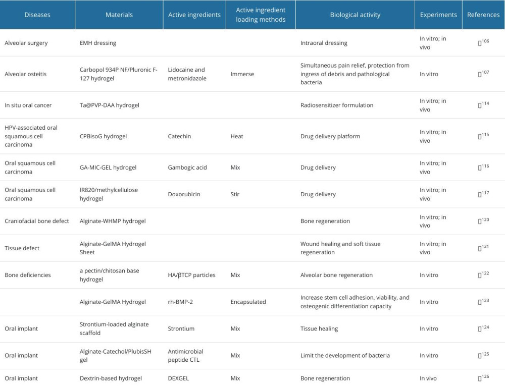 Table 4. Hydrogels in other oral diseases.Table 1. Hydrogels in periodontics.Table 2. Hydrogels in endodontics.Table 3. Hydrogel in oral mucosal disease.Table 4. Hydrogels in other oral diseases.
 In Press
Clinical Research
Association Between Total Cholesterol–to–High-Density Lipoprotein Ratio and Gestational Hypertension: A Cas...Med Sci Monit In Press; DOI: 10.12659/MSM.952395
Review article
Clinical Use of Endotracheal Intubation Without Neuromuscular Blockade: The Current Stage of KnowledgeMed Sci Monit In Press; DOI: 10.12659/MSM.951765
Clinical Research
Effect of Dexmedetomidine Hydrochloride Nasal Spray on Anxiety and Sleep in Patients Undergoing Gynecologic...Med Sci Monit In Press; DOI: 10.12659/MSM.952465
Clinical Research
Prognostic Value of Mortality Scoring Systems in Patients With Severe Burns: Identifying Key Predictors of ...Med Sci Monit In Press; DOI: 10.12659/MSM.951713
Most Viewed Current Articles
17 Jan 2024 : Review article 14,176,173
Vaccination Guidelines for Pregnant Women: Addressing COVID-19 and the Omicron VariantDOI :10.12659/MSM.942799
Med Sci Monit 2024; 30:e942799
13 Nov 2021 : Clinical Research 3,757,769
Acceptance of COVID-19 Vaccination and Its Associated Factors Among Cancer Patients Attending the Oncology ...DOI :10.12659/MSM.932788
Med Sci Monit 2021; 27:e932788
14 Dec 2022 : Clinical Research 2,466,143
Prevalence and Variability of Allergen-Specific Immunoglobulin E in Patients with Elevated Tryptase LevelsDOI :10.12659/MSM.937990
Med Sci Monit 2022; 28:e937990
16 May 2023 : Clinical Research 708,797
Electrophysiological Testing for an Auditory Processing Disorder and Reading Performance in 54 School Stude...DOI :10.12659/MSM.940387
Med Sci Monit 2023; 29:e940387